Advertisement

Main menu

  • Home
  • Content
    • Current Issue
    • Accepted Manuscripts
    • Article Preview
    • Past Issue Archive
    • Video Articles
    • AJNR Case Collection
    • Case of the Week Archive
    • Case of the Month Archive
    • Classic Case Archive
  • Special Collections
    • AJNR Awards
    • ASNR Foundation Special Collection
    • Most Impactful AJNR Articles
    • Photon-Counting CT
    • Spinal CSF Leak Articles (Jan 2020-June 2024)
  • Multimedia
    • AJNR Podcasts
    • AJNR SCANtastic
    • Trainee Corner
    • MRI Safety Corner
    • Imaging Protocols
  • For Authors
    • Submit a Manuscript
    • Submit a Video Article
    • Submit an eLetter to the Editor/Response
    • Manuscript Submission Guidelines
    • Statistical Tips
    • Fast Publishing of Accepted Manuscripts
    • Graphical Abstract Preparation
    • Imaging Protocol Submission
    • Author Policies
  • About Us
    • About AJNR
    • Editorial Board
    • Editorial Board Alumni
  • More
    • Become a Reviewer/Academy of Reviewers
    • Subscribers
    • Permissions
    • Alerts
    • Feedback
    • Advertisers
    • ASNR Home

User menu

  • Alerts
  • Log in

Search

  • Advanced search
American Journal of Neuroradiology

American Journal of Neuroradiology

ASHNR American Society of Functional Neuroradiology ASHNR American Society of Pediatric Neuroradiology ASSR
  • Alerts
  • Log in

Advanced Search

  • Home
  • Content
    • Current Issue
    • Accepted Manuscripts
    • Article Preview
    • Past Issue Archive
    • Video Articles
    • AJNR Case Collection
    • Case of the Week Archive
    • Case of the Month Archive
    • Classic Case Archive
  • Special Collections
    • AJNR Awards
    • ASNR Foundation Special Collection
    • Most Impactful AJNR Articles
    • Photon-Counting CT
    • Spinal CSF Leak Articles (Jan 2020-June 2024)
  • Multimedia
    • AJNR Podcasts
    • AJNR SCANtastic
    • Trainee Corner
    • MRI Safety Corner
    • Imaging Protocols
  • For Authors
    • Submit a Manuscript
    • Submit a Video Article
    • Submit an eLetter to the Editor/Response
    • Manuscript Submission Guidelines
    • Statistical Tips
    • Fast Publishing of Accepted Manuscripts
    • Graphical Abstract Preparation
    • Imaging Protocol Submission
    • Author Policies
  • About Us
    • About AJNR
    • Editorial Board
    • Editorial Board Alumni
  • More
    • Become a Reviewer/Academy of Reviewers
    • Subscribers
    • Permissions
    • Alerts
    • Feedback
    • Advertisers
    • ASNR Home
  • Follow AJNR on Twitter
  • Visit AJNR on Facebook
  • Follow AJNR on Instagram
  • Join AJNR on LinkedIn
  • RSS Feeds

AJNR is seeking candidates for the AJNR Podcast Editor. Read the position description.

Research ArticleHEAD AND NECK

Synovial Chondromatosis in the Temporomandibular Joint Complicated by Displacement and Calcification of the Articular Disk: Report of Two Cases

Jun-ichi Koyama, Jusuke Ito, Takafumi Hayashi and Fukiko Kobayashi
American Journal of Neuroradiology June 2001, 22 (6) 1203-1206;
Jun-ichi Koyama
  • Find this author on Google Scholar
  • Find this author on PubMed
  • Search for this author on this site
Jusuke Ito
  • Find this author on Google Scholar
  • Find this author on PubMed
  • Search for this author on this site
Takafumi Hayashi
  • Find this author on Google Scholar
  • Find this author on PubMed
  • Search for this author on this site
Fukiko Kobayashi
  • Find this author on Google Scholar
  • Find this author on PubMed
  • Search for this author on this site
  • Article
  • Figures & Data
  • Info & Metrics
  • Responses
  • References
  • PDF
Loading

Abstract

Summary: Two cases of synovial chondromatosis of the temporomandibular joint (TMJ) are presented, including correlation of CT and MR imaging characteristics with surgical and pathologic findings. The usefulness of CT and MR imaging in the diagnosis of TMJ disorders is discussed.

Synovial chondromatosis is a benign tumorlike disorder of the joint characterized by chondrometaplasia of the synovial membrane, in which cartilaginous nodules form and may become pedunculated and/or detach from the synovial membrane, becoming loose bodies within the joint space. These may also calcify. The chief clinical features of this disorder in the temporomandibular joint (TMJ) are preauricular swelling and pain, and restricted movement. Synovial chondromatosis of the TMJ, first described by Auhausen in 1933, is rare, with only about 40 cases reported in the literature. We present two cases of this disorder with correlation of the imaging and pathologic findings.

Case Reports

Case 1

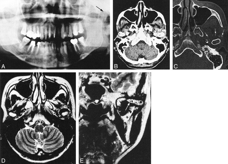
A 47-year-old woman was referred to our institution for deviation of the mandible to the right, pain in the left TMJ on opening the mouth, and left preauricular tenderness. Physical examination revealed an asymmetric face with slight diffuse swelling of the left preauricular region, inability to open the mouth more than 14 mm because of pain, slight deviation of the mandibular midline, and open bite of the bilateral molar regions. The patient had no history of trauma or rheumatoid arthrosis. A panoramic radiograph and CT scans revealed a small calcification anterior to the neck of the mandible, bilateral anterior disk displacement, and a partially calcified and protruded disk on the left side (Fig 1A–E). Clinical and radiologic findings prompted a diagnosis of left temporomandibular arthritis due to fracture of the condylar osteophyte.

fig 1.
  • Download figure
  • Open in new tab
  • Download powerpoint
fig 1.

Case 1: 47-year-old woman with left TMJ swelling and pain.

A, Panoramic radiograph shows an ovoid calcified body (arrow) anterior to the left mandibular neck.

B, Axial CT scan with soft tissue display shows semilunar soft tissue masses (arrowheads), suggesting bilateral anterior disk displacement.

C, Axial CT scan with bone display at same level as B shows irregularly bordered calcified body in zonal area (arrowheads), thought to be partial calcification of the disk.

D and E, Axial (D) and coronal (E) CT scans show ovoid calcified body (arrow), about 2 mm in diameter, which was visible on the panoramic radiograph (A).

F, Histologic section of loose body shows cartilaginous tissue (A), transitional zone (B), bone tissue (C), and thin connective tissue (D) (hematoxylin-eosin, original magnification ×300).

G, Histologic section of the synovial membrane shows a partial transformation from the fibrous connective tissue to the cartilaginous tissue, or chondrometaplasia (hematoxylin-eosin, original magnification × 300).

Exploration of the left TMJ was performed under general anesthesia. When the joint capsule was opened, the articular disk was seen to be displaced anteriorly to the condylar head, and a comparatively large perforation of the retrodiskal pad was noticed. In one calcified body, verified by radiologic findings, a diskectomy was performed, which revealed white calcification anterior to the condylar neck. The perforated retrodiskal pad was removed. Histologic examination disclosed that the small calcification was a loose body (Fig 1F) and also revealed chondrometaplasia of the synovial membrane and a partially calcified disk (Fig 1G). These histopathologic findings were consistent with a diagnosis of synovial chondromatosis.

Case 2

A 47-year-old woman was referred to our institution for pain in the left TMJ region on opening the mouth and preauricular tenderness. Her face was symmetrical, with slight trismus. The mouth opening was 31 mm and the midline of the mandible was deviated slightly to the right. The patient had no history of trauma. While the panoramic radiograph (Fig 2A) revealed one small calcification posterosuperior to the mandibular condyle, CT scans (Fig 2B and C) disclosed many small calcifications circling the mandibular condyle anteriorly. MR images (Fig 2D and E) showed a zonal area containing many small calcifications encircling the expanded joint space. A series of clinical and radiologic findings supported a diagnosis of synovial chondromatosis of the left TMJ.

fig 2.
  • Download figure
  • Open in new tab
  • Download powerpoint
fig 2.

47-year-old woman with left TMJ pain.

A, Panoramic radiograph shows one small calcification (arrow) posterosuperior to the mandibular condyle.

B, Axial CT scan with soft tissue display shows many small calcifications (arrows) scattered throughout the left joint capsule.

C, Axial CT scan with bone display shows many small calcifications (arrows) anterior to the mandibular condyle.

D and E, Axial (D) and coronal (E) MR images show a zonal region (arrow) anterior to the mandibular head with many calcified bodies scattered inside.

Exploration of the left TMJ was performed under general anesthesia. A preauricular inverted “hockey stick” incision was selected. Immediately after opening the joint capsule, some transparent and viscous fluid issued from the upper joint space. Many calcifications of various size were floating while others were scattered throughout the upper space. All the calcifications were removed, along with the irregularly expanded synovial membrane, and a diskectomy was subsequently performed. Histologic examination revealed chondrometaplasia of the synovial membrane of the TMJ, and the calcifications proved to be a total of 95 loose bodies. The histologic findings confirmed the diagnosis of synovial chondromatosis of the left TMJ.

Discussion

Synovial chondromatosis usually occurs in large joints, such as the knee or shoulder. It is a benign, chronic, and progressive condition that does not seem to undergo spontaneous resolution. Synovial chondromatosis of other joints has been reported to occur twice as often in males as in females, with a mean age of onset in the fifth decade (1). Within the TMJ, it occurs more often in females by a ratio of 4:1, and is usually located on the right side (right-to-left ratio of 4:1) (2). Synovial chondromatosis is a rare pathologic condition, particularly in the TMJ. Calcification of the surrounding soft tissue, such as the ligament or tendon, and calcified deposits of the meniscus have been reported in several cases (3). To our knowledge, our case 1 is the first reported example of synovial chondromatosis accompanied by a partially calcified, anteriorly displaced disk of the TMJ. Few cases of synovial chondromatosis have been reported in which there were as many as 95 loose bodies, as seen in our case 2 (4).

Histologically, synovial chondromatosis may be divided into three stages of development: 1) metaplasia found in the synovial membrane without the presence of detached particles, 2) metaplasia found in the synovial membrane with the presence of detached particles, and 3) only detached particles, which may vary in size from less than 1 mm to greater than 10 mm (5). Our cases are representative of the second developmental stage. A definitive diagnosis may be made after histologic examination of the particles or the synovial membrane.

Noyek and coworkers (1) pointed out radiologic features of synovial chondromatosis in the TMJ, namely 1) widening of the joint space, 2) limitation of motion, 3) irregularity of the joint surface, 4) presence of calcified loose bodies (cartilage), and 5) sclerosis or hyperostosis (overgrowth) of the glenoid fossa and mandibular condyle. These radiologic features, however, are also commonly seen in osteoarthrosis involving the TMJ, except for the presence of calcified loose bodies. On the other hand, other causes of loose bodies within a joint include osteochondritis dissecans, intracapsular fracture, avascular necrosis, osteoarthritis, tuberculous or pyogenic arthritis, rheumatoid arthritis, and neurotrophic arthritis (6, 7), with osteochondritis dissecans being the most common cause (7). Therefore, when these radiologic features are not observed in their entirety, there may be some difficulty in establishing an accurate imaging diagnosis of synovial chondromatosis. In both our cases, calcified loose bodies were seen on conventional radiographs, although this technique is not particularly sensitive to the presence of calcified loose bodies, since they have been detected in less than 45% of reported cases.

Recent reports have suggested that CT, MR imaging, and arthroscopy facilitate the clinical diagnosis of synovial chondromatosis (5, 8–12); however, to establish the radiologic diagnosis of this disease it is mandatory to verify pathologically a transitional change from fibrous connective tissue to cartilaginous tissue, or chondrometaplasia (13). Chondrometaplasia was confirmed histologically in both our cases. It is sometimes difficult to differentiate calcified loose bodies from a partially calcified anteriorly displaced disk, but in our case 1, loose bodies were clearly differentiated with bone display on CT scans; and, in case 2, MR images clearly showed calcified loose bodies embedded in a soft tissue mass as small dots of signal void.

Synovial chondromatosis is a local, nonneoplastic, self-limiting process. Removal of the loose bodies and of the involved synovial membrane is sufficient to control the disease in the majority of cases. Multiple and often short-interval recurrences should alert the physician to the possibility of synovial chondrosarcoma (14).

Conclusion

We have described two cases of synovial chondromatosis, one with anterior displacement and calcification of the left articular disk and the other with numerous loose bodies in the joint space. CT and MR imaging characteristics were correlated with surgical and histopathologic findings.

Footnotes

  • 1 Presented in part at the annual meeting of the Japan Society for Oral and Maxillofacial Radiology, Kitanihon Section, July 1995.

References

  1. ↵
    Noyek AM, Holgate RC, Fireman SM, Rosen P, Pritzker KPH. The radiological findings in synovial chondromatosis (chondrometaplasia) of the temporomandibular joint. J Otolaryngol 1977;6:45-48
  2. ↵
    Fee WE Jr, Windhorst P, Wiggins R, Pang L. Synovial chondromatosis of the temporomandibular joint. Otolaryngol Head Neck Surg 1979;87:741-748
    PubMed
  3. ↵
    Ogilvie-Harris DJ, Saleh K. Generalized synovial chondromatosis of the knee: a comparison of removal of the loose bodies alone with arthroscopic synovectomy. Arthroscopy 1994;10:166-170
    PubMed
  4. ↵
    van Ingen JM, de Man K, Bakri I. CT diagnosis of synovial chondromatosis of the temporomandibular joint. Br J Oral Maxillofac Surg 1990;28:164-167
    CrossRefPubMed
  5. ↵
    Doran EA, Vogler JB, Angelillo JC. Synovial chondromatosis of the temporo-mandibular joint diagnosed by magnetic resonance imaging. J Oral Maxillofac Surg 1989;47:411-413
    PubMed
  6. ↵
    Blenkinsopp PT. Loose bodies of the temporomandibular joint, synovial chondromatosis or osteoarthritis. Br J Oral Surg 1978;16:12-20
    CrossRefPubMed
  7. ↵
    Olley SF, Leopard PJ. Osteochondritis dissecans affecting the temporomandibular joint. Br J Oral Surg 1978;16:21-25
    CrossRefPubMed
  8. Munk PL, Helms CA. Temporomandibular joint synovial chondromatosis: CT manifestations. J Can Assoc Radiol 1989;40:274-276
  9. Herzog S, Mafee M. Synovial chondromatosis of the TMJ: MR and CT findings. AJNR Am J Neuroradiol 1990;11:742-745
    FREE Full Text
  10. Nitzan DW, Marmary Y, et al. The diagnostic value of computed tomography in temporomandibular joint synovial chondromatosis. Comput Med Imaging Graph 1991;15:53-56
    PubMed
  11. Mendonca-Caridad JJ, Schwartz HC. Synovial chondromatosis of the temporomandibular joint: arthroscopic diagnosis and treatment of a case. J Oral Maxillofac Surg 1994;52:624-625
    PubMed
  12. Wise DP, Ruskin JD. Arthroscopic diagnosis and treatment of temporomandibular joint synovial chondromatosis: report of a case. J Oral Maxillofac Surg 1994;52:90-93
    PubMed
  13. ↵
    Jeffreys TE. Synovial chondromatosis. J Bone Joint Surg Br 1967;49:530-534
  14. ↵
    Hamilton A, Davis RI, Hayes D, Mollan RAB. Chondrosarcoma developing in synovial chondromatosis. J Bone Joint Surg Br 1987;69:137-140
  • Received September 26, 2000.
  • Copyright © American Society of Neuroradiology
View Abstract
PreviousNext
Back to top

In this issue

American Journal of Neuroradiology: 22 (6)
American Journal of Neuroradiology
Vol. 22, Issue 6
1 Jun 2001
  • Table of Contents
  • Index by author
Advertisement
Print
Download PDF
Email Article

Thank you for your interest in spreading the word on American Journal of Neuroradiology.

NOTE: We only request your email address so that the person you are recommending the page to knows that you wanted them to see it, and that it is not junk mail. We do not capture any email address.

Enter multiple addresses on separate lines or separate them with commas.
Synovial Chondromatosis in the Temporomandibular Joint Complicated by Displacement and Calcification of the Articular Disk: Report of Two Cases
(Your Name) has sent you a message from American Journal of Neuroradiology
(Your Name) thought you would like to see the American Journal of Neuroradiology web site.
CAPTCHA
This question is for testing whether or not you are a human visitor and to prevent automated spam submissions.
Cite this article
Jun-ichi Koyama, Jusuke Ito, Takafumi Hayashi, Fukiko Kobayashi
Synovial Chondromatosis in the Temporomandibular Joint Complicated by Displacement and Calcification of the Articular Disk: Report of Two Cases
American Journal of Neuroradiology Jun 2001, 22 (6) 1203-1206;

Citation Manager Formats

  • BibTeX
  • Bookends
  • EasyBib
  • EndNote (tagged)
  • EndNote 8 (xml)
  • Medlars
  • Mendeley
  • Papers
  • RefWorks Tagged
  • Ref Manager
  • RIS
  • Zotero
0 Responses
Respond to this article
Share
Bookmark this article
Synovial Chondromatosis in the Temporomandibular Joint Complicated by Displacement and Calcification of the Articular Disk: Report of Two Cases
Jun-ichi Koyama, Jusuke Ito, Takafumi Hayashi, Fukiko Kobayashi
American Journal of Neuroradiology Jun 2001, 22 (6) 1203-1206;
del.icio.us logo Twitter logo Facebook logo Mendeley logo
  • Tweet Widget
  • Facebook Like
  • Google Plus One

Jump to section

  • Article
    • Abstract
    • Case Reports
    • Discussion
    • Conclusion
    • Footnotes
    • References
  • Figures & Data
  • Info & Metrics
  • Responses
  • References
  • PDF

Related Articles

  • No related articles found.
  • PubMed
  • Google Scholar

Cited By...

  • Synovial chondromatosis of the temporomandibular joint: MRI findings with pathological comparison
  • Synovial chondromatosis in the temporomandibular joint: a case with typical imaging features and pathological findings
  • Ultrasonographic and arthrographic diagnoses of synovial chondromatosis
  • Synovial chondromatosis of the temporomandibular joint: CT and MRI findings
  • Crossref
  • Google Scholar

This article has not yet been cited by articles in journals that are participating in Crossref Cited-by Linking.

More in this TOC Section

  • Correlation of Apparent Diffusion Coefficient at 3T with Prognostic Parameters of Retinoblastoma
  • Parathyroid Lesions: Characterization with Dual-Phase Arterial and Venous Enhanced CT of the Neck
  • Kimura Disease: CT and MR Imaging Findings
Show more HEAD AND NECK

Similar Articles

Advertisement

Indexed Content

  • Current Issue
  • Accepted Manuscripts
  • Article Preview
  • Past Issues
  • Editorials
  • Editor's Choice
  • Fellows' Journal Club
  • Letters to the Editor
  • Video Articles

Cases

  • Case Collection
  • Archive - Case of the Week
  • Archive - Case of the Month
  • Archive - Classic Case

Special Collections

  • AJNR Awards
  • ASNR Foundation Special Collection
  • Most Impactful AJNR Articles
  • Photon-Counting CT
  • Spinal CSF Leak Articles (Jan 2020-June 2024)

More from AJNR

  • Trainee Corner
  • Imaging Protocols
  • MRI Safety Corner

Multimedia

  • AJNR Podcasts
  • AJNR Scantastics

Resources

  • Turnaround Time
  • Submit a Manuscript
  • Submit a Video Article
  • Submit an eLetter to the Editor/Response
  • Manuscript Submission Guidelines
  • Statistical Tips
  • Fast Publishing of Accepted Manuscripts
  • Graphical Abstract Preparation
  • Imaging Protocol Submission
  • Evidence-Based Medicine Level Guide
  • Publishing Checklists
  • Author Policies
  • Become a Reviewer/Academy of Reviewers
  • News and Updates

About Us

  • About AJNR
  • Editorial Board
  • Editorial Board Alumni
  • Alerts
  • Permissions
  • Not an AJNR Subscriber? Join Now
  • Advertise with Us
  • Librarian Resources
  • Feedback
  • Terms and Conditions
  • AJNR Editorial Board Alumni

American Society of Neuroradiology

  • Not an ASNR Member? Join Now

© 2025 by the American Society of Neuroradiology All rights, including for text and data mining, AI training, and similar technologies, are reserved.
Print ISSN: 0195-6108 Online ISSN: 1936-959X

Powered by HighWire