Advertisement

Main menu

  • Home
  • Content
    • Current Issue
    • Accepted Manuscripts
    • Article Preview
    • Past Issue Archive
    • Video Articles
    • AJNR Case Collection
    • Case of the Week Archive
    • Case of the Month Archive
    • Classic Case Archive
  • Special Collections
    • AJNR Awards
    • ASNR Foundation Special Collection
    • Most Impactful AJNR Articles
    • Photon-Counting CT
    • Spinal CSF Leak Articles (Jan 2020-June 2024)
  • Multimedia
    • AJNR Podcasts
    • AJNR SCANtastic
    • Trainee Corner
    • MRI Safety Corner
    • Imaging Protocols
  • For Authors
    • Submit a Manuscript
    • Submit a Video Article
    • Submit an eLetter to the Editor/Response
    • Manuscript Submission Guidelines
    • Statistical Tips
    • Fast Publishing of Accepted Manuscripts
    • Graphical Abstract Preparation
    • Imaging Protocol Submission
    • Author Policies
  • About Us
    • About AJNR
    • Editorial Board
    • Editorial Board Alumni
  • More
    • Become a Reviewer/Academy of Reviewers
    • Subscribers
    • Permissions
    • Alerts
    • Feedback
    • Advertisers
    • ASNR Home

User menu

  • Alerts
  • Log in

Search

  • Advanced search
American Journal of Neuroradiology

American Journal of Neuroradiology

ASHNR American Society of Functional Neuroradiology ASHNR American Society of Pediatric Neuroradiology ASSR
  • Alerts
  • Log in

Advanced Search

  • Home
  • Content
    • Current Issue
    • Accepted Manuscripts
    • Article Preview
    • Past Issue Archive
    • Video Articles
    • AJNR Case Collection
    • Case of the Week Archive
    • Case of the Month Archive
    • Classic Case Archive
  • Special Collections
    • AJNR Awards
    • ASNR Foundation Special Collection
    • Most Impactful AJNR Articles
    • Photon-Counting CT
    • Spinal CSF Leak Articles (Jan 2020-June 2024)
  • Multimedia
    • AJNR Podcasts
    • AJNR SCANtastic
    • Trainee Corner
    • MRI Safety Corner
    • Imaging Protocols
  • For Authors
    • Submit a Manuscript
    • Submit a Video Article
    • Submit an eLetter to the Editor/Response
    • Manuscript Submission Guidelines
    • Statistical Tips
    • Fast Publishing of Accepted Manuscripts
    • Graphical Abstract Preparation
    • Imaging Protocol Submission
    • Author Policies
  • About Us
    • About AJNR
    • Editorial Board
    • Editorial Board Alumni
  • More
    • Become a Reviewer/Academy of Reviewers
    • Subscribers
    • Permissions
    • Alerts
    • Feedback
    • Advertisers
    • ASNR Home
  • Follow AJNR on Twitter
  • Visit AJNR on Facebook
  • Follow AJNR on Instagram
  • Join AJNR on LinkedIn
  • RSS Feeds

AJNR is seeking candidates for the AJNR Podcast Editor. Read the position description.

Research ArticlePediatrics

CT and MRI of Pediatric Skull Lesions with Fluid-Fluid Levels

S.A. Nabavizadeh, L.T. Bilaniuk, T. Feygin, K.V. Shekdar, R.A. Zimmerman and A. Vossough
American Journal of Neuroradiology March 2014, 35 (3) 604-608; DOI: https://doi.org/10.3174/ajnr.A3712
S.A. Nabavizadeh
aFrom the Department of Radiology, The Children's Hospital of Philadelphia, University of Pennsylvania, Philadelphia, Pennsylvania.
  • Find this author on Google Scholar
  • Find this author on PubMed
  • Search for this author on this site
L.T. Bilaniuk
aFrom the Department of Radiology, The Children's Hospital of Philadelphia, University of Pennsylvania, Philadelphia, Pennsylvania.
  • Find this author on Google Scholar
  • Find this author on PubMed
  • Search for this author on this site
T. Feygin
aFrom the Department of Radiology, The Children's Hospital of Philadelphia, University of Pennsylvania, Philadelphia, Pennsylvania.
  • Find this author on Google Scholar
  • Find this author on PubMed
  • Search for this author on this site
K.V. Shekdar
aFrom the Department of Radiology, The Children's Hospital of Philadelphia, University of Pennsylvania, Philadelphia, Pennsylvania.
  • Find this author on Google Scholar
  • Find this author on PubMed
  • Search for this author on this site
R.A. Zimmerman
aFrom the Department of Radiology, The Children's Hospital of Philadelphia, University of Pennsylvania, Philadelphia, Pennsylvania.
  • Find this author on Google Scholar
  • Find this author on PubMed
  • Search for this author on this site
A. Vossough
aFrom the Department of Radiology, The Children's Hospital of Philadelphia, University of Pennsylvania, Philadelphia, Pennsylvania.
  • Find this author on Google Scholar
  • Find this author on PubMed
  • Search for this author on this site
  • Article
  • Figures & Data
  • Info & Metrics
  • Responses
  • References
  • PDF
Loading

Abstract

SUMMARY: Fluid-fluid levels can occur whenever different fluid densities are contained within a cystic or compartmentalized lesion, usually related to the evolution of hematoma or necrosis. Review of the literature demonstrated that throughout the skeletal system, the most common etiology for fluid-fluid levels is aneurysmal bone cyst, but there are no dedicated studies of the pediatric calvaria, to our knowledge. In this report, we present clinicopathologic characteristics and CT and MR imaging of 11 patients with pediatric skull mass lesions demonstrating fluid-fluid levels. MR imaging demonstrated more fluid-fluid levels compared with CT in all cases. The etiologies of skull lesions with fluid-fluid levels were Langerhans cell histiocytosis in 4 (36.6%), aneurysmal bone cysts in 3 (27.2%), cephalohematoma in 3 (27.2%), and metastatic neuroblastoma in 1 (9%). Radiologists should be aware of the other etiologies of calvarial lesions with fluid-fluid levels in the pediatric skull.

ABBREVIATIONS:

ABC
aneurysmal bone cyst
FFL
fluid-fluid level
LCH
Langerhans cell histiocytosis

Fluid-fluid levels (FFLs) may occur whenever different fluid densities are contained within a cystic or compartmentalized structure, usually related to hemorrhage or necrosis.1,2 Image acquisition near perpendicular to the plane of the FFL is required to optimally demonstrate it.1,2 FFLs in skeletal lesions have been a well-known imaging feature of aneurysmal bone cyst (ABC)3,4 for many decades. To our knowledge, there is no dedicated study focusing on the different etiologies of FFLs in the pediatric population and there is no study focused on fluid-fluid levels in calvarial lesions. However, 2 large studies primarily in adult patients have demonstrated that ABCs are the most common etiology of osseous lesions with FFLs throughout the body.5,6 Despite these data, the presence of FFLs remains a nonspecific finding and can be seen in a wide range of benign and malignant bone lesions, including fibrous dysplasia, telangiectatic osteosarcoma, osteoblastoma, simple bone cyst, lymphatic malformations, Ewing sarcoma, brown tumor, giant cell tumor, and chondroblastoma.5⇓⇓⇓–9

In this study, head CT and MR imaging of pediatric patients having skull lesions with FFLs were retrospectively reviewed with the aim of characterizing these lesions.

Case Series

Patients

The study was performed with approval of the institutional review board and was compliant with guidelines of the Health Insurance Portability and Accountability Act. We performed a retrospective review of records of pediatric patients with skull masses diagnosed between 2002 and 2012. Cases were ascertained from teaching files of the authors and a computerized search of the PACS for patients younger than 18 years of age who were diagnosed with a skull lesion showing fluid-fluid levels on CT or MR imaging. The search terms included various combinations and derivatives of “fluid-fluid,” “blood-fluid,” “fluid levels,” “skull,” and “calvarium.” Medical records were reviewed for patient age, sex, history of recent trauma to the lesion, history of coagulopathy, types of treatment, and histopathologic diagnosis. Patients with a history of surgery on the skull were excluded. The etiologic diagnosis was made by histopathologic examination of resected lesions in 6 patients and of biopsied specimens in 1 patient. In 3 patients who were classified as having cephalohematoma, the diagnosis was made on the basis of characteristic clinical presentation, history of traumatic vaginal delivery, and/or spontaneous resolution of the lesion by consensus of pediatric neuroradiologists and by review of medical records. Finally, 1 patient with primary neuroblastoma was diagnosed as having metastatic involvement of the skull on the basis of typical imaging findings of neuroblastoma both on CT and MR imaging and considerable improvement of the lesion after targeted chemotherapy.

CT and MR Imaging

CT was performed on various multidetector CT scanners (Siemens, Erlangen, Germany), and MR imaging was performed on 1.5T or 3T magnets (Siemens) with standard clinical protocols used at the time of the study. Axial CT sections were reconstructed at 3- or 5-mm thicknesses. MR imaging sequences varied among patients but generally included pre- and postcontrast T1-weighted, T2-weighted, fluid-attenuated inversion recovery, echo-planar spin-echo diffusion-weighted imaging, and either T2* gradient-echo susceptibility or susceptibility-weighted imaging.

The CT and MR imaging studies of these patients were reviewed by 2 pediatric neuroradiologists by consensus, and the findings were tabulated. For each scan, lesion location, number of fluid-fluid levels, and the presence or absence of soft-tissue components were evaluated. Diagnosis of hemorrhage was based on hyperattenuation on noncontrast CT or typical MR imaging findings (mainly hyperintensity on T1-weighted, hypointensity on T2-weighted, and/or susceptibility on gradient-echo or susceptibility-weighted images).

Clinicopathologic and Imaging Findings

Eleven patients (6 male, 5 female) with skull lesions showing fluid-fluid levels met the inclusion and exclusion criteria (Table). The mean age of patients at the time of primary diagnosis was 6.2 ± 6.1 years (range, 8 days to 16 years). The etiologies of the lesions included Langerhans cell histiocytosis (LCH) (n = 4 [36.6%]), aneurysmal bone cyst (n = 3 [27.2%]), cephalohematoma (n = 3 [27.2%]), and neuroblastoma (n = 1 [9%]).

View this table:
  • View inline
  • View popup

Demographic, clinical, imaging, and histopathologic findings of pediatric patients with FFLs in skull lesions

In 7 patients, both CT (4 noncontrast, 3 contrast-enhanced) and MR imaging with and without contrast were performed. MR imaging with and without contrast was the only imaging technique in another 4 patients. In the 7 patients in whom a CT had been performed, FFLs were identified in 6 patients (85%). The mean number of fluid-fluid levels detected on CT was 3.5 ± 3.2 (range, 1–9). FFLs were present on MR imaging in all 11 patients (100%). The mean number of detected fluid-fluid levels on MR imaging was 9.2 ± 9.1 (range, 1–29). The number of observed FFLs was significantly higher on MR imaging compared with CT (Z = −2.36, Wilcoxon signed-rank test, P = .018).

Three of 4 patients with LCH demonstrated prominent soft-tissue components (75%, Fig 1). The 3 patients with LCH who had undergone CT showed sharply marginated bone destruction. None of the patients with LCH showed substantial cystic bone expansion. Two patients with LCH (patients 1 and 7) had a history of minor recent trauma to the lesion before imaging, having hit their head against a fish tank and television remote, respectively. There was no evidence of an abnormal coagulation profile in any of the patients. In patients with ABCs, 2 had primary ABCs and 1 had ABC in the setting of fibrous dysplasia. All patients with ABCs showed bone expansion (Fig 2).

Fig 1.
  • Download figure
  • Open in new tab
  • Download powerpoint
Fig 1.

MR imaging in a patient with Langerhans cell histiocytosis. Axial T1- (A) and T2-weighted (B) images reveal multiple FFLs. An enhancing solid soft-tissue component is seen medially on postcontrast axial (C) and coronal (D) T1-weighted images.

Fig 2.
  • Download figure
  • Open in new tab
  • Download powerpoint
Fig 2.

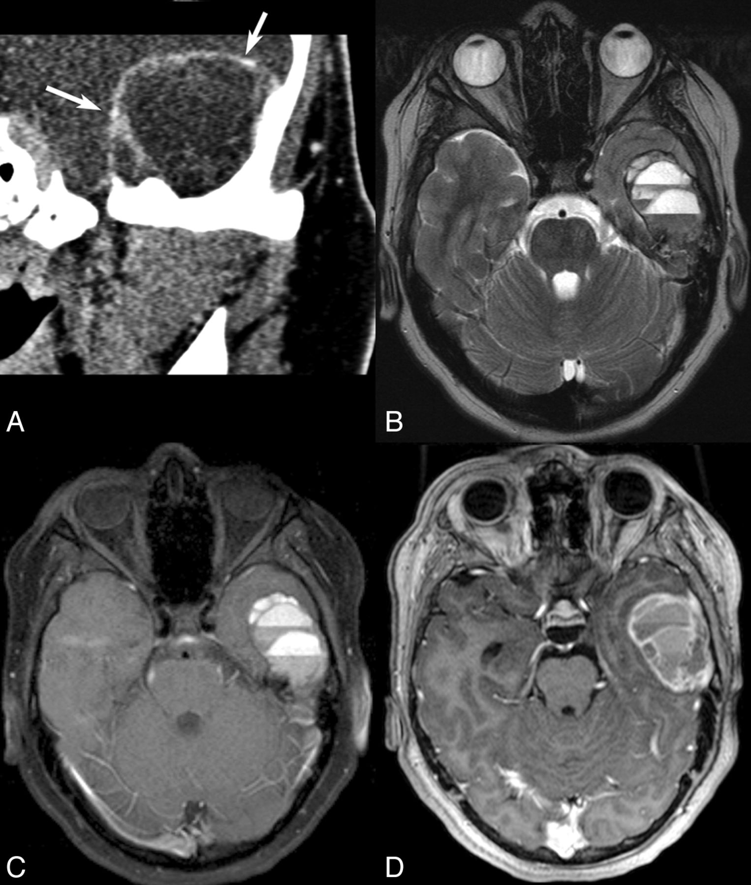
CT and MR imaging findings in a patient with an aneurysmal bone cyst. Coronal CT scan (A) reveals marked bone expansion by a well-defined cystic-appearing mass projecting into the left temporal fossa with very thin remnants of the bony cortex seen in the wall (arrows). T2-weighted (B) and precontrast T1-weighted (C) images reveal multiple FFLs. Peripheral and septal enhancement is noted on CT (A) and postcontrast T1-weighted images (D), but no large enhancing solid soft-tissue component is identified.

All 3 patients with ABCs underwent surgical resection. Among the patients with LCH, 3 underwent surgical resection and 1 (patient 9) underwent chemotherapy after biopsy. Most interesting, follow-up imaging of this latter patient 3.5 months after completion of chemotherapy revealed a considerable increase in the number of FFLs, from 14 to >50 (Fig 3). The 1 patient with metastatic neuroblastoma also underwent treatment with chemotherapy, with a substantial decrease in the size of the lesion (Fig 4). None of the patients with cephalohematoma had a detectable solid or enhancing soft-tissue component at the time of the study, and no frank bone destruction was seen on CT or MR imaging (Fig 5). All patients with cephalohematoma were managed conservatively.

Fig 3.
  • Download figure
  • Open in new tab
  • Download powerpoint
Fig 3.

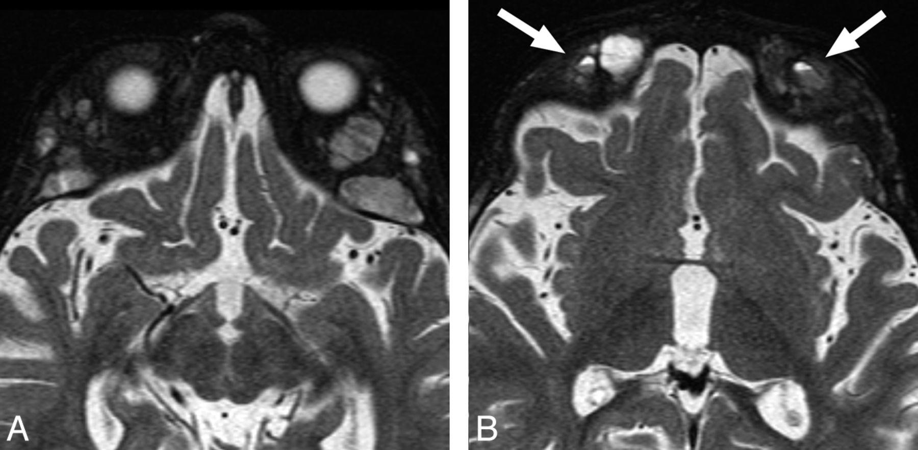
CT and MR imaging findings in a patient with Langerhans cell histiocytosis. Noncontrast CT (A) reveals a lytic lesion with a prominent soft-tissue component and well-defined bone destruction. An FFL is faintly seen (arrow). T2-weighted image (B) and postcontrast T1-weighted image (C) show a few FFLs and enhancement of soft-tissue components, respectively. Axial T2-weighted image (D) following chemotherapy now reveals an increase in the number of FFLs.

Fig 4.
  • Download figure
  • Open in new tab
  • Download powerpoint
Fig 4.

MR imaging findings in a patient with neuroblastoma demonstrate typical bone marrow infiltration and expansion by metastases (A). Multiple small FFLs are seen in the lesions (B).

Fig 5.
  • Download figure
  • Open in new tab
  • Download powerpoint
Fig 5.

MR imaging of a 3-month-old patient found to have a bump on the head on physical examination. T2-weighted axial image shows an FFL within a cephalohematoma (arrow), confirmed to be within the bone on multiple planes and sequences (not shown).

Discussion

Generally, a bone lesion with FFLs is considered more likely to represent an ABC; therefore, it was surprising to us that our review of pediatric skull lesions revealed that LCH was the most common etiology of skull lesions with FFLs, followed by ABC. There is no dedicated study on the most common etiology of osseous lesions with FFLs in pediatric patients, to our knowledge. In addition, large studies that may have included some pediatric patients had not published their detailed results based on age groups,5⇓–7 and the current literature is mainly limited to published case reports.9⇓⇓⇓⇓–14

There are only 2 case reports describing the presence of fluid-fluid level in LCH.12,14 In both of these cases, the FFL was demonstrated on both CT and MR imaging. The FFL in LCH represents hemorrhage, which may be triggered by trauma or may be spontaneous hemorrhage. Albadr et al12 reported sudden spontaneous scalp swelling in a patient with LCH of the skull, which was then proved to be secondary to hemorrhage. Chen et al15 reported a patient with LCH of the skull complicated by a spontaneous epidural hematoma. Another potential hypothesis for hemorrhage in LCH is development of aneurysmal bone cyst features, which have been reported to be observed on histology in LCH.16,17 In our patients with LCH, 2 had a history of recent minor trauma, but the other 2 did not. Most interesting, the number of FFLs dramatically increased in 1 patient following chemotherapy (Fig 3), though the overall size of the lesions did decrease. Chemotherapy was used in this patient because there was orbital osseous involvement.18 No evidence of secondary ABC formation was demonstrated on histology in any of the 4 patients with LCH in this study.

We believe that several factors may contribute to the relatively high prevalence of LCH with FFL in this study. First, LCH is relatively more common in the pediatric population compared with adults, and the skull is the most frequently involved site in LCH.19 On the other hand, while ABCs can also occur in young patients, they are most often found in the metaphyses of long bones and rarely affect the skull.3,4,13 Despite the presence of FFLs, we believe that LCH may still potentially be differentiated from ABC on the basis of the presence of characteristic imaging findings such as sharp, nonsclerotic borders with a punched out appearance, lack of demonstrable bone expansion, and uneven destruction of the outer and inner cranial tables with a beveled edge contour on CT, though this may not be possible in all cases. The distinction may be more difficult on MR imaging. Another differentiating feature of LCH can be the presence of a prominent soft-tissue component, which was demonstrated in most of the patients in this study, though occasionally an obvious soft-tissue component may not be present.

ABCs have been reported in many locations, including the orbit, occipital bone, frontal bone, temporal bone, parietal bone, and skull base.20⇓–22 In this study, we found 2 primary ABCs and a secondary ABC in the setting of fibrous dysplasia. The coexistence of an aneurysmal bone cyst and a companion lesion is well-known and is consistent with the concept of local hemodynamic changes secondary to precursor pathology. Trauma, benign lesions of the skeleton (eg, chondroblastoma, chondromyxoid fibroma, osteoblastoma, giant cell tumor, fibrous dysplasia), and even some malignant tumors (eg, osteosarcoma, chondrosarcoma, hemangioendothelioma) have been associated with development of aneurysmal bone cysts.10,17,23 In the setting of fibrous dysplasia, secondary ABC formation can occur in addition to the more common evolving fibrous dysplasia with lytic areas or cystic degeneration.24

Cephalohematoma is a subperiosteal hemorrhage that results from tearing of delicate vessels that traverse through the bone into the scalp. It has been reported in 0.2%–3% of all births,25⇓–27 though most are small and resolve spontaneously. Cephalohematomas are confined to suture borders where the periosteum is tightly adherent to the membranous tissue of the sutures.28 The most common location is over the parietal bone followed by the occipital bone.26

Multiple risk factors have been associated with the development of cephalohematoma, including birth trauma, fetal scalp monitors, instrumentation, greater birth weight, and nulliparity.25,29,30 Cephalohematoma is a benign condition, which usually does not require specific therapy. Most cephalohematomas resolve within a few weeks to months, depending on their size.29,31 Many patients with typical history and clinical examination for cephalohematoma will not be imaged. On the infrequent occasion when the hematoma fails to resorb, progressive subpericranial osteogenesis results in a calcified cephalohematoma. The incidence of calcification has been reported to occur in 3%–5% of all cephalohematomas.32 In these cases, there is usually a history of a mass present since birth that is initially boggy and fluctuant and then gradually hardens, typical of an ossifying cephalohematoma.31 In this series, we found 3 cases of cephalohematoma, one in a neonate, another in a young infant, and the other, a partially calcified cephalohematoma, in a 10-year-old patient. All of the lesions were in the parietal area, which is consistent with findings in the literature.26

Neuroblastoma is the most common extracranial solid tumor. Metastatic involvement of the head and neck is common and manifests most often as osseous metastases with periosteal reactions involving the calvaria, orbit, or skull base33,34 and is also commonly seen both at presentation and at the time of tumor recurrence. One of the patients in this study had metastatic neuroblastoma involving the sphenoid bone. We are not aware of any report in the English literature describing FFLs in metastatic neuroblastoma to the skull.

The main limitation of our study is the relatively small number of patients, though there has been no focused study of osseous lesions of the skull with FFL, particularly in pediatric patients. Future larger studies may shed more light on the various etiologies of these lesions.

Conclusions

Radiologists should be aware that besides ABCs, there are other lesions in the pediatric skull that can demonstrate FFLs. In our series, LCH was the most common etiology followed by ABC. Cephalohematoma should also be considered in the appropriate clinical setting. As would be expected, MR imaging was more sensitive in detecting FFLs within the lesions.

REFERENCES

  1. 1.↵
    1. Tsai J,
    2. Dalinka M,
    3. Fallon M,
    4. et al
    . Fluid-fluid level: a non-specific finding in tumors of bone and soft tissue. Radiology 1990;175:779–82
    PubMed
  2. 2.↵
    1. Davies AM,
    2. Cassar-Pullicino VN,
    3. Grimer RJ
    . The incidence and significance of fluid-fluid levels on computed tomography of osseous lesions. Br J Radiol 1992;65:193–98
    Abstract/FREE Full Text
  3. 3.↵
    1. Hudson T
    . Fluid levels in aneurysmal bone cysts: a CT feature. AJR Am J Roentgenol 1984;141:1001–04
  4. 4.↵
    1. Hertzanu Y,
    2. Mendelsohn D,
    3. Gottschalk F
    . Aneurysmal bone cyst of the calcaneus. Radiology 1984;151:51–52
    PubMed
  5. 5.↵
    1. Van Dyck P,
    2. Vanhoenacker FM,
    3. Vogel J,
    4. et al
    . Prevalence, extension and characteristics of fluid-fluid levels in bone and soft tissue tumors. Eur Radiol 2006;16:2644–51
    CrossRefPubMed
  6. 6.↵
    1. Alyas F,
    2. Saifuddin A
    . Fluid-fluid levels in bone neoplasms: variation of T1-weighted signal intensity of the superior to inferior layers—diagnostic significance on magnetic resonance imaging. Eur Radiol 2008;18:2642–51
    CrossRefPubMed
  7. 7.↵
    1. O'Donnell P,
    2. Saifuddin A
    . The prevalence and diagnostic significance of fluid-fluid levels in focal lesions of bone. Skeletal Radiol 2004;33:330–36
    CrossRefPubMed
  8. 8.↵
    1. Kobayashi Y,
    2. Murakami R,
    3. Toba M,
    4. et al
    . Chondroblastoma of the temporal bone. Skeletal Radiol 2001;30:714–18
    CrossRefPubMed
  9. 9.↵
    1. Sharma A,
    2. Garg A,
    3. Mishra NK,
    4. et al
    . Primary Ewing's sarcoma of the sphenoid bone with unusual imaging features: a case report. Clin Neurol Neurosurg 2005;107:528–31
    CrossRefPubMed
  10. 10.↵
    1. Haddad GF,
    2. Hambali F,
    3. Mufarrij A,
    4. et al
    . Concomitant fibrous dysplasia and aneurysmal bone cyst of the skull base: case report and review of the literature. Pediatr Neurosurg 1998;28:147–53
    CrossRefPubMed
  11. 11.↵
    1. Wang YC,
    2. Huang JS,
    3. Wu CJ,
    4. et al
    . A huge osteoblastoma with aneurysmal bone cyst in skull base. Clin Imaging 2001;25:247–50
    CrossRefPubMed
  12. 12.↵
    1. Albadr FB,
    2. Elgamal EA,
    3. Alorainy IA,
    4. et al
    . Fluid-fluid level in Langerhans cell histiocytosis of the skull. Neurosciences 2006;11:47–49
  13. 13.↵
    1. Sawin PD,
    2. Muhonen MG,
    3. Sato Y,
    4. et al
    . Aneurysmal bone cyst of the temporal bone presenting as hearing loss in a child. Int J Pediatr Otorhinolaryngol 1995;33:275–84
    CrossRefPubMed
  14. 14.↵
    1. Hindman BW,
    2. Thomas RD,
    3. Young LW,
    4. et al
    . Langerhans' cell histiocytosis: unusual skeletal manifestations observed in thirty four cases. Skeletal Radiol 1998;27:177–81
    CrossRefPubMed
  15. 15.↵
    1. Chen HC,
    2. Shen WC,
    3. Chou DY,
    4. et al
    . Langerhans cell histiocytosis of the skull complicated with an epidural hematoma. AJNR Am J Neuroradiol 2002;23:493–95
    Abstract/FREE Full Text
  16. 16.↵
    1. Mirra JM
    . Bone Tumors: Clinical, Radiologic and Pathologic Correlations. Philadelphia: Lea & Febiger; 1989:1269
  17. 17.↵
    1. Roncaroli F,
    2. Consales A,
    3. Galassi E,
    4. et al
    . Occipital aneurysmal bone cyst secondary to eosinophilic granuloma. Pediatr Neurosurg 2001;35:103–06
    CrossRefPubMed
  18. 18.↵
    Langerhans Cell Histiocytosis Treatment. National Cancer Institute. http://www.cancer.gov/cancertopics/pdq/treatment/lchistio/HealthProfessional/page7. Accessed June 15, 2013
  19. 19.↵
    1. Bollini G,
    2. Jouve JL,
    3. Gentet JC,
    4. et al
    . Bone lesions in histiocytosis X. J Pediatr Orthopaed 1991;11:469–77
    CrossRef
  20. 20.↵
    1. Calliauw L,
    2. Roels H,
    3. Caemaert J
    . Aneurysmal bone cysts in the cranial vault and base of skull. Surg Neurol 1985;23:193–98
    CrossRefPubMed
  21. 21.↵
    1. Lichtenstein L
    . Aneurysmal bone cyst: observations on fifty cases. Bone Joint Surg Am 1957;39-A:873–82
  22. 22.↵
    1. Tillman BP,
    2. Dahlin DC,
    3. Lipscomb PR,
    4. et al
    . Aneurysmal bone cyst: an analysis of ninety-five cases. Mayo Clin Proc 1968;43:478–95
    PubMed
  23. 23.↵
    1. Ito H,
    2. Kizu O,
    3. Yamada K,
    4. et al
    . Secondary aneurysmal bone cyst derived from a giant-cell tumour of the skull base. Neuroradiology 2003;45:616–17
    CrossRefPubMed
  24. 24.↵
    1. Faul S,
    2. Link J,
    3. Behrendt S,
    4. et al
    . MRI features of craniofacial fibrous dysplasia. Orbit 1998;17:125–32
    CrossRefPubMed
  25. 25.↵
    1. Tan KL
    . Cephalohaematoma. Aust N Z J Obstet Gynaecol 1970;10:101–06
    CrossRefPubMed
  26. 26.↵
    1. Ingram MD,
    2. Hamilton WM
    . Cephalhematoma in the newborn. Radiology 1950;55:502–07
    PubMed
  27. 27.↵
    1. LeBlanc CM,
    2. Allen UD,
    3. Ventureyra E
    . Cephalhematomas revisited: when should a diagnostic tap be performed? Clin Pediatr (Phila) 1995;34:86–89
    Abstract/FREE Full Text
  28. 28.↵
    1. Merlob P,
    2. Grunebaum M,
    3. Reisner SH
    . Crossed sagittal-suture cephalhaematoma. Br J Radiol 1985;58:1007–08
    Abstract/FREE Full Text
  29. 29.↵
    1. Hartley JB,
    2. Burnett CW
    . An inquiry into the causation and characteristics of cephalhematoma. Br J Radiol 1944;17:33–41
    Abstract/FREE Full Text
  30. 30.↵
    1. Petersen JD,
    2. Becker DB,
    3. Fundakowski JL,
    4. et al
    . A novel management for calcifying cephalhematoma. Plast Reconstr Surg 2004;113:1404–09
    CrossRefPubMed
  31. 31.↵
    1. Morgan JE
    . Calcification in cephalhematomata of the newborn infant. Am J Obstet Gynecol 1944;48:702
  32. 32.↵
    1. Wilkins RH,
    2. Rengachary SS
    1. Bruce DA
    . Cephalhematoma and subgaleal hematoma. In: Wilkins RH, Rengachary SS, eds. Neurosurgery. 2nd ed. New York: McGraw-Hill; 1995:2739–40
  33. 33.↵
    1. Chirathivat S,
    2. Post MJ
    . CT demonstration of dural metastases in neuroblastoma. J Comput Assist Tomogr 1980;4:316–19
    CrossRefPubMed
  34. 34.↵
    1. Zimmerman RA,
    2. Bilaniuk L
    . CT of primary and secondary craniocerebral neuroblastoma. AJR Am J Roentgenol 1980;135:1239–42
    CrossRefPubMed
  • Received May 10, 2013.
  • Accepted after revision June 20, 2013.
  • © 2014 by American Journal of Neuroradiology
View Abstract
PreviousNext
Back to top

In this issue

American Journal of Neuroradiology: 35 (3)
American Journal of Neuroradiology
Vol. 35, Issue 3
1 Mar 2014
  • Table of Contents
  • Index by author
  • Complete Issue (PDF)
Advertisement
Print
Download PDF
Email Article

Thank you for your interest in spreading the word on American Journal of Neuroradiology.

NOTE: We only request your email address so that the person you are recommending the page to knows that you wanted them to see it, and that it is not junk mail. We do not capture any email address.

Enter multiple addresses on separate lines or separate them with commas.
CT and MRI of Pediatric Skull Lesions with Fluid-Fluid Levels
(Your Name) has sent you a message from American Journal of Neuroradiology
(Your Name) thought you would like to see the American Journal of Neuroradiology web site.
CAPTCHA
This question is for testing whether or not you are a human visitor and to prevent automated spam submissions.
Cite this article
S.A. Nabavizadeh, L.T. Bilaniuk, T. Feygin, K.V. Shekdar, R.A. Zimmerman, A. Vossough
CT and MRI of Pediatric Skull Lesions with Fluid-Fluid Levels
American Journal of Neuroradiology Mar 2014, 35 (3) 604-608; DOI: 10.3174/ajnr.A3712

Citation Manager Formats

  • BibTeX
  • Bookends
  • EasyBib
  • EndNote (tagged)
  • EndNote 8 (xml)
  • Medlars
  • Mendeley
  • Papers
  • RefWorks Tagged
  • Ref Manager
  • RIS
  • Zotero
0 Responses
Respond to this article
Share
Bookmark this article
CT and MRI of Pediatric Skull Lesions with Fluid-Fluid Levels
S.A. Nabavizadeh, L.T. Bilaniuk, T. Feygin, K.V. Shekdar, R.A. Zimmerman, A. Vossough
American Journal of Neuroradiology Mar 2014, 35 (3) 604-608; DOI: 10.3174/ajnr.A3712
del.icio.us logo Twitter logo Facebook logo Mendeley logo
  • Tweet Widget
  • Facebook Like
  • Google Plus One
Purchase

Jump to section

  • Article
    • Abstract
    • ABBREVIATIONS:
    • Case Series
    • Discussion
    • Conclusions
    • REFERENCES
  • Figures & Data
  • Info & Metrics
  • Responses
  • References
  • PDF

Related Articles

  • No related articles found.
  • PubMed
  • Google Scholar

Cited By...

  • Point-of-Care Ultrasound Findings Associated With Langerhans Cell Histiocytosis in the Pediatric Emergency Department
  • Crossref (25)
  • Google Scholar

This article has been cited by the following articles in journals that are participating in Crossref Cited-by Linking.

  • Skull Vault Lesions: A Review
    Lucie Colas, Sabine Caron, Anne Cotten
    American Journal of Roentgenology 2015 205 4
  • Pediatric Cranial Fasciitis: Discussion of Cases and Systematic Review of the Literature
    Mohammed Alshareef, Gibson Klapthor, Ahmed Alshareef, Zayed Almadidy, Zachary Wright, Libby Infinger, Ramin Eskandari
    World Neurosurgery 2019 125
  • Craniospinal Polyostotic Fibrous Dysplasia, Aneurysmal Bone Cysts, and Chiari Type 1 Malformation Coexistence in a Patient with McCune-Albright Syndrome
    Kamran Urgun, Baran Yılmaz, Zafer Orkun Toktaş, Akın Akakın, Deniz Konya, Mustafa Kemal Demir, Türker Kılıç
    Pediatric Neurosurgery 2016 51 5
  • Imaging of focal calvarial lesions
    I. Mitra, M. Duraiswamy, J. Benning, H.M. Joy
    Clinical Radiology 2016 71 4
  • Eosinophilic Granuloma of Skull with Fluid Level and Epidural Hematoma: A Case Report and Review of the Literature
    Nishanth Sadashiva, Satyakam Baruah, Shilpa Rao, Anita Mahadevan, Kannepalli Narasinga Rao, Vikas Vazhayil, Sampath Somanna
    Pediatric Neurosurgery 2017 52 1
  • Calcified cephalohematoma as an unusual cause of EEG anomalies: case report
    Vera Vigo, Domenica Immacolata Battaglia, Paolo Frassanito, Gianpiero Tamburrini, Massimo Caldarelli, Luca Massimi
    Journal of Neurosurgery: Pediatrics 2017 19 1
  • Fluid-fluid Levels in Musculoskeletal Tumor Imaging
    Shinji Tsukamoto, Costantino Errani, Fabio Facchini, Panayiotis Papagelopoulos, Andreas F. Mavrogenis
    Current Medical Imaging Formerly Current Medical Imaging Reviews 2021 17 2
  • Langerhans Cell Histiocytosis with Multiple Fluid–fluid Levels in the Parietal Bone
    Kazuki Matsushita, Taro Shimono, Yukio Miki
    Magnetic Resonance in Medical Sciences 2020 19 1
  • A clinical approach to parasellar lesions in the transition age
    Emilia Sbardella, Giulia Puliani, Tiziana Feola, Riccardo Pofi, Rosa Pirchio, Franz Sesti, Federica Verdecchia, Daniele Gianfrilli, Daniel Moffat, Andrea M. Isidori, Ashley B. Grossman
    Journal of Neuroendocrinology 2021 33 6
  • Pediatric focal calvarial lesions: an illustrated review
    Tyler McDermott, Mohammad Amarneh, Yutaka Sato, Pankaj Watal, Vijapura Charmi, Michaelangelo Fuortes, Sami Faruqui, T. Shawn Sato
    Pediatric Radiology 2023 53 13

More in this TOC Section

  • SyMRI & MR Fingerprinting in Brainstem Myelination
  • Comparison of Image Quality and Radiation Dose in Pediatric Temporal Bone CT Using Photon-Counting Detector CT and Energy-Integrating Detector CT
  • Dual-Layer Detector CT for PEDS Image Quality
Show more Pediatrics

Similar Articles

Advertisement

Indexed Content

  • Current Issue
  • Accepted Manuscripts
  • Article Preview
  • Past Issues
  • Editorials
  • Editor's Choice
  • Fellows' Journal Club
  • Letters to the Editor
  • Video Articles

Cases

  • Case Collection
  • Archive - Case of the Week
  • Archive - Case of the Month
  • Archive - Classic Case

Special Collections

  • AJNR Awards
  • ASNR Foundation Special Collection
  • Most Impactful AJNR Articles
  • Photon-Counting CT
  • Spinal CSF Leak Articles (Jan 2020-June 2024)

More from AJNR

  • Trainee Corner
  • Imaging Protocols
  • MRI Safety Corner

Multimedia

  • AJNR Podcasts
  • AJNR Scantastics

Resources

  • Turnaround Time
  • Submit a Manuscript
  • Submit a Video Article
  • Submit an eLetter to the Editor/Response
  • Manuscript Submission Guidelines
  • Statistical Tips
  • Fast Publishing of Accepted Manuscripts
  • Graphical Abstract Preparation
  • Imaging Protocol Submission
  • Evidence-Based Medicine Level Guide
  • Publishing Checklists
  • Author Policies
  • Become a Reviewer/Academy of Reviewers
  • News and Updates

About Us

  • About AJNR
  • Editorial Board
  • Editorial Board Alumni
  • Alerts
  • Permissions
  • Not an AJNR Subscriber? Join Now
  • Advertise with Us
  • Librarian Resources
  • Feedback
  • Terms and Conditions
  • AJNR Editorial Board Alumni

American Society of Neuroradiology

  • Not an ASNR Member? Join Now

© 2025 by the American Society of Neuroradiology All rights, including for text and data mining, AI training, and similar technologies, are reserved.
Print ISSN: 0195-6108 Online ISSN: 1936-959X

Powered by HighWire