Advertisement

Main menu

  • Home
  • Content
    • Current Issue
    • Accepted Manuscripts
    • Article Preview
    • Past Issue Archive
    • Video Articles
    • AJNR Case Collection
    • Case of the Week Archive
    • Case of the Month Archive
    • Classic Case Archive
  • Special Collections
    • AJNR Awards
    • ASNR Foundation Special Collection
    • Most Impactful AJNR Articles
    • Photon-Counting CT
    • Spinal CSF Leak Articles (Jan 2020-June 2024)
  • Multimedia
    • AJNR Podcasts
    • AJNR SCANtastic
    • Trainee Corner
    • MRI Safety Corner
    • Imaging Protocols
  • For Authors
    • Submit a Manuscript
    • Submit a Video Article
    • Submit an eLetter to the Editor/Response
    • Manuscript Submission Guidelines
    • Statistical Tips
    • Fast Publishing of Accepted Manuscripts
    • Graphical Abstract Preparation
    • Imaging Protocol Submission
    • Author Policies
  • About Us
    • About AJNR
    • Editorial Board
    • Editorial Board Alumni
  • More
    • Become a Reviewer/Academy of Reviewers
    • Subscribers
    • Permissions
    • Alerts
    • Feedback
    • Advertisers
    • ASNR Home

User menu

  • Alerts
  • Log in

Search

  • Advanced search
American Journal of Neuroradiology

American Journal of Neuroradiology

ASHNR American Society of Functional Neuroradiology ASHNR American Society of Pediatric Neuroradiology ASSR
  • Alerts
  • Log in

Advanced Search

  • Home
  • Content
    • Current Issue
    • Accepted Manuscripts
    • Article Preview
    • Past Issue Archive
    • Video Articles
    • AJNR Case Collection
    • Case of the Week Archive
    • Case of the Month Archive
    • Classic Case Archive
  • Special Collections
    • AJNR Awards
    • ASNR Foundation Special Collection
    • Most Impactful AJNR Articles
    • Photon-Counting CT
    • Spinal CSF Leak Articles (Jan 2020-June 2024)
  • Multimedia
    • AJNR Podcasts
    • AJNR SCANtastic
    • Trainee Corner
    • MRI Safety Corner
    • Imaging Protocols
  • For Authors
    • Submit a Manuscript
    • Submit a Video Article
    • Submit an eLetter to the Editor/Response
    • Manuscript Submission Guidelines
    • Statistical Tips
    • Fast Publishing of Accepted Manuscripts
    • Graphical Abstract Preparation
    • Imaging Protocol Submission
    • Author Policies
  • About Us
    • About AJNR
    • Editorial Board
    • Editorial Board Alumni
  • More
    • Become a Reviewer/Academy of Reviewers
    • Subscribers
    • Permissions
    • Alerts
    • Feedback
    • Advertisers
    • ASNR Home
  • Follow AJNR on Twitter
  • Visit AJNR on Facebook
  • Follow AJNR on Instagram
  • Join AJNR on LinkedIn
  • RSS Feeds

AJNR is seeking candidates for the AJNR Podcast Editor. Read the position description.

Research ArticleINTERVENTIONAL

Therapeutic Internal Carotid Artery Occlusion for Large and Giant Aneurysms: A Single Center Cohort of 146 Patients

R.S. Bechan, C.B. Majoie, M.E. Sprengers, J.P. Peluso, M. Sluzewski and W.J. van Rooij
American Journal of Neuroradiology January 2016, 37 (1) 125-129; DOI: https://doi.org/10.3174/ajnr.A4487
R.S. Bechan
aFrom Sint Elisabeth Ziekenhuis (R.S.B., J.P.P., M.S., W.J.v.R.), Tilburg, the Netherlands
  • Find this author on Google Scholar
  • Find this author on PubMed
  • Search for this author on this site
  • ORCID record for R.S. Bechan
C.B. Majoie
bAcademisch Medisch Centrum (C.B.M., M.E.S.), Amsterdam, the Netherlands.
  • Find this author on Google Scholar
  • Find this author on PubMed
  • Search for this author on this site
  • ORCID record for C.B. Majoie
M.E. Sprengers
bAcademisch Medisch Centrum (C.B.M., M.E.S.), Amsterdam, the Netherlands.
  • Find this author on Google Scholar
  • Find this author on PubMed
  • Search for this author on this site
  • ORCID record for M.E. Sprengers
J.P. Peluso
aFrom Sint Elisabeth Ziekenhuis (R.S.B., J.P.P., M.S., W.J.v.R.), Tilburg, the Netherlands
  • Find this author on Google Scholar
  • Find this author on PubMed
  • Search for this author on this site
  • ORCID record for J.P. Peluso
M. Sluzewski
aFrom Sint Elisabeth Ziekenhuis (R.S.B., J.P.P., M.S., W.J.v.R.), Tilburg, the Netherlands
  • Find this author on Google Scholar
  • Find this author on PubMed
  • Search for this author on this site
  • ORCID record for M. Sluzewski
W.J. van Rooij
aFrom Sint Elisabeth Ziekenhuis (R.S.B., J.P.P., M.S., W.J.v.R.), Tilburg, the Netherlands
  • Find this author on Google Scholar
  • Find this author on PubMed
  • Search for this author on this site
  • ORCID record for W.J. van Rooij
  • Article
  • Figures & Data
  • Info & Metrics
  • Responses
  • References
  • PDF
Loading

Abstract

BACKGROUND AND PURPOSE: At our institution, patients with large or giant ICA aneurysms are preferably treated with endovascular ICA balloon occlusion. Alternative treatment or conservative treatment is offered only for patients who cannot tolerate permanent ICA occlusion. In this observational study, we report the clinical and imaging results of ICA occlusion for aneurysms in a large single-center patient cohort.

MATERIALS AND METHODS: Between January 1995 and January 2015, occlusion of the ICA was considered in 146 patients with large or giant ICA aneurysms. Ninety-six patients (66%) passed the angiographic test occlusion, and, in 88 of these 96 patients (92%), the ICA was permanently occluded. In 11 of 88 patients with angiographic tolerance, ICA occlusion was performed with the patient under general anesthesia without clinical testing.

RESULTS: There was 1 hypoperfusion infarction after hypovolemic shock from a large retroperitoneal hematoma (complication rate 1.1% [95% CI, 1%–6.8%]). The mean imaging and clinical follow-up was 35 months (median 18 months; range, 3–180 months). On the latest MR imaging, 87 of 88 aneurysms (99%) were completely occluded and 61 of 80 aneurysms (76%) were decreased in size or completely obliterated. Of 62 patients who presented with cranial nerve dysfunction by mass effect of the aneurysm, 30 (48%) were cured, 25 (40%) improved, 6 (10%) were unchanged, and 1 patient (2%) was hemiplegic after a complication.

CONCLUSIONS: ICA occlusion for large and giant aneurysms after angiographic test occlusion was safe and effective. Two-thirds of eligible patients passed the angiographic test. Most aneurysms shrunk, and most cranial nerve dysfunctions were cured or improved.

Large and giant aneurysms of the internal carotid artery can be located intradurally from the ophthalmic segment upward or extradurally in the cavernous sinus. Intradural aneurysms may be symptomatic by SAH or decreased visual acuity by mass effect on the optic nerve or chiasm. Large and giant cavernous sinus aneurysms may be symptomatic by mass effect on cranial nerves III–VII or by carotid cavernous fistula, epistaxis, or, rarely, SAH when ruptured.1⇓–3

In general, treatment of large and giant ICA aneurysms is indicated after rupture to prevent recurrent SAH. Treatment is also indicated in symptomatic or asymptomatic unruptured intradural aneurysms to prevent first-time SAH or to alleviate symptoms of mass effect. Cavernous sinus aneurysms generally exhibit a benign clinical course. For both symptomatic and asymptomatic cavernous aneurysms, the risk profile of treatment should be balanced against the benign natural history.

Treatment of large and giant ICA aneurysms can be surgical, endovascular, or a combination. Surgery consists of direct clipping or bypass construction followed by parent ICA occlusion.4,5 During the past decades, endovascular techniques have largely replaced surgery for these aneurysms. Endovascular treatment can consist of ICA balloon occlusion, selective coiling with or without balloon or stent assistance, or parent ICA reconstruction with flow diverters.6⇓⇓–9

At the Sint Elisabeth Ziekenhuis, therapeutic ICA occlusion has been the preferred treatment for large and giant ICA aneurysms since 1995, despite the availability in the last decade of new endovascular devices, such as stents and flow diverters, intended to spare the parent ICA. We report the clinical and imaging results of ICA occlusion for aneurysms in a large single-center patient cohort. Although some data of our cohort have been published previously, in this article we intend to give a comprehensive and complete overview of our results with narrowed confidence intervals.

Materials and Methods

General

The indication for treatment of large and giant ICA aneurysms was discussed jointly with neurosurgeons and neurologists. Treatment was tailored to the individual patient by accounting for clinical presentation, aneurysm characteristics, patient age, comorbidity, and the patient's wishes. At our institution, patients with large or giant ICA aneurysms were preferably treated with endovascular ICA balloon occlusion. Selective coiling, flow-diverter treatment, bypass surgery, or conservative therapy was offered only for patients who could not tolerate permanent ICA occlusion. Large or giant ICA aneurysms in patients who presented with SAH were generally coiled first to prevent secondary hemorrhage, and, in a later stage, ICA occlusion was considered as definitive therapy. ICA occlusion in the acute phase of SAH was preferably not performed because vasospasm could induce ischemic events by decreasing reserve capacity after ICA occlusion.

ICA Occlusion

The protocol for therapeutic ICA occlusion has been described previously.10,11 In short, during ICA balloon test occlusion, angiography of the contralateral ICA and/or vertebral artery was performed to assess collateral flow via the anterior and posterior communicating arteries. Apart from clinical tolerance in awake patients, synchronous opacification of the cortical cerebral veins in the territories of the examined and occluded vessels was considered indicative of tolerance to permanent occlusion. We considered synchronicity if the relative delay in contrast filling of the cortical veins was ≤1 second (2 frames in an angiographic run of 2 frames/second). This was best appreciated in forward and backward cine-loop with enhanced image contrast. After tolerance was determined, the ICA was permanently occluded proximal to the aneurysm with detachable balloons (Gold Valve no. 16 balloon; Nycomed, Paris, France, or Balt, Montmorency, France) or, in a short period when balloons were not available, with detachable coils. Aneurysm trapping was never performed. In the second half of the study period, therapeutic ICA occlusion was also performed in selected patients under general anesthesia without clinical testing. After permanent ICA occlusion, the patient was monitored for 24 hours in a medium care unit with special attention to blood pressure and fluid balance.

Patients Without Tolerance for ICA Occlusion

Patients who could not tolerate ICA occlusion were discussed again in a joint meeting with neurosurgeons and neurologists. Bypass surgery, selective coiling with or without stent assistance, or flow-diverter treatment was offered in symptomatic patients or asymptomatic patients with intradural aneurysms. In selected patients with asymptomatic or symptomatic cavernous sinus aneurysms, conservative treatment was recommended.

Follow-Up Assessment

After ICA occlusion, MR imaging was performed within 72 hours to evaluate thrombosis of the aneurysm and to detect (clinically silent) ischemic events in the watershed areas. In addition to T1- and T2-weighted images, diffusion-weighted images were included in the protocol in later years. A clinical outpatient visit and MR imaging were scheduled at 3 months and, in many patients, at various intervals thereafter. A substantial proportion of patients were the subject of several long-term MR imaging follow-up studies.12⇓⇓–15

Statistical Analysis

Quantitative variables were expressed as mean (standard deviation), and categoric variables were expressed as frequencies or percentages. Statistical analysis was performed with MedCalc statistical software (version 14.12.0; MedCalc Software, Mariakerke, Belgium).

Results

Patients

Between January 1995 and January 2015 occlusion of the ICA was considered in 146 patients with large or giant ICA aneurysms. There were 126 women (86%) and 20 men (14%), with a mean age of 57.8 years (median, 59 years; range, 16–91 years). The locations of the 146 ICA aneurysms are displayed in the Table. There were 87 extradural aneurysms (60%) and 59 intradural aneurysms (40%).

View this table:
  • View inline
  • View popup

Location of 146 large and giant ICA aneurysms in 146 patients

Clinical presentation was oculomotor dysfunction in 74 (50%); SAH in 22 (15%); decreased visual acuity in 19 (13%); carotid cavernous fistula in 9 (6%); hemiplegia in 2 (1%); and trigeminal neuralgia, panhypopituitarism, and epistaxis each in 1 patient. In 17 patients (12%), the aneurysm was an incidental finding.

Patients with Tolerance for ICA Occlusion

Of 146 patients who underwent ICA test occlusion, venous filling was synchronous in 96 (66%). In 88 of these 96 patients (92%), the ICA was permanently occluded. In 11 of 88 patients with angiographic tolerance, ICA occlusion was performed with the patient under general anesthesia without clinical testing. Six patients with SAH were treated with coiling first and were later treated with ICA occlusion for the same aneurysm. Of the remaining 8 aneurysms in 8 patients, 6 were selectively coiled, 1 was treated with a flow diverter, and 1 was left untreated. In 7 of these 8 patients (6 with ruptured cavernous sinus aneurysms and 1 with carotid cavernous fistula), ICA test occlusion was performed before parent vessel sparing treatment in order to be informed beforehand about tolerance and the possibility of carotid sacrifice as a potential bailout during treatment.

Patients with Nontolerance for ICA Occlusion

In 50 of 146 patients (34%), cortical venous filling during ICA test occlusion was not synchronous. Nineteen of 42 patients (45%) who were awake during test occlusion had neurologic symptoms. Twenty-one patients were treated with selective coiling, in 13 with stent or balloon assistance. Five patients had uncomplicated bypass surgery before ICA occlusion. One patient was treated with a flow diverter. One patient with severe head trauma and carotid cavernous fistula died soon after test occlusion. Twenty-two patients (with cavernous sinus aneurysms) were treated conservatively.

Procedural and Postprocedural Complications

There were no complications of the test occlusion in 146 patients (0% [95% CI, 0%–3%]). Of 88 patients treated by ICA occlusion, 6 (6.8%) had small hypoperfusion infarctions in the watershed zone between the anterior and middle cerebral artery on MR imaging 1–3 days after the occlusion. In 3 patients, this was subclinical; the other 3 patients had a mild transient hemiparesis. There were no thromboembolic ischemic complications. In another patient, a detachable balloon detached prematurely, and the empty balloon migrated with the blood flow into a left M3 branch. MR imaging after ICA occlusion demonstrated a localized ischemic area in the insula without clinical symptoms. There was 1 permanent neurologic complication in a 78-year-old woman who developed hypoperfusion infarction with hemiparesis as a result of hypovolemic shock due to a large retroperitoneal hematoma. The permanent complication rate was 1 of 88 (1.1% [95% CI, 1%–6.8%]).

Imaging Follow-Up

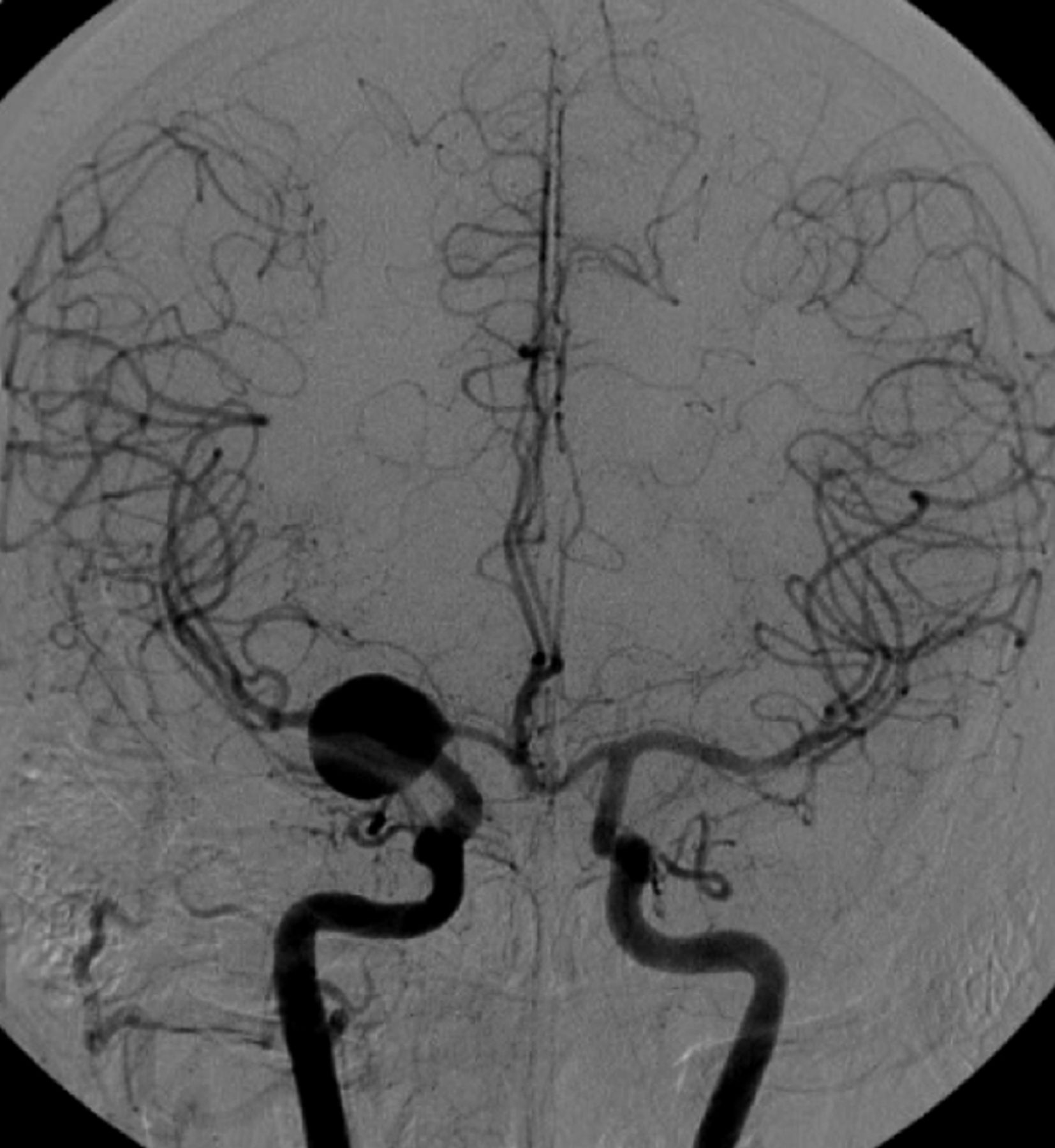
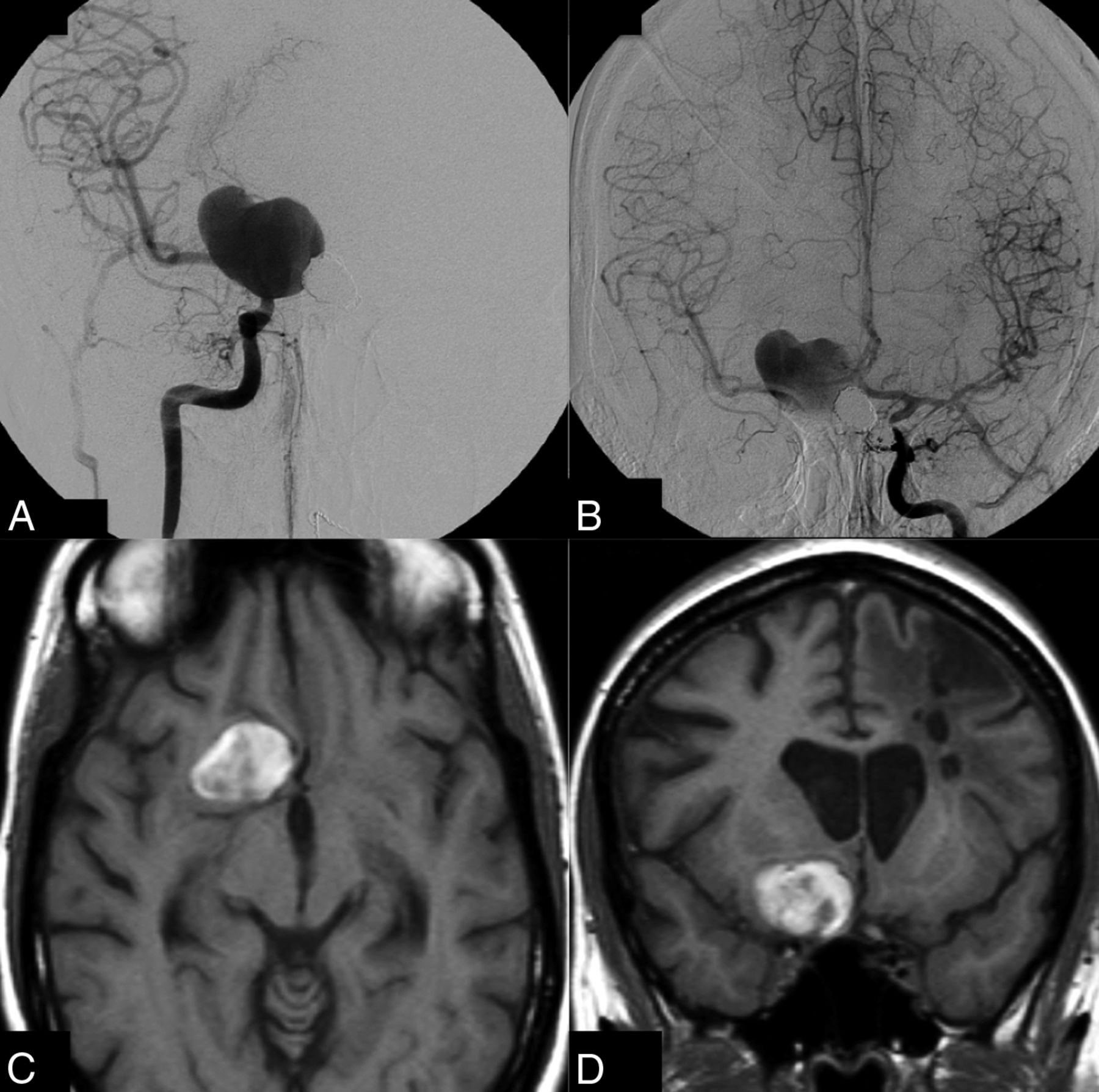
Mean imaging and clinical follow-up was 35 months (median, 18 months; range, 3–180 months). Angiographic and MR imaging follow-up showed complete occlusion of 86 of 88 aneurysms (97.7%) treated by ICA occlusion. In 2 patients (1.4%), the aneurysm was not completely occluded on short-term follow-up. One patient with a supraclinoid ICA aneurysm had persistent filling of the aneurysm via the posterior communicating artery; this patient later underwent bypass surgery and clipping (Fig 1). The other patient had persistent filling of the giant carotid tip aneurysm at the 10-month follow-up angiogram, but MR imaging 3 months later demonstrated complete thrombosis (Fig 2).

Fig 1.
  • Download figure
  • Open in new tab
  • Download powerpoint
Fig 1.

Large carotid tip aneurysm incidentally found in a 45-year-old woman. This aneurysm failed to thrombose after right ICA occlusion and was surgically treated 10 months later.

Fig 2.
  • Download figure
  • Open in new tab
  • Download powerpoint
Fig 2.

Giant unruptured right carotid tip aneurysm in a 36-year-old woman with multiple aneurysms. A, Right ICA angiogram shows the giant aneurysm; test occlusion showed ample collateral circulation via the anterior communicating artery. B, Left ICA angiogram 6 months after right ICA occlusion demonstrates that the aneurysm is still not occluded. C and D, T1-weighted MR imaging 10 months after right ICA occlusion reveals complete thrombosis of the aneurysm lumen.

At the latest MR imaging, 61 of 80 aneurysms (76%) were decreased in size or were completely obliterated.

Recovery of Cranial Nerve Function after ICA Occlusion

Of the 62 patients who presented with cranial nerve dysfunction by mass effect of the aneurysm and were treated with ICA occlusion, 30 (48%) were cured, 25 (40%) improved, 6 (10%) were unchanged, and 1 patient (2%) was hemiplegic after a complication.

Discussion

In this large cohort of consecutive patients with large and giant ICA aneurysms and therapeutic carotid artery occlusion after angiographic testing, the risk of permanent neurologic complications was extremely low. This study confirmed previous data from the same cohort and demonstrated that good clinical and anatomic results were sustained over time in a growing cohort of patients. These new data were more robust, with incremental narrowing of confidence intervals.

The only permanent neurologic complication was the result of hypovolemic shock due to an undetected retroperitoneal hematoma during ambulance transportation back to the referring hospital. Thus, the angiographic test occlusion as a predictor for tolerance to ICA occlusion proved to be very accurate, with no false-positives. The negative predictive value of the angiographic test occlusion could not be assessed because no ICA occlusion was performed when the test indicated nontolerance. Data from the era of surgical ICA clamp occlusion without previous tolerance testing indicate that approximately three-fourths of patients can tolerate ICA occlusion.16⇓–18 In our cohort, the angiographic test indicated tolerance to permanent ICA occlusion in two-thirds of patients, which indicates that the proportion of false-negatives is probably limited.

After ICA occlusion, all aneurysms thrombosed completely except one, which makes the treatment very effective. The one exception was a large carotid tip aneurysm in a patient with a patent ipsilateral posterior communicating artery aneurysm that was not appreciated on angiography during test occlusion. Apparently, hemodynamic changes in the circle of Willis after ICA occlusion induced increased flow over the posterior communicating artery that prevented intraluminal thrombosis of the aneurysm. In aneurysms located distal to the posterior communicating artery, one should be aware of this phenomenon. We never encountered persistent aneurysm filling through collaterals via the ophthalmic artery. The effectiveness of our protocol of ICA balloon occlusion proximal to the aneurysm is comparable with the protocol of trapping the aneurysm with coils used by Labeyrie et al.19 However, we are in favor of our protocol because it is much easier to perform and definitely much cheaper. In addition, trapping in aneurysms located close to the ICA bifurcation is not possible because the distal ICA segment is too short to accommodate the coils.

During follow-up of at least 3 months in all the patients and much longer in most, three-fourths of the aneurysms decreased in size and almost 90% of the patients who presented with cranial nerve dysfunction were cured or improved. These favorable results on aneurysm size and symptoms of mass effect are in concordance with other studies.7,20,21

Our data confirmed that ICA occlusion for large and giant carotid artery aneurysms was a very safe and effective therapy in patients with adequate collateral circulation. The angiographic test occlusion accurately predicted tolerance to ICA occlusion with probably only a few false-negatives but no false-positives. The angiographic test occlusion obviates the need for clinical testing and can be safely performed in patients under general anesthesia. After ICA occlusion and confirmation of aneurysm thrombosis on MR imaging, the aneurysm can be considered cured, and further imaging follow-up is not necessary. In a previous 3T MRA follow-up study in a selection of 26 patients of the present cohort, there were no de novo aneurysms after a mean follow-up of >50 months. In conjunction with other follow-up studies of patients with clipped aneurysms, there was no reason to believe that there is an increased risk of developing de novo aneurysms in patients with aneurysms treated by ICA occlusion compared with patients with clipped aneurysms.13 In another follow-up study by using MR arterial spin-labeling in 11 patients from this cohort after a mean 39-month follow-up, CBF values were within normal range and there was no significant CBF difference between hemispheres ipsilateral and contralateral to carotid sacrifice.15

Several investigators advocate ICA sparing therapy with flow diverters for ICA aneurysms, also in patients who can tolerate ICA occlusion.22 However, recent meta-analyses that concern flow-diverter stents show that the combined morbidity and mortality rate is approximately 10%, regardless of the type of aneurysm.23⇓⇓⇓⇓⇓–29 Mortality was 4% in the study of Briganti et al24 in 76 patients with cavernous aneurysms treated with flow diverters. Many fatal complications with stent-assisted techniques are due to major hemorrhagic or ischemic complications, which are completely absent from carotid occlusion series. Because long-term imaging follow-up studies after flow-diverter treatment of ICA aneurysms are not yet available, complication rates may be underestimated: delayed in-stent occlusion may go clinically undetected in the three-fourths of patients who can tolerate ICA occlusion.30 In our opinion, supported by others,19 the safety profile of flow diverters is not good enough to justify treatment of ICA aneurysms in patients who can tolerate carotid occlusion, especially not in patients with cavernous aneurysms that generally exhibit a benign natural history.

ICA occlusion according to our protocol is also cost effective: angiographic test occlusion, followed by permanent ICA occlusion, is a straightforward procedure that takes 45–120 minutes. Occlusion balloons are cheap, and the patient is discharged home the next day. Prolonged imaging follow-up beyond the 3-month interval is usually not needed.

For patients with large and giant ICA aneurysms who cannot tolerate ICA occlusion, parent vessel sparing therapies should be considered with caution. In balancing the risks and benefits of these alternative therapies, it should be kept in mind that complications that lead to acute or delayed ICA occlusion in these patients certainly will cause neurologic deficit or even death. With the use of stents or flow diverters, this risk is not negligible: in-stent stenosis and occlusion occurs in approximately 10% of cases in the short term and the midterm.23 With bypass surgery, the risk of occlusion of the bypass is certainly present.4 The safest option to treat an aneurysm in a patient who cannot tolerate ICA occlusion seems to be selective coiling with or without balloon assistance. In patients with cavernous sinus aneurysms, conservative therapy can be the best option in a substantial proportion.

Conclusions

In patients with large and giant ICA aneurysms, ICA occlusion, when tolerated, remains a very safe and effective therapy. Tolerance to ICA occlusion can be reliably predicted by the angiographic test occlusion, also in patients under general anesthesia. In patients who cannot tolerate ICA occlusion, alternative treatments sparing the ICA should be offered only after careful benefit to risk analysis because complication rates of these therapies are substantially higher.

Footnotes

  • Disclosures: Charles Majoie—UNRELATED: Grants/Grants Pending: Dutch Heart Foundation*; Payment for Lectures (including service on speakers bureaus): Stryker.* *Money paid to the institution.

References

  1. 1.↵
    1. van Rooij WJ
    . Endovascular treatment of cavernous sinus aneurysms. AJNR Am J Neuroradiol 2012;33:323–26 doi:10.3174/ajnr.A2759 pmid:22033713
    Abstract/FREE Full Text
  2. 2.↵
    1. van der Schaaf IC,
    2. Brilstra EH,
    3. Buskens E, et al
    . Endovascular treatment of aneurysms in the cavernous sinus: a systematic review on balloon occlusion of the parent vessel and embolization with coils. Stroke 2002;33:313–38 doi:10.1161/hs0102.101479 pmid:11779933
    Abstract/FREE Full Text
  3. 3.↵
    1. van Rooij WJ,
    2. Sluzewski M,
    3. Beute GN
    . Ruptured cavernous sinus aneurysms causing carotid cavernous fistula: incidence, clinical presentation, treatment, and outcome. AJNR Am J Neuroradiol 2006;27:185–89 pmid:16418380
    Abstract/FREE Full Text
  4. 4.↵
    1. Brilstra EH,
    2. Rinkel GJ,
    3. Klijn CJ, et al
    . Excimer laser-assisted bypass in aneurysm treatment: short-term outcomes. J Neurosurg 2002;97:1029–35 doi:10.3171/jns.2002.97.5.1029 pmid:12450023
    CrossRefPubMed
  5. 5.↵
    1. Langer DJ,
    2. Van Der Zwan A,
    3. Vajkoczy P, et al
    . Excimer laser-assisted nonocclusive anastomosis: an emerging technology for use in the creation of intracranial-intracranial and extracranial-intracranial cerebral bypass. Neurosurg Focus 2008;24:E6 doi:10.3171/FOC/2008/24/2/E6 pmid:18275301
    CrossRefPubMed
  6. 6.↵
    1. van Rooij WJ,
    2. Sluzewski M
    . Endovascular treatment of large and giant aneurysms. AJNR Am J Neuroradiol 2009;30:12–18 doi:10.3174/ajnr.A1267 pmid:18719032
    Abstract/FREE Full Text
  7. 7.↵
    1. Lubicz B,
    2. Gauvrit JY,
    3. Leclerc X, et al
    . Giant aneurysms of the internal carotid artery: endovascular treatment and long-term follow-up. Neuroradiology 2003;45:650–55 doi:10.1007/s00234-003-1047-4 pmid:12923666
    CrossRefPubMed
  8. 8.↵
    1. Lanzino G,
    2. Crobeddu E,
    3. Cloft HJ, et al
    . Efficacy and safety of flow diversion for paraclinoid aneurysms: a matched-pair analysis compared with standard endovascular approaches. AJNR Am J Neuroradiol 2012;33:2158–61 doi:10.3174/ajnr.A3207 pmid:22790243
    Abstract/FREE Full Text
  9. 9.↵
    1. D'Urso PI,
    2. Karadeli HH,
    3. Kallmes DF, et al
    . Coiling for paraclinoid aneurysms: time to make way for flow diverters? AJNR Am J Neuroradiol 2012;33:1470–74 doi:10.3174/ajnr.A3009 pmid:22403773
    Abstract/FREE Full Text
  10. 10.↵
    1. van Rooij WJ,
    2. Sluzewski M,
    3. Slob MJ, et al
    . Predictive value of angiographic testing for tolerance to therapeutic occlusion of the carotid artery. AJNR Am J Neuroradiol 2005;1:175–78 pmid:15661722
    PubMed
  11. 11.↵
    1. van Rooij WJ,
    2. Sluzewski M,
    3. Metz NH, et al
    . Carotid balloon occlusion for large and giant aneurysms: evaluation of a new test occlusion protocol. Neurosurgery 2000;47:116–21; discussion 122 doi:10.1227/00006123-200007000-00025 pmid:10917354
    CrossRefPubMed
  12. 12.↵
    1. van Rooij WJ,
    2. Sluzewski M
    . Unruptured large and giant carotid artery aneurysms presenting with cranial nerve palsy: comparison of clinical recovery after selective aneurysm coiling and therapeutic carotid artery occlusion. AJNR Am J Neuroradiol 2008;29:997–1002 doi:10.3174/ajnr.A1023 pmid:18296545
    Abstract/FREE Full Text
  13. 13.↵
    1. de Gast AN,
    2. Sprengers ME,
    3. van Rooij WJ, et al
    . Long-term 3T MR angiography follow-up after therapeutic occlusion of the internal carotid artery to detect possible de novo aneurysm formation. AJNR Am J Neuroradiol 2007;28:508–10 pmid:17353324
    Abstract/FREE Full Text
  14. 14.↵
    1. de Gast AN,
    2. Sprengers ME,
    3. van Rooij WJ, et al
    . Midterm clinical and magnetic resonance imaging follow-up of large and giant carotid artery aneurysms after therapeutic carotid artery occlusion. Neurosurgery 2007;60:1025–29; discussion 1029–31 doi:10.1227/01.NEU.0000255468.30011.AA pmid:17538375
    CrossRefPubMed
  15. 15.↵
    1. Gevers S,
    2. Heijtel D,
    3. Ferns SP, et al
    . Cerebral perfusion long term after therapeutic occlusion of the internal carotid artery in patients who tolerated angiographic balloon test occlusion. AJNR Am J Neuroradiol 2012;33:329–35 doi:10.3174/ajnr.A2776 pmid:22081677
    Abstract/FREE Full Text
  16. 16.↵
    1. Linskey ME,
    2. Jungreis CA,
    3. Yonas H, et al
    . Stroke risk after abrupt internal carotid artery sacrifice: accuracy of preoperative assessment with balloon test occlusion and stable xenon-enhanced CT. AJNR Am J Neuroradiol 1994;15:829–43 pmid:8059649
    Abstract/FREE Full Text
  17. 17.↵
    1. Drake CG,
    2. Peerless SJ,
    3. Ferguson GG
    . Hunterian proximal arterial occlusion for giant aneurysms of the carotid circulation. J Neurosurg 1994;81:656–65 doi:10.3171/jns.1994.81.5.0656 pmid:7931611
    CrossRefPubMed
  18. 18.↵
    1. Fox AJ,
    2. Viñuela F,
    3. Pelz DM, et al
    . Use of detachable balloons for proximal artery occlusion in the treatment of unclippable cerebral aneurysms. J Neurosurg 1987;66:40–46 doi:10.3171/jns.1987.66.1.0040 pmid:3783258
    CrossRefPubMed
  19. 19.↵
    1. Labeyrie MA,
    2. Lenck S,
    3. Bresson D, et al
    . Parent artery occlusion in large, giant, or fusiform aneurysms of the carotid siphon: clinical and imaging results. AJNR Am J Neuroradiol 2015;36:140–45 doi:10.3174/ajnr.A4064 pmid:25082818
    Abstract/FREE Full Text
  20. 20.↵
    1. Abud DG,
    2. Spelle L,
    3. Piotin M, et al
    . Venous phase timing during balloon test occlusion as a criterion for permanent internal carotid artery sacrifice. AJNR Am J Neuroradiol 2005;26:2602–09 pmid:16286409
    Abstract/FREE Full Text
  21. 21.↵
    1. Zhang Z,
    2. Lv X,
    3. Wu Z, et al
    . Clinical and angiographic outcome of endovascular and conservative treatment for giant cavernous carotid artery aneurysms. Interv Neuroradiol 2014;20:29–36 doi:10.15274/INR-2014-10005 pmid:24556297
    Abstract/FREE Full Text
  22. 22.↵
    1. Tanweer O,
    2. Raz E,
    3. Brunswick A, et al
    . Cavernous carotid aneurysms in the era of flow diversion: a need to revisit treatment paradigms. AJNR Am J Neuroradiol 2014;35:2334–40 doi:10.3174/ajnr.A4081 pmid:25147199
    Abstract/FREE Full Text
  23. 23.↵
    1. Velioglu M,
    2. Kizilkilic O,
    3. Selcuk H, et al
    . Early and midterm results of complex cerebral aneurysms treated with Silk stent. Neuroradiology 2012;54:1355–65 doi:10.1007/s00234-012-1051-7 pmid:22695740
    CrossRefPubMed
  24. 24.↵
    1. Briganti F,
    2. Napoli M,
    3. Tortora F, et al
    . Italian multicenter experience with flow-diverter devices for intracranial unruptured aneurysm treatment with periprocedural complications–a retrospective data analysis. Neuroradiology 2012;54:1145–52 doi:10.1007/s00234-012-1047-3 pmid:22569955
    CrossRefPubMed
  25. 25.↵
    1. Berge J,
    2. Biondi A,
    3. Machi P, et al
    . Flow-diverter Silk stent for the treatment of intracranial aneurysms: 1-year follow-up in a multicenter study. AJNR Am J Neuroradiol 2012;33:1150–55 doi:10.3174/ajnr.A2907 pmid:22300924
    Abstract/FREE Full Text
  26. 26.↵
    1. Nelson PK,
    2. Sahlein D,
    3. Shapiro M, et al
    . Recent steps toward a reconstructive endovascular solution for the orphaned, complex-neck aneurysm. Neurosurgery 2006;59(5 suppl 3):S77–92; discussion S3-13 doi:10.1227/01.NEU.0000240664.00611.BB pmid:17053621
    CrossRefPubMed
  27. 27.↵
    1. Kallmes DF,
    2. Hanel R,
    3. Lopes D, et al
    . International retrospective study of the Pipeline embolization device: a multicenter aneurysm treatment study. AJNR Am J Neuroradiol 2015;36:108–15 doi:10.3174/ajnr.A4111 pmid:25355814
    Abstract/FREE Full Text
  28. 28.↵
    1. Arrese I,
    2. Sarabia R,
    3. Pintado R, et al
    . Flow-diverter devices for intracranial aneurysms: systematic review and meta-analysis. Neurosurgery 2013;73:193–99; discussion 199–200 doi:10.1227/01.neu.0000430297.17961.f1 pmid:23624409
    CrossRefPubMed
  29. 29.↵
    1. Brinjikji W,
    2. Murad MH,
    3. Lanzino G, et al
    . Endovascular treatment of intracranial aneurysms with flow diverters: a meta-analysis. Stroke 2013;44:442–47 doi:10.1161/STROKEAHA.112.678151 pmid:23321438
    Abstract/FREE Full Text
  30. 30.↵
    1. Fiorella D,
    2. Hsu D,
    3. Woo HH, et al
    . Very late thrombosis of a Pipeline embolization device construct: case report. Neurosurgery 2010;67(suppl operative):onsE313–34; discussion onsE314 doi:10.1227/01.NEU.0000383875.08681.23 pmid:20679914
    CrossRefPubMed
  • Received May 5, 2015.
  • Accepted after revision June 8, 2015.
  • © 2016 by American Journal of Neuroradiology
View Abstract
PreviousNext
Back to top

In this issue

American Journal of Neuroradiology: 37 (1)
American Journal of Neuroradiology
Vol. 37, Issue 1
1 Jan 2016
  • Table of Contents
  • Index by author
  • Complete Issue (PDF)
Advertisement
Print
Download PDF
Email Article

Thank you for your interest in spreading the word on American Journal of Neuroradiology.

NOTE: We only request your email address so that the person you are recommending the page to knows that you wanted them to see it, and that it is not junk mail. We do not capture any email address.

Enter multiple addresses on separate lines or separate them with commas.
Therapeutic Internal Carotid Artery Occlusion for Large and Giant Aneurysms: A Single Center Cohort of 146 Patients
(Your Name) has sent you a message from American Journal of Neuroradiology
(Your Name) thought you would like to see the American Journal of Neuroradiology web site.
CAPTCHA
This question is for testing whether or not you are a human visitor and to prevent automated spam submissions.
Cite this article
R.S. Bechan, C.B. Majoie, M.E. Sprengers, J.P. Peluso, M. Sluzewski, W.J. van Rooij
Therapeutic Internal Carotid Artery Occlusion for Large and Giant Aneurysms: A Single Center Cohort of 146 Patients
American Journal of Neuroradiology Jan 2016, 37 (1) 125-129; DOI: 10.3174/ajnr.A4487

Citation Manager Formats

  • BibTeX
  • Bookends
  • EasyBib
  • EndNote (tagged)
  • EndNote 8 (xml)
  • Medlars
  • Mendeley
  • Papers
  • RefWorks Tagged
  • Ref Manager
  • RIS
  • Zotero
0 Responses
Respond to this article
Share
Bookmark this article
Therapeutic Internal Carotid Artery Occlusion for Large and Giant Aneurysms: A Single Center Cohort of 146 Patients
R.S. Bechan, C.B. Majoie, M.E. Sprengers, J.P. Peluso, M. Sluzewski, W.J. van Rooij
American Journal of Neuroradiology Jan 2016, 37 (1) 125-129; DOI: 10.3174/ajnr.A4487
del.icio.us logo Twitter logo Facebook logo Mendeley logo
  • Tweet Widget
  • Facebook Like
  • Google Plus One
Purchase

Jump to section

  • Article
    • Abstract
    • Materials and Methods
    • Results
    • Discussion
    • Conclusions
    • Footnotes
    • References
  • Figures & Data
  • Info & Metrics
  • Responses
  • References
  • PDF

Related Articles

  • No related articles found.
  • PubMed
  • Google Scholar

Cited By...

  • Flow Diversion for ICA Aneurysms with Compressive Neuro-Ophthalmologic Symptoms: Predictors of Morbidity, Mortality, and Incomplete Aneurysm Occlusion
  • Recovery from Cranial Nerve Symptoms after Flow Diversion without Coiling for Unruptured Very Large and Giant ICA Aneurysms
  • Carotid cavernous fistula after Pipeline placement: a single-center experience and review of the literature
  • Crossref (26)
  • Google Scholar

This article has been cited by the following articles in journals that are participating in Crossref Cited-by Linking.

  • Flow Diverter Therapy Using a Pipeline Embolization Device for 100 Unruptured Large and Giant Internal Carotid Artery Aneurysms in a Single Center in a Japanese Population
    Hidenori OISHI, Kosuke TERANISHI, Kenji YATOMI, Takashi FUJII, Munetaka YAMAMOTO, Hajime ARAI
    Neurologia medico-chirurgica 2018 58 11
  • Carotid cavernous fistula after Pipeline placement: a single-center experience and review of the literature
    Anil K Roy, Jonathan A Grossberg, Joshua W Osbun, Susana L Skukalek, Brian M Howard, Faiz U Ahmad, Frank Tong, Jacques E Dion, Charles M Cawley
    Journal of NeuroInterventional Surgery 2017 9 2
  • Comparison of Safety and Effectiveness of Endovascular Treatments for Unruptured Intracranial Large or Giant Aneurysms in Internal Carotid Artery
    Peng Yan, Yupeng Zhang, Fei Liang, Chao Ma, Shikai Liang, Feng Guo, Chuhan Jiang
    World Neurosurgery 2019 125
  • Endovascular parent-artery occlusion of large or giant unruptured internal carotid artery aneurysms. A long-term single-center experience
    Kampei Shimizu, Hirotoshi Imamura, Yohei Mineharu, Hidemitsu Adachi, Chiaki Sakai, Shoichi Tani, Koichi Arimura, Mikiya Beppu, Nobuyuki Sakai
    Journal of Clinical Neuroscience 2017 37
  • The Rate of Symptomatic Ischemic Events after Passing Balloon Test Occlusion of the Major Intracranial Arteries: Meta-Analysis
    John T. Butterfield, Clark C. Chen, Andrew W. Grande, Bharathi Jagadeesan, Ramachandra Tummala, Andrew S. Venteicher
    World Neurosurgery 2021 146
  • A Hybrid Strategy for Patients With Complex Cerebral Aneurysm: STA–MCA Bypass in Combination With Endovascular Embolization
    Gang Wang, Xi'an Zhang, Yanxia Gou, Yunyu Wen, Guozhong Zhang, Mingzhou Li, Shichao Zhang, Yanyi Yin, Siyuan Chen, Songtao Qi, Wenfeng Feng
    Frontiers in Neurology 2021 11
  • Recent Trends in Neuro-endovascular Treatment for Acute Ischemic Stroke, Cerebral Aneurysms, Carotid Stenosis, and Brain Arteriovenous Malformations
    Yuji MATSUMARU, Eiichi ISHIKAWA, Tetsuya YAMAMOTO, Akira MATSUMURA
    Neurologia medico-chirurgica 2017 57 6
  • Flow Diverter Devices for Treatment of Intracranial Aneurysms in Small Parent Vessels—A Systematic Review of Literature
    Nisha Dabhi, Danyas Sarathy, M. Harrison Snyder, Ryan T. Kellogg, Min S. Park
    World Neurosurgery 2022 162
  • Flow Diversion for ICA Aneurysms with Compressive Neuro-Ophthalmologic Symptoms: Predictors of Morbidity, Mortality, and Incomplete Aneurysm Occlusion
    D.P.O. Kaiser, G. Boulouis, S. Soize, V. Maus, S. Fischer, D. Lobsien, J. Klisch, H. Styczen, C. Deuschl, N. Abdullayev, C. Kabbasch, A. Jamous, D. Behme, K. Janot, G. Bellanger, C. Cognard, L. Pierot, M. Gawlitza
    American Journal of Neuroradiology 2022 43 7
  • Combining Internal Carotid Ligation with Low-Flow Bypass for Treating Large-Giant Cavernous Sinus Segment Aneurysms: A Report of Four Cases
    Chuan Chen, Bo Hou, Wen-Sheng Li, Ying Guo
    World Neurosurgery 2017 100

More in this TOC Section

  • SAVE vs. Solumbra Techniques for Thrombectomy
  • Contrast-Induced Encephalopathy after NeuroIR
  • CT Perfusion&Reperfusion in Acute Ischemic Stroke
Show more Interventional

Similar Articles

Advertisement

Indexed Content

  • Current Issue
  • Accepted Manuscripts
  • Article Preview
  • Past Issues
  • Editorials
  • Editor's Choice
  • Fellows' Journal Club
  • Letters to the Editor
  • Video Articles

Cases

  • Case Collection
  • Archive - Case of the Week
  • Archive - Case of the Month
  • Archive - Classic Case

Special Collections

  • AJNR Awards
  • ASNR Foundation Special Collection
  • Most Impactful AJNR Articles
  • Photon-Counting CT
  • Spinal CSF Leak Articles (Jan 2020-June 2024)

More from AJNR

  • Trainee Corner
  • Imaging Protocols
  • MRI Safety Corner

Multimedia

  • AJNR Podcasts
  • AJNR Scantastics

Resources

  • Turnaround Time
  • Submit a Manuscript
  • Submit a Video Article
  • Submit an eLetter to the Editor/Response
  • Manuscript Submission Guidelines
  • Statistical Tips
  • Fast Publishing of Accepted Manuscripts
  • Graphical Abstract Preparation
  • Imaging Protocol Submission
  • Evidence-Based Medicine Level Guide
  • Publishing Checklists
  • Author Policies
  • Become a Reviewer/Academy of Reviewers
  • News and Updates

About Us

  • About AJNR
  • Editorial Board
  • Editorial Board Alumni
  • Alerts
  • Permissions
  • Not an AJNR Subscriber? Join Now
  • Advertise with Us
  • Librarian Resources
  • Feedback
  • Terms and Conditions
  • AJNR Editorial Board Alumni

American Society of Neuroradiology

  • Not an ASNR Member? Join Now

© 2025 by the American Society of Neuroradiology All rights, including for text and data mining, AI training, and similar technologies, are reserved.
Print ISSN: 0195-6108 Online ISSN: 1936-959X

Powered by HighWire