Advertisement

Main menu

  • Home
  • Content
    • Current Issue
    • Accepted Manuscripts
    • Article Preview
    • Past Issue Archive
    • Video Articles
    • AJNR Case Collection
    • Case of the Week Archive
    • Case of the Month Archive
    • Classic Case Archive
  • Special Collections
    • AJNR Awards
    • ASNR Foundation Special Collection
    • Most Impactful AJNR Articles
    • Photon-Counting CT
    • Spinal CSF Leak Articles (Jan 2020-June 2024)
  • Multimedia
    • AJNR Podcasts
    • AJNR SCANtastic
    • Trainee Corner
    • MRI Safety Corner
    • Imaging Protocols
  • For Authors
    • Submit a Manuscript
    • Submit a Video Article
    • Submit an eLetter to the Editor/Response
    • Manuscript Submission Guidelines
    • Statistical Tips
    • Fast Publishing of Accepted Manuscripts
    • Graphical Abstract Preparation
    • Imaging Protocol Submission
    • Author Policies
  • About Us
    • About AJNR
    • Editorial Board
    • Editorial Board Alumni
  • More
    • Become a Reviewer/Academy of Reviewers
    • Subscribers
    • Permissions
    • Alerts
    • Feedback
    • Advertisers
    • ASNR Home

User menu

  • Alerts
  • Log in

Search

  • Advanced search
American Journal of Neuroradiology

American Journal of Neuroradiology

ASHNR American Society of Functional Neuroradiology ASHNR American Society of Pediatric Neuroradiology ASSR
  • Alerts
  • Log in

Advanced Search

  • Home
  • Content
    • Current Issue
    • Accepted Manuscripts
    • Article Preview
    • Past Issue Archive
    • Video Articles
    • AJNR Case Collection
    • Case of the Week Archive
    • Case of the Month Archive
    • Classic Case Archive
  • Special Collections
    • AJNR Awards
    • ASNR Foundation Special Collection
    • Most Impactful AJNR Articles
    • Photon-Counting CT
    • Spinal CSF Leak Articles (Jan 2020-June 2024)
  • Multimedia
    • AJNR Podcasts
    • AJNR SCANtastic
    • Trainee Corner
    • MRI Safety Corner
    • Imaging Protocols
  • For Authors
    • Submit a Manuscript
    • Submit a Video Article
    • Submit an eLetter to the Editor/Response
    • Manuscript Submission Guidelines
    • Statistical Tips
    • Fast Publishing of Accepted Manuscripts
    • Graphical Abstract Preparation
    • Imaging Protocol Submission
    • Author Policies
  • About Us
    • About AJNR
    • Editorial Board
    • Editorial Board Alumni
  • More
    • Become a Reviewer/Academy of Reviewers
    • Subscribers
    • Permissions
    • Alerts
    • Feedback
    • Advertisers
    • ASNR Home
  • Follow AJNR on Twitter
  • Visit AJNR on Facebook
  • Follow AJNR on Instagram
  • Join AJNR on LinkedIn
  • RSS Feeds

AJNR is seeking candidates for the AJNR Podcast Editor. Read the position description.

Research ArticleINTERVENTIONAL

Validity of the Meyer Scale for Assessment of Coiled Aneurysms and Aneurysm Recurrence

A. Rouchaud, W. Brinjikji, T. Gunderson, J. Caroff, J.-C. Gentric, G. Lanzino, H.J. Cloft and D.F. Kallmes
American Journal of Neuroradiology May 2016, 37 (5) 844-848; DOI: https://doi.org/10.3174/ajnr.A4616
A. Rouchaud
aFrom the Departments of Radiology (A.R., W.B., G.L., H.J.C., D.F.K.)
dDepartment of Interventional Neuroradiology (A.R., J.C.), Bicetre Hospital, Clichy, France
  • Find this author on Google Scholar
  • Find this author on PubMed
  • Search for this author on this site
  • ORCID record for A. Rouchaud
W. Brinjikji
aFrom the Departments of Radiology (A.R., W.B., G.L., H.J.C., D.F.K.)
  • Find this author on Google Scholar
  • Find this author on PubMed
  • Search for this author on this site
  • ORCID record for W. Brinjikji
T. Gunderson
cHealth Sciences Research, Division of Biomedical Statistics and Informatics (T.G.), Mayo Clinic, Rochester, Minnesota
  • Find this author on Google Scholar
  • Find this author on PubMed
  • Search for this author on this site
  • ORCID record for T. Gunderson
J. Caroff
dDepartment of Interventional Neuroradiology (A.R., J.C.), Bicetre Hospital, Clichy, France
  • Find this author on Google Scholar
  • Find this author on PubMed
  • Search for this author on this site
  • ORCID record for J. Caroff
J.-C. Gentric
eDepartment of Interventional Neuroradiology (J.-C.G.), Notre-Dame Hospital, Montreal, Quebec, Canada
fDepartment of Interventional Neuroradiology (J.-C.G.), CHU Cavale Blanche, Brest, France.
  • Find this author on Google Scholar
  • Find this author on PubMed
  • Search for this author on this site
  • ORCID record for J.-C. Gentric
G. Lanzino
aFrom the Departments of Radiology (A.R., W.B., G.L., H.J.C., D.F.K.)
bNeurosurgery (G.L., H.J.C., D.F.K.)
  • Find this author on Google Scholar
  • Find this author on PubMed
  • Search for this author on this site
  • ORCID record for G. Lanzino
H.J. Cloft
aFrom the Departments of Radiology (A.R., W.B., G.L., H.J.C., D.F.K.)
bNeurosurgery (G.L., H.J.C., D.F.K.)
  • Find this author on Google Scholar
  • Find this author on PubMed
  • Search for this author on this site
  • ORCID record for H.J. Cloft
D.F. Kallmes
aFrom the Departments of Radiology (A.R., W.B., G.L., H.J.C., D.F.K.)
bNeurosurgery (G.L., H.J.C., D.F.K.)
  • Find this author on Google Scholar
  • Find this author on PubMed
  • Search for this author on this site
  • ORCID record for D.F. Kallmes
  • Article
  • Figures & Data
  • Info & Metrics
  • Responses
  • References
  • PDF
Loading

Abstract

BACKGROUND AND PURPOSE: Both the Meyer and Raymond scales are commonly used to report angiographic outcomes following coil embolization of intracranial aneurysms. The objectives of this study were the following: 1) to assess the interobserver agreement of the Meyer and Raymond scales, and 2) to evaluate and compare their performance in predicting major recurrence at follow-up.

MATERIALS AND METHODS: A retrospective series of 120 coiled aneurysms was included. Four investigators independently graded DSA images immediately posttreatment and at follow-up according to the Meyer and Raymond scales. On follow-up DSA, readers also evaluated recurrence outcome. Interobserver agreement was assessed via the intraclass correlation coefficient. The ability of posttreatment Meyer and Raymond scales to predict major recurrence was modeled by using logistic regression and assessed by using receiver operating characteristic analysis.

RESULTS: For the Meyer scale, interobserver intraclass correlation coefficients were 0.58 (95% CI, 0.46–0.68) on posttreatment and 0.78 (95% CI, 0.72–0.83) on follow-up evaluations. For the Raymond scale, interobserver intraclass correlation coefficients were 0.50 (95% CI, 0.39–0.61) and 0.69 (95% CI, 0.62–0.76), respectively, for posttreatment and follow-up. The areas under the curve for the receiver operating characteristic analyses regarding the performance to predict major recurrence at follow-up were 0.69 (95% CI, 0.60–0.79) for the Meyer and 0.70 (95% CI, 0.61–0.78) for the Raymond scale.

CONCLUSIONS: The Meyer scale appears consistent and reliable with observer agreement as high or higher than that of the Raymond scale. Performance of both scales in predicting the risk of major recurrence at follow-up is adequate, with no statistical difference between the scales.

ABBREVIATIONS:

ICC
intraclass correlation coefficient
ROC
receiver operating characteristic

The most commonly used grading scale to assess aneurysm occlusion after coiling is the Raymond and Roy scale.1 This grading scale divides the arteriographic outcomes of endovascularly treated saccular aneurysms into 3 categories: 1) complete occlusion, 2) residual neck, and 3) residual aneurysm.1 However, despite its simplicity, its application in large clinical registries demonstrated important interobserver variability, which may limit its application and efficacy as an outcome-assessment tool.2 Moreover, important variability in aneurysm morphology and occlusion may occur within the same category by using this 3-category scale. To standardize the reporting on the degree to which volumetric occlusion has been achieved, a new consensual 6-point grading system, the Meyer scale, has been proposed jointly by multiple societies across neurointerventional radiology, neurosurgery, and neurology.3⇓⇓–6 This proposed scale, published in 2012, was put forth to standardize the reporting of the degree of volumetric occlusion and to properly assess technical outcomes in a standardized report. The rationale for this new scale was that the commonly used Raymond scale demonstrated important interobserver variability that may limit its application and efficacy as an outcome-assessment tool.2 However, despite the necessity for high reproducibility and interobserver rates, this new grading scale has never been evaluated, to our knowledge. As stated by the consensus writing group, there is no literature support for the proposed grading system, though this writing group believes it is the best available grading scale and offers a greater likelihood of reporting additional degrees of aneurysm recurrence.3⇓⇓–6

In addition, none of the scales have been validated as predictive of aneurysm recurrence.7,8 Possible risk factors for reopening of a coiled aneurysm with time, such as the initial degree of occlusion,9 seem to have an influence on the likelihood of recurrence, with subtotal initial treatment leading to remnant regrowth.10,11

Using a large retrospective series of patients with coiled aneurysms, the objectives of this current study were the following: 1) to assess interobserver agreement of the Meyer scale, and 2) to evaluate its performance in predicting recurrence at follow-up and compare the results with those of the Raymond scale.

Materials and Methods

Study Cases

Mayo Clinic institutional review board approval was obtained for this retrospective study. Records for patients treated with endovascular coil embolization for either ruptured or unruptured aneurysms at our institution were evaluated. Some of the cases were used in a prior publication, but there is no overlap in scope with the current article.12 These images of 120 cases of coiled aneurysms were compiled, de-identified, and placed into a 120-page PDF file. Each page included a DSA of the initial uncoiled aneurysm if available, the final postcoiling working-projection DSA, and the follow-up DSA. The PDF file was then distributed to 4 interventional neuroradiologists in different international centers. Two readers had 6 years' experience, and 2 readers had 4 years' experience.

Angiographic Evaluations

The Meyer scale is a 6-point grading scale based on the percentage of the aneurysm filled by contrast on DSA. Grade zero indicates complete and total aneurysm occlusion without remnant or interstitial filling within the aneurysm. Grade 1 represents >90% volumetric occlusion of the aneurysm based on planar imaging assessment; grade 2, 70%–89% aneurysm occlusion; grade 3, 50%–69%; grade 4, 25%–49%; and grade 5, <25% volumetric aneurysm occlusion. Two other “modifier” criteria are combined with this scale: The modifier “I” may be used to describe interstitial opacification within the confines of the coil mass and the modifier “G” may be used to describe interval growth in the overall dimensions of the aneurysm as a separate phenomenon from coil compaction with recurrence of the aneurysm.

The Raymond scale is a commonly used outcome grading scale that divides the angiographic outcomes of endovascularly treated saccular aneurysms into 3 categories: complete occlusion, residual neck, and residual aneurysm.1 Complete occlusion and residual neck are considered adequate occlusion; and residual aneurysm, inadequate occlusion.13⇓⇓–16

The 4 investigators independently and retrospectively examined each set of images of the posttreatment and follow-up DSAs to grade occlusion status according to the Raymond scale, the Meyer scale, and the modifier criteria. The investigators also evaluated the recurrence status of the aneurysm at follow-up DSA as follows: no recurrence, minor recurrence, or major recurrence. Major recurrence was defined as a theoretically or technically retreatable aneurysm and was considered an unfavorable outcome.

To evaluate the performance of posttreatment DSA evaluation to predict the risk for major recurrence, we used a consensus rating based on the most common grade among all readers. In case of disagreement among readers, 2 readers (A.R., W.B.) did a consensus reading.

Statistical Analysis

Reader agreement was assessed via the intraclass correlation coefficient (ICC) by using the ICC method (2,1), as specified by Shrout and Fleiss,17 for each of the 7 outcomes (posttreatment and follow-up Raymond scale, Meyer scale, Meyer modifier criteria, and recurrence status at follow-up). For the ICC description, we used the verbal description proposed by Portney and Watkins18: 0–.2, poor agreement; 0.3–0.4, fair agreement; 0.5–0.6, moderate agreement; 0.6–0.7, substantial agreement; 0.7–0.8, strong agreement; and >0.8, almost perfect agreement.

Aneurysm recurrence at follow-up was modeled as a function of either the Raymond scale or the Meyer scale (including the Meyer modifier criteria). Univariable (for Raymond) or multivariable (for Meyer) logistic regression by using the Firth penalized likelihood was performed by using the consensus rating of favorable or unfavorable recurrence at follow-up as the outcome. Aneurysm recurrence was modeled and receiver operating characteristic (ROC) curves were constructed both for each of the 4 raters individually and by using a consensus rating for each scale.

Statistical analyses for ICC were performed in SAS (Version 9.3; SAS, Cary, North Carolina). All other analyses were performed in R (Version 3.1.1; http://www.r-project.org/). Penalized logistic regression was performed by using the logistf package (version 1.21; https://cran.r-project.org/web/packages/logistf/logistf.pdf). ROC, area under the curve, and confidence interval estimates were obtained by using the pROC package (Version 1.7.3; http://www.rdocumentation.org/packages/pROC/functions/pROC-package).

Results

Posttreatment Results

With the Raymond scale, we classified 165 (34%) of 480 readings as complete occlusion, 163 (34%) as neck remnants, and 152 (32%) as aneurysms remnants. The ICC for the Raymond scale was 0.50 (95% CI, 0.39–0.61), indicating moderate agreement among readers. Using the Meyer scale, we classified 159 (33%) of 480 readings as complete occlusion, 152 (32%) as grade 1, 112 (23%) as grade 2, 43 (9%) as grade 3, 11 (2%) as grade 4, and 3 (1%) as grade 5. The ICC for the Meyer scale was 0.58 (95% CI, 0.46–0.68), indicating moderate agreement among readers. Using the modifier criteria, we classified 213 (44%) of 480 readings as having interstitial opacification (modifier I). The ICC was 0.36 (95% CI, 0.26–0.47), indicating fair agreement among readers (range, 0.24–0.56).

Follow-Up Results

The mean time between treatment and follow-up DSA was 37.8 months, with a median of 30.5 months (range, 0.37–166.0 months). Using the Raymond scale, we classified 138 (29%) of 480 readings as complete occlusion, 147 (31%) as neck remnants, and 195 (41%) as aneurysms remnants. The ICC for the Raymond scale was 0.69 (95% CI, 0.62–0.76), indicating substantial agreement among readers. Using the Meyer scale, we classified 126 (26%) of 480 readings as complete occlusion, 107 (22%) as grade 1, 128 (27%) as grade 2, 82 (17%) as grade 3, 29 (6%) as grade 4, and 8 (2%) as grade 5. The ICC for the Meyer scale was 0.78 (95% CI, 0.72–0.83), indicating strong agreement among readers. Using the modifier criteria, we classified 113 (24%) of 480 readings with the interstitial opacification modifier (modifier I) and 35 (7%) with interval aneurysm growth (modifier G). The ICC was 0.37 (95% CI, 0.27–0.47), indicating fair agreement among readers.

Assessment of Recurrence Status

The evaluation of the recurrence status yielded 43.8% (n = 210/480 readings) of cases with no recurrence, 20.6% (n = 99) with minor recurrence, and 35.6% (n = 171) with major recurrence (unfavorable outcome). The ICC was 0.72 (95% CI, 0.66–0.79), indicating strong agreement among readers. ICCs are presented in the Table.

View this table:
  • View inline
  • View popup

Interreader agreements

After consensual assessment, dichotomized evaluation yielded 65.0% (n = 78/120 cases) good outcomes (no or minor recanalization) and 35.0% (n = 42/120) bad outcomes (major recanalization).

Prediction of Recurrence According to the Posttreatment Angiographic Evaluation

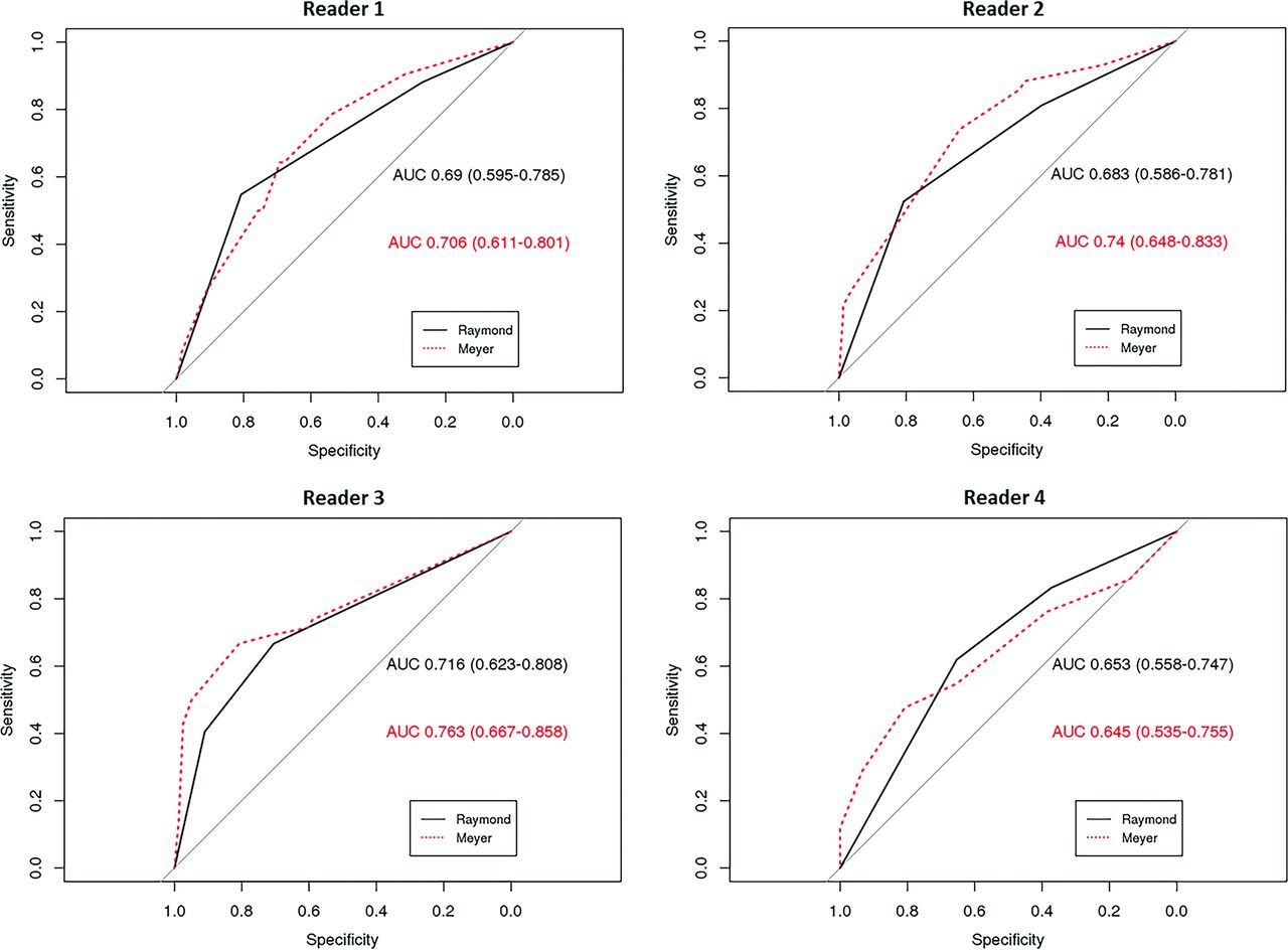
For the Raymond and Meyer evaluations, individual readers' ROC evaluations with areas under the curve are presented in Fig 1, and the ROC curve for the model by using the consensus reading is presented in Fig 2. According to the consensus reading, the area under the curve for the model using the Raymond scale was 0.70 (95% CI, 0.61–0.78). The area under the curve for the model using the Meyer scale was 0.69 (95% CI, 0.60–0.79). The confidence interval estimates overlap, and both of the scales have a fair performance to predict major recurrence.

Fig 1.
  • Download figure
  • Open in new tab
  • Download powerpoint
Fig 1.

ROC curves per reader for the prediction of unfavorable outcome at follow-up (major recurrence) according to the posttreatment Raymond and Meyer scale evaluations.

Fig 2.
  • Download figure
  • Open in new tab
  • Download powerpoint
Fig 2.

ROC curves for the prediction of unfavorable outcome at follow-up (major recurrence) according to the consensus posttreatment Raymond and Meyer scales evaluations.

Discussion

Our current study demonstrated that the Meyer scale has moderate interreader agreement immediately after treatment and strong agreement at follow-up, and for both immediate posttreatment and follow-up, the agreement for the Meyer scale is comparable with that of the Raymond scale. Furthermore, the performance of the Meyer and Raymond scales for predicting major recurrence risk, based on immediate posttreatment results, was fair and similar between scales. These data indicate that the Meyer and Raymond scales have similar performance and consistency levels for aneurysm occlusion evaluation and prediction of a major recurrence.

The interobserver agreement in our study for the Raymond scale is higher than that previously reported by Tollard et al,19 who reported a fair κ statistic at 0.276. Interobserver agreement statistics for the Raymond scale ranged from 0.28 to 0.83 in prior studies.19⇓⇓⇓⇓–24 Most interesting, the Meyer scale agreement score was higher than that of the Raymond scale despite the higher chance for better agreement when grading with fewer responses.8 This outcome is likely because the Meyer score allows a little more flexibility in the assessment of angiographic occlusion. For example, in a case in which an aneurysm is well-occluded with the exception of filling in the interstices, some may mark the status as complete occlusion on the Raymond scale and others may mark the status as aneurysm remnant. However, with the Meyer scale, a visual assessment of the percentage of angiographic filling may be more reproducible. The interstitial opacification criteria have previously been taken into account in the recently introduced modified Raymond scale, with a higher performance in predicting recurrence at follow-up.20

Beyond assessing an adequate or inadequate operator result, grading aneurysm occlusion at the end of the coiling is of interest for predicting the long-term outcome and managing follow-up. Subtotal initial treatment leads to a higher likelihood of potential bleeding risk.10,11 Raymond et al22 reported major recurrence rates of 9% for aneurysms initially completely occluded, 23% for aneurysms with a residual neck, and 47% for aneurysms with residual aneurysm. Thornton et al25 reported a recurrence rate of 1.8% for aneurysms that were 100% occluded and 26% for aneurysms with a residual neck. To our knowledge, this study is the first to evaluate the Meyer scale for predicting the risk of major recurrence at follow-up. Both the Raymond and the Meyer scales, combined with the interstitial opacification criteria, have a fair performance level in predicting the risk of unfavorable outcome at follow-up. This is important because in cases of an inadequate posttreatment angiographic results, given the higher risk for major recurrence, physicians could plan to perform surveillance angiography earlier or more frequently.12,19,26,27 Further studies are needed to determine a cutoff value for adequate-versus-inadequate posttreatment occlusion according to the Meyer scale. In our study, we focused on posttreatment angiographic grading, but this single criterion is not the only one that influences the recurrence outcome. Other factors such as aneurysm size,9,22 smoking status,28,29 and rupture status22 should be taken into account to have a model for predicting recurrence.

Limitations

Our study is limited by its retrospective nature and the use of only selected images for the DSA readers' assessment. Readers did not have access to the complete angiographic run when assessing angiographic occlusion. Another limitation of this study is that DSA was performed at different time points, which can modify the outcomes, depending on the length of follow-up.22

Conclusions

This study highlights the consistency and the reliability of both the Meyer and Raymond occlusion scales for DSA evaluation of coiled aneurysms. Both scales have substantial interobserver agreement and fair performance levels in predicting major recurrence at follow-up. These findings are very important in managing patients at follow-up.

Footnotes

  • Disclosures: Giuseppe Lanzino—UNRELATED: Consultancy: Covidien.* David F. Kallmes—UNRELATED: Grant: Sequent Medical,* Comments: preclinical research; Support for Travel to Meetings for the Study or Other Purposes: Sequent Medical,* Comments: travel to training for a clinical trial; UNRELATED: Board Membership: GE Healthcare,* Comments: Cost-Effectiveness Board; Consultancy: ev3/Medtronic,* Comments: Steering Committee membership for a clinical trial; Grants/Grants Pending: MicroVention,* Surmodics,* Sequent Medical,* Codman,* NeuroSigma*; Royalties: University of Virginia Patent Foundation (Spine Fusion); Travel/Accommodations/Meeting Expenses Unrelated to Activities Listed: ev3/Medtronic,* Comments: travel to FDA panel meeting. *Money paid to the institution.

References

  1. 1.↵
    1. Roy D,
    2. Milot G,
    3. Raymond J
    . Endovascular treatment of unruptured aneurysms. Stroke 2001;32:1998–2004 doi:10.1161/hs0901.095600 pmid:11546888
    Abstract/FREE Full Text
  2. 2.↵
    1. Sluzewski M,
    2. van Rooij WJ
    . Questionable interpretation of results of ACTIVE study on Matrix coils by Boston Scientific. AJNR Am J Neuroradiol 2005;26:1882–83 pmid:16091550
    FREE Full Text
  3. 3.↵
    1. Meyers PM,
    2. Schumacher HC,
    3. Higashida RT, et al
    ; Joint Writing Group of the Technology Assessment Committee, Society of Neurolnterventional Surgery, Society of Interventional Radiology, Joint Section on Cerebrovascular Neurosurgery of the American Association of Neurological Surgeons and Congress of Neurological Surgeons, Section of Stroke and Interventional Neurology of the American Academy of Neurology. Reporting standards for endovascular repair of saccular intracranial cerebral aneurysms. J Neurointerv Surg 2010;2:312–23 doi:10.1136/jnis.2010.002337 pmid:21990640
    Abstract/FREE Full Text
  4. 4.↵
    1. Meyers PM,
    2. Schumacher HC,
    3. Higashida RT, et al
    . Reporting standards for endovascular repair of saccular intracranial cerebral aneurysms. AJNR Am J Neuroradiol 2010;31:E12–24 pmid:20075104
    Abstract/FREE Full Text
  5. 5.↵
    1. Meyers PM,
    2. Schumacher HC,
    3. Higashida RT, et al
    ; American Society of Interventional Neuroradiology, Society of Interventional Radiology. Reporting standards for endovascular repair of saccular intracranial cerebral aneurysms. J Vasc Interv Radiol 2009;20:S435–50 doi:10.1016/j.jvir.2009.03.004 pmid:19560031
    CrossRefPubMed
  6. 6.↵
    1. Meyers PM,
    2. Schumacher HC,
    3. Higashida RT, et al
    . Reporting standards for endovascular repair of saccular intracranial cerebral aneurysms. Stroke 2009;40:e366–79 doi:10.1161/STROKEAHA.108.527572 pmid:19246711
    Abstract/FREE Full Text
  7. 7.↵
    1. Sluzewski M,
    2. van Rooij WJ
    . Early rebleeding after coiling of ruptured cerebral aneurysms: incidence, morbidity, and risk factors. AJNR Am J Neuroradiol 2005;26:1739–43 pmid:16091523
    Abstract/FREE Full Text
  8. 8.↵
    1. Cloft HJ,
    2. Kaufmann T,
    3. Kallmes DF
    . Observer agreement in the assessment of endovascular aneurysm therapy and aneurysm recurrence. AJNR Am J Neuroradiol 2007;28:497–500 pmid:17353321
    Abstract/FREE Full Text
  9. 9.↵
    1. Ferns SP,
    2. Sprengers ME,
    3. van Rooij WJ, et al
    . Coiling of intracranial aneurysms: a systematic review on initial occlusion and reopening and retreatment rates. Stroke 2009;40:e523–29 doi:10.1161/STROKEAHA.109.553099 pmid:19520984
    Abstract/FREE Full Text
  10. 10.↵
    1. Grunwald IQ,
    2. Papanagiotou P,
    3. Struffert T, et al
    . Recanalization after endovascular treatment of intracerebral aneurysms. Neuroradiology 2007;49:41–47 doi:10.1007/s00234-006-0153-5 pmid:17094000
    CrossRefPubMed
  11. 11.↵
    1. Li MH,
    2. Gao BL,
    3. Fang C, et al
    . Angiographic follow-up of cerebral aneurysms treated with Guglielmi detachable coils: an analysis of 162 cases with 173 aneurysms. AJNR Am J Neuroradiol 2006;27:1107–12 pmid:16687553
    Abstract/FREE Full Text
  12. 12.↵
    1. McDonald JS,
    2. Carter RE,
    3. Layton KF, et al
    . Interobserver variability in retreatment decisions of recurrent and residual aneurysms. AJNR Am J Neuroradiol 2013;34:1035–39 doi:10.3174/ajnr.A3326 pmid:23099500
    Abstract/FREE Full Text
  13. 13.↵
    1. Pierot L,
    2. Cognard C,
    3. Anxionnat R, et al
    ; CLARITY Investigators. Endovascular treatment of ruptured intracranial aneurysms: factors affecting midterm quality anatomic results: analysis in a prospective, multicenter series of patients (CLARITY). AJNR Am J Neuroradiol 2012;33:1475–80 doi:10.3174/ajnr.A3003 pmid:22517279
    Abstract/FREE Full Text
  14. 14.↵
    1. Johnston SC,
    2. Dowd CF,
    3. Higashida RT, et al
    ; CARAT Investigators. Predictors of rehemorrhage after treatment of ruptured intracranial aneurysms: the Cerebral Aneurysm Rerupture After Treatment (CARAT) study. Stroke 2008;39:120–25 doi:10.1161/STROKEAHA.107.495747 pmid:18048860
    Abstract/FREE Full Text
  15. 15.↵
    1. Mortimer AM,
    2. Bradley MD,
    3. Mews P, et al
    . Endovascular treatment of 300 consecutive middle cerebral artery aneurysms: clinical and radiologic outcomes. AJNR Am J Neuroradiol 2014;35:706–14 doi:10.3174/ajnr.A3776 pmid:24231847
    Abstract/FREE Full Text
  16. 16.↵
    1. Ng P,
    2. Khangure MS,
    3. Phatouros CC, et al
    . Endovascular treatment of intracranial aneurysms with Guglielmi detachable coils: analysis of midterm angiographic and clinical outcomes. Stroke 2002;33:210–17 doi:10.1161/hs0102.100486 pmid:11779912
    Abstract/FREE Full Text
  17. 17.↵
    1. Shrout PE,
    2. Fleiss JL
    . Intraclass correlations: uses in assessing rater reliability. Psychol Bull 1979;86:420–28 doi:10.1037/0033-2909.86.2.420 pmid:18839484
    CrossRefPubMed
  18. 18.↵
    1. Portney LG,
    2. Watkins MP
    . Foundations of Clinical Research Applications to Practice. New Jersey: Pearson Prentice; 2009
  19. 19.↵
    1. Tollard É,
    2. Darsaut TE,
    3. Bing F, et al
    . Outcomes of endovascular treatments of aneurysms: observer variability and implications for interpreting case series and planning randomized trials. AJNR Am J Neuroradiol 2012;33:626–31 doi:10.3174/ajnr.A2848 pmid:22194386
    Abstract/FREE Full Text
  20. 20.↵
    1. Mascitelli JR,
    2. Moyle H,
    3. Oermann EK, et al
    . An update to the Raymond-Roy Occlusion Classification of intracranial aneurysms treated with coil embolization. J Neurointerv Surg 2015;7:496–502 doi:10.1136/neurintsurg-2014-011258 pmid:24898735
    Abstract/FREE Full Text
  21. 21.↵
    1. Ogilvy CS,
    2. Chua MH,
    3. Fusco MR, et al
    . Validation of a system to predict recanalization after endovascular treatment of intracranial aneurysms. Neurosurgery 2015;77:168–73; discussion 173–74 doi:10.1227/NEU.0000000000000744 pmid:25850603
    CrossRefPubMed
  22. 22.↵
    1. Raymond J,
    2. Guilbert F,
    3. Weill A, et al
    . Long-term angiographic recurrences after selective endovascular treatment of aneurysms with detachable coils. Stroke 2003;34:1398–403 doi:10.1161/01.STR.0000073841.88563.E9 pmid:12775880
    Abstract/FREE Full Text
  23. 23.↵
    1. Singla A,
    2. Villwock MR,
    3. Jacobsen W, et al
    . Aneurysm embolization grade: a predictive tool for aneurysm recurrence after coil embolization. Acta Neurochir (Wien) 2013;155:231–36 doi:10.1007/s00701-012-1554-3 pmid:23151771
    CrossRefPubMed
  24. 24.↵
    1. Urbach H,
    2. Dorenbeck U,
    3. von Falkenhausen M, et al
    . Three-dimensional time-of-flight MR angiography at 3 T compared to digital subtraction angiography in the follow-up of ruptured and coiled intracranial aneurysms: a prospective study. Neuroradiology 2008;50:383–89 doi:10.1007/s00234-007-0355-5 pmid:18196229
    CrossRefPubMed
  25. 25.↵
    1. Thornton J,
    2. Debrun GM,
    3. Aletich VA, et al
    . Follow-up angiography of intracranial aneurysms treated with endovascular placement of Guglielmi detachable coils. Neurosurgery 2002;50:239–49; discussion 249–50 doi:10.1097/00006123-200202000-00003, doi:10.1227/00006123-200202000-00003 pmid:11844258
    CrossRefPubMed
  26. 26.↵
    1. Daugherty WP,
    2. Rad AE,
    3. White JB, et al
    . Observer agreement regarding the necessity of retreatment of previously coiled recurrent cerebral aneurysms. AJNR Am J Neuroradiol 2011;32:566–69 doi:10.3174/ajnr.A2336 pmid:21252043
    Abstract/FREE Full Text
  27. 27.↵
    1. Raymond J,
    2. White PM,
    3. Molyneux AJ
    . Scales, agreement, outcome measures, and progress in aneurysm therapy. AJNR Am J Neuroradiol 2007;28:501–02 pmid:17353322
    FREE Full Text
  28. 28.↵
    1. Ortiz R,
    2. Stefanski M,
    3. Rosenwasser R, et al
    . Cigarette smoking as a risk factor for recurrence of aneurysms treated by endosaccular occlusion. J Neurosurg 2008;108:672–75 doi:10.3171/JNS/2008/108/4/0672 pmid:18377244
    CrossRefPubMed
  29. 29.↵
    1. Brinjikji W,
    2. Lingineni RK,
    3. Gu CN, et al
    . Smoking is not associated with recurrence and retreatment of intracranial aneurysms after endovascular coiling. J Neurosurg 2015;122:95–100 doi:10.3171/2014.10.JNS141035 pmid:25380112
    CrossRefPubMed
  • Received July 2, 2015.
  • Accepted after revision September 18, 2015.
  • © 2016 by American Journal of Neuroradiology
View Abstract
PreviousNext
Back to top

In this issue

American Journal of Neuroradiology: 37 (5)
American Journal of Neuroradiology
Vol. 37, Issue 5
1 May 2016
  • Table of Contents
  • Index by author
  • Complete Issue (PDF)
Advertisement
Print
Download PDF
Email Article

Thank you for your interest in spreading the word on American Journal of Neuroradiology.

NOTE: We only request your email address so that the person you are recommending the page to knows that you wanted them to see it, and that it is not junk mail. We do not capture any email address.

Enter multiple addresses on separate lines or separate them with commas.
Validity of the Meyer Scale for Assessment of Coiled Aneurysms and Aneurysm Recurrence
(Your Name) has sent you a message from American Journal of Neuroradiology
(Your Name) thought you would like to see the American Journal of Neuroradiology web site.
CAPTCHA
This question is for testing whether or not you are a human visitor and to prevent automated spam submissions.
Cite this article
A. Rouchaud, W. Brinjikji, T. Gunderson, J. Caroff, J.-C. Gentric, G. Lanzino, H.J. Cloft, D.F. Kallmes
Validity of the Meyer Scale for Assessment of Coiled Aneurysms and Aneurysm Recurrence
American Journal of Neuroradiology May 2016, 37 (5) 844-848; DOI: 10.3174/ajnr.A4616

Citation Manager Formats

  • BibTeX
  • Bookends
  • EasyBib
  • EndNote (tagged)
  • EndNote 8 (xml)
  • Medlars
  • Mendeley
  • Papers
  • RefWorks Tagged
  • Ref Manager
  • RIS
  • Zotero
0 Responses
Respond to this article
Share
Bookmark this article
Validity of the Meyer Scale for Assessment of Coiled Aneurysms and Aneurysm Recurrence
A. Rouchaud, W. Brinjikji, T. Gunderson, J. Caroff, J.-C. Gentric, G. Lanzino, H.J. Cloft, D.F. Kallmes
American Journal of Neuroradiology May 2016, 37 (5) 844-848; DOI: 10.3174/ajnr.A4616
del.icio.us logo Twitter logo Facebook logo Mendeley logo
  • Tweet Widget
  • Facebook Like
  • Google Plus One
Purchase

Jump to section

  • Article
    • Abstract
    • ABBREVIATIONS:
    • Materials and Methods
    • Results
    • Discussion
    • Conclusions
    • Footnotes
    • References
  • Figures & Data
  • Info & Metrics
  • Responses
  • References
  • PDF

Related Articles

  • No related articles found.
  • PubMed
  • Google Scholar

Cited By...

  • No citing articles found.
  • Crossref (4)
  • Google Scholar

This article has been cited by the following articles in journals that are participating in Crossref Cited-by Linking.

  • Establishment of an optimal occupant behavior considering the energy consumption and indoor environmental quality by region
    Jimin Kim, Taehoon Hong, Jaemin Jeong, Myeonghwi Lee, Minhyun Lee, Kwangbok Jeong, Choongwan Koo, Jaewook Jeong
    Applied Energy 2017 204
  • The impact of LEED certification on energy performance and occupant satisfaction: A case study of residential college buildings
    Sorena Vosoughkhosravi, Lesheena Dixon-Grasso, Amirhosein Jafari
    Journal of Building Engineering 2022 59
  • The clinical value of ceMRA versus DSA for follow-up of intracranial aneurysms treated by coil embolization: an assessment of occlusion classifications and impact on treatment decisions
    Maximilian Patzig, Robert Forbrig, Margaretha Gruber, Thomas Liebig, Franziska Dorn
    European Radiology 2021 31 6
  • Efficacy and safety of fetal posterior cerebral artery stented coil embolization for fetal posterior cerebral aneurysms
    Michiyasu Fuga, Toshihide Tanaka, Rintaro Tachi, Koreaki Irie, Ikki Kajiwara, Akihiko Teshigawara, Toshihiro Ishibashi, Yuzuru Hasegawa, Yuichi Murayama
    Interventional Neuroradiology 2023

More in this TOC Section

  • SAVE vs. Solumbra Techniques for Thrombectomy
  • Contrast-Induced Encephalopathy after NeuroIR
  • CT Perfusion&Reperfusion in Acute Ischemic Stroke
Show more Interventional

Similar Articles

Advertisement

Indexed Content

  • Current Issue
  • Accepted Manuscripts
  • Article Preview
  • Past Issues
  • Editorials
  • Editor's Choice
  • Fellows' Journal Club
  • Letters to the Editor
  • Video Articles

Cases

  • Case Collection
  • Archive - Case of the Week
  • Archive - Case of the Month
  • Archive - Classic Case

Special Collections

  • AJNR Awards
  • ASNR Foundation Special Collection
  • Most Impactful AJNR Articles
  • Photon-Counting CT
  • Spinal CSF Leak Articles (Jan 2020-June 2024)

More from AJNR

  • Trainee Corner
  • Imaging Protocols
  • MRI Safety Corner

Multimedia

  • AJNR Podcasts
  • AJNR Scantastics

Resources

  • Turnaround Time
  • Submit a Manuscript
  • Submit a Video Article
  • Submit an eLetter to the Editor/Response
  • Manuscript Submission Guidelines
  • Statistical Tips
  • Fast Publishing of Accepted Manuscripts
  • Graphical Abstract Preparation
  • Imaging Protocol Submission
  • Evidence-Based Medicine Level Guide
  • Publishing Checklists
  • Author Policies
  • Become a Reviewer/Academy of Reviewers
  • News and Updates

About Us

  • About AJNR
  • Editorial Board
  • Editorial Board Alumni
  • Alerts
  • Permissions
  • Not an AJNR Subscriber? Join Now
  • Advertise with Us
  • Librarian Resources
  • Feedback
  • Terms and Conditions
  • AJNR Editorial Board Alumni

American Society of Neuroradiology

  • Not an ASNR Member? Join Now

© 2025 by the American Society of Neuroradiology All rights, including for text and data mining, AI training, and similar technologies, are reserved.
Print ISSN: 0195-6108 Online ISSN: 1936-959X

Powered by HighWire