Advertisement

Main menu

  • Home
  • Content
    • Current Issue
    • Accepted Manuscripts
    • Article Preview
    • Past Issue Archive
    • Video Articles
    • AJNR Case Collection
    • Case of the Week Archive
    • Case of the Month Archive
    • Classic Case Archive
  • Special Collections
    • AJNR Awards
    • ASNR Foundation Special Collection
    • Most Impactful AJNR Articles
    • Photon-Counting CT
    • Spinal CSF Leak Articles (Jan 2020-June 2024)
  • Multimedia
    • AJNR Podcasts
    • AJNR SCANtastic
    • Trainee Corner
    • MRI Safety Corner
    • Imaging Protocols
  • For Authors
    • Submit a Manuscript
    • Submit a Video Article
    • Submit an eLetter to the Editor/Response
    • Manuscript Submission Guidelines
    • Statistical Tips
    • Fast Publishing of Accepted Manuscripts
    • Graphical Abstract Preparation
    • Imaging Protocol Submission
    • Author Policies
  • About Us
    • About AJNR
    • Editorial Board
    • Editorial Board Alumni
  • More
    • Become a Reviewer/Academy of Reviewers
    • Subscribers
    • Permissions
    • Alerts
    • Feedback
    • Advertisers
    • ASNR Home

User menu

  • Alerts
  • Log in

Search

  • Advanced search
American Journal of Neuroradiology

American Journal of Neuroradiology

ASHNR American Society of Functional Neuroradiology ASHNR American Society of Pediatric Neuroradiology ASSR
  • Alerts
  • Log in

Advanced Search

  • Home
  • Content
    • Current Issue
    • Accepted Manuscripts
    • Article Preview
    • Past Issue Archive
    • Video Articles
    • AJNR Case Collection
    • Case of the Week Archive
    • Case of the Month Archive
    • Classic Case Archive
  • Special Collections
    • AJNR Awards
    • ASNR Foundation Special Collection
    • Most Impactful AJNR Articles
    • Photon-Counting CT
    • Spinal CSF Leak Articles (Jan 2020-June 2024)
  • Multimedia
    • AJNR Podcasts
    • AJNR SCANtastic
    • Trainee Corner
    • MRI Safety Corner
    • Imaging Protocols
  • For Authors
    • Submit a Manuscript
    • Submit a Video Article
    • Submit an eLetter to the Editor/Response
    • Manuscript Submission Guidelines
    • Statistical Tips
    • Fast Publishing of Accepted Manuscripts
    • Graphical Abstract Preparation
    • Imaging Protocol Submission
    • Author Policies
  • About Us
    • About AJNR
    • Editorial Board
    • Editorial Board Alumni
  • More
    • Become a Reviewer/Academy of Reviewers
    • Subscribers
    • Permissions
    • Alerts
    • Feedback
    • Advertisers
    • ASNR Home
  • Follow AJNR on Twitter
  • Visit AJNR on Facebook
  • Follow AJNR on Instagram
  • Join AJNR on LinkedIn
  • RSS Feeds

AJNR is seeking candidates for the AJNR Podcast Editor. Read the position description.

Research ArticleAdult Brain

Diffusion-Weighted Zonal Oblique Multislice–EPI Enhances the Detection of Small Lesions with Diffusion Restriction in the Brain Stem and Hippocampus: A Clinical Report of Selected Cases

T. Sartoretti, E. Sartoretti, C. Binkert, A. Gutzeit, C. Reischauer, D. Czell, M. Wyss, E. Brüllmann and S. Sartoretti-Schefer
American Journal of Neuroradiology July 2018, 39 (7) 1255-1259; DOI: https://doi.org/10.3174/ajnr.A5635
T. Sartoretti
aFrom the Institut für Radiologie (T.S., E.S., C.B., M.W., S.S.-S.), Kantonsspital Winterthur, Winterthur, Switzerland
  • Find this author on Google Scholar
  • Find this author on PubMed
  • Search for this author on this site
  • ORCID record for T. Sartoretti
E. Sartoretti
aFrom the Institut für Radiologie (T.S., E.S., C.B., M.W., S.S.-S.), Kantonsspital Winterthur, Winterthur, Switzerland
  • Find this author on Google Scholar
  • Find this author on PubMed
  • Search for this author on this site
  • ORCID record for E. Sartoretti
C. Binkert
aFrom the Institut für Radiologie (T.S., E.S., C.B., M.W., S.S.-S.), Kantonsspital Winterthur, Winterthur, Switzerland
  • Find this author on Google Scholar
  • Find this author on PubMed
  • Search for this author on this site
  • ORCID record for C. Binkert
A. Gutzeit
bInstitut für Radiologie und Nuklearmedizin (A.G., C.R.), Hirslanden Klinik St. Anna, Luzern, Switzerland
  • Find this author on Google Scholar
  • Find this author on PubMed
  • Search for this author on this site
  • ORCID record for A. Gutzeit
C. Reischauer
bInstitut für Radiologie und Nuklearmedizin (A.G., C.R.), Hirslanden Klinik St. Anna, Luzern, Switzerland
  • Find this author on Google Scholar
  • Find this author on PubMed
  • Search for this author on this site
  • ORCID record for C. Reischauer
D. Czell
cMedizinische Klinik (D.C.), Abteilung für Neurologie, Uznach, Switzerland
  • Find this author on Google Scholar
  • Find this author on PubMed
  • Search for this author on this site
  • ORCID record for D. Czell
M. Wyss
aFrom the Institut für Radiologie (T.S., E.S., C.B., M.W., S.S.-S.), Kantonsspital Winterthur, Winterthur, Switzerland
dPhilips HealthSystems (M.W., E.B.), Zürich, Switzerland.
  • Find this author on Google Scholar
  • Find this author on PubMed
  • Search for this author on this site
  • ORCID record for M. Wyss
E. Brüllmann
dPhilips HealthSystems (M.W., E.B.), Zürich, Switzerland.
  • Find this author on Google Scholar
  • Find this author on PubMed
  • Search for this author on this site
  • ORCID record for E. Brüllmann
S. Sartoretti-Schefer
aFrom the Institut für Radiologie (T.S., E.S., C.B., M.W., S.S.-S.), Kantonsspital Winterthur, Winterthur, Switzerland
  • Find this author on Google Scholar
  • Find this author on PubMed
  • Search for this author on this site
  • ORCID record for S. Sartoretti-Schefer
  • Article
  • Figures & Data
  • Info & Metrics
  • Responses
  • References
  • PDF
Loading

Abstract

SUMMARY: Diffusion restriction is the morphologic hallmark of acute ischemic infarcts and excitotoxic brain injury in various cerebral pathologies. Diffusion restriction is visible as hyperintensity on DWI and as hypointensity on ADC maps. Due to the vicinity of multiple anatomic structures in the brain stem and hippocampus, very small lesions with diffusion restriction may result in severe clinical symptomatology, but these small lesions easily go undetected on standard cerebral DWI due to insufficient spatial resolution, T2* blurring, and image artifacts caused by susceptibility-related image distortions. Diffusion-weighted zonal oblique multislice–EPI with reduced FOV acquisition permits a considerable increase in spatial resolution and enhances the visualization of very small pathologic lesions in the brain stem and hippocampus. Improved performance in the depiction of different pathologic lesions with diffusion restriction in the brain stem and hippocampus using this sequence compared with standard DWI in selected cases is presented.

ABBREVIATIONS:

CN III
oculomotor nerve nucleus area
CN VI
abducens nerve nucleus area
DW
diffusion-weighted
sshot SE
single-shot spin-echo
TGA
transient global amnesia
ZOOM
zonal oblique multislice

Diffusion restriction is caused by different cerebral pathologies leading to cytotoxic edema by energy failure, altered intra- and extracellular ion and water concentrations, and excitotoxic brain injury due to extracellular increase of glutamate.1,2 These mechanisms may overlap in certain pathologies. Diffusion restriction is the main characteristic feature of an acute ischemic event2; therefore, DWI is the only reliable sequence for detecting an acute ischemic event.

DWI is routinely used in brain protocols for the evaluation of various cerebral pathologies characterized by diffusion restriction. The limited spatial resolution of a standard single-shot spin-echo (sshot SE) EPI DWI, called “standard DWI” in our work, is an enormous disadvantage if the clinical symptomatology indicates a lesion in the brain stem and hippocampus.

This clinical report shows how diffusion-weighted (DW) zonal oblique multislice (ZOOM)-EPI can improve the detection of DWI abnormalities in pathologies in the brain stem and hippocampus by increasing spatial resolution, allowing the visualization of small lesions with diffusion restriction not visible on standard DWI.

Materials and Methods

In this clinical report, the MR images of 7 patients of a group with 44 patients (23 women and 21 men; 40–89 years of age; mean age, 76.4 years) are depicted. Informed and written consent for publication of patient data and images was obtained by all patients described in this study. All patients with various acute clinical symptoms all indicating a brain stem pathology (mainly characterized by nausea, vertigo, ataxia, nystagmus, gaze disorders) or hippocampal pathology with amnestic syndrome were examined by both standard transverse sshot SE EPI DWI (called “standard DWI”) and transverse DW ZOOM-EPI during the same examination. The acquisition of DW ZOOM-EPI followed immediately after the acquisition of the standard DWI on the same MR imaging scanner with an identical field strength. Identical image plane angulation was used in both transverse diffusion-weighted sequences.

Patient examinations were performed from October 2016 to August 2017 on a 1.5T Ingenia (Philips Healthcare, Best, the Netherlands) or a 3T Achieva (Philips Healthcare) scanner. Imaging parameters of DW ZOOM-EPI and of standard DWI are shown in Tables 1 and 2. In all patients, 3D FLAIR imaging and transverse T2 TSE sequences were added for possible confirmation of the suspected pathology.

View this table:
  • View inline
  • View popup
Table 1:

Imaging parameters of DW ZOOM-EPI

View this table:
  • View inline
  • View popup
Table 2:

Imaging parameters of standard DWI

In each patient, the presence or absence of acute ischemic lesions in the brain stem on standard DWI and on DW ZOOM-EPI was determined retrospectively. In 27 patients, no pathologic lesion was visible on both standard DWI and DW ZOOM-EPI. In 9 patients both standard DWI and DW ZOOM-EPI showed an acute ischemic infarct with diffusion restriction, involving both the brain stem and cerebellum in 6 patients, only the cerebellum in 2 patients, and only the brain stem in 1 patient. All infarcts visible on both standard DWI and DW ZOOM-EPI had a minimum size of 1 cm. In 8 patients, mostly punctate acute ischemic foci, either in the brain stem, cerebellum, or hippocampi, were only visible on DW ZOOM-EPI.

Despite the acquisition time of the DW ZOOM-EPI (3 minutes to 5 minutes and 30 seconds), no visually evident movement artifacts in the patients examined were observed, but a quantification of movement artifacts was not performed.

Case Series

Transient Global Amnesia.

A 65-year-old female patient presented with antegrade and retrograde amnesia due to clinically suspected transient global amnesia (TGA) lasting for 18 hours (Fig 1). Punctate diffusion restriction was demonstrated on DW ZOOM-EPI with ADC maps in the lateral hippocampal body as a specific TGA-associated lesion. No abnormality was visible on standard DWI or on FLAIR.

Fig 1.
  • Download figure
  • Open in new tab
  • Download powerpoint
Fig 1.

TGA. MR imaging was performed on a 1.5T scanner 24 hours after the beginning of the clinical symptomatology. On DWI ZOOM-EPI (white arrow in A). a punctate hyperintensity and, on DWI ZOOM-EPI ADC map (white arrow in B), a punctate hypointensity with an ADC of 0.915 × 10⁻³ mm2/second were demonstrated in the left lateral hippocampal body as a specific TGA-associated lesion. ADC in the normal right hippocampus was 1.093 × 10⁻³ mm2/second. Standard DWI (C) did not show the abnormality nor was any signal intensity change visible on transverse FLAIR (D).

Hippocampal Ischemia.

An 83-year-old woman with atrial fibrillation presented with acute hemiparesis on the left side and antegrade amnesia (Fig 2). Multiple punctate acute embolic ischemic infarcts with diffusion restriction were visible on DW ZOOM-EPI with ADC maps, namely in the mesencephalon along the pyramidal tract, in the left medial occipitotemporal gyrus, in the left hippocampus, and bilaterally in the pre- and postcentral area on the right. The punctate hippocampal infarct was not visible on standard DWI or FLAIR.

Fig 2.
  • Download figure
  • Open in new tab
  • Download powerpoint
Fig 2.

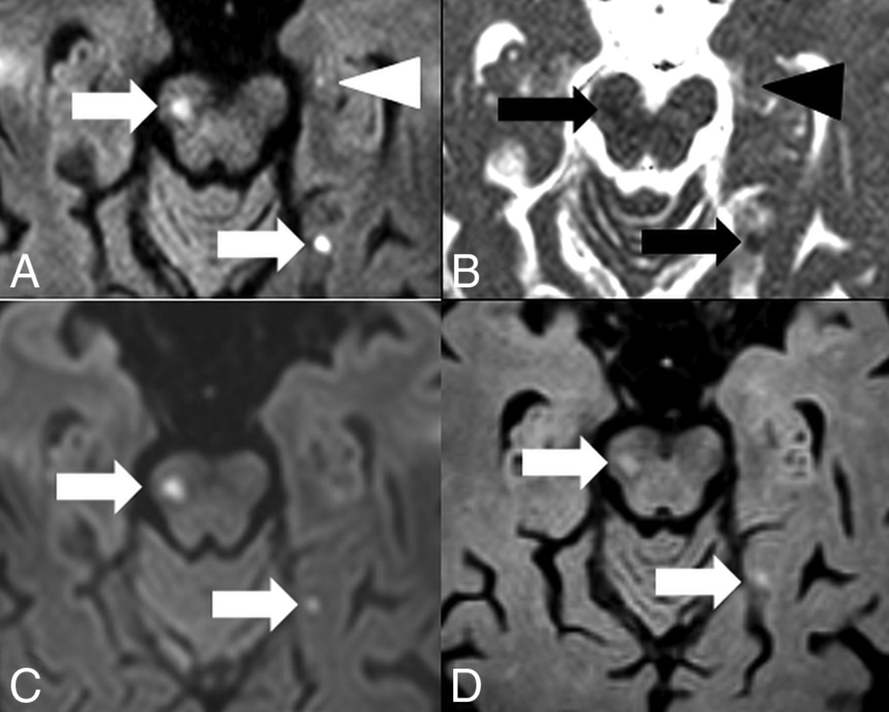
Hippocampal embolic ischemia. MR imaging was performed on a 1.5T scanner. On DW ZOOM-EPI, 3 acute ischemic infarcts in the right mesencephalon along the pyramidal tract (white arrow), in the left medial occipitotemporal gyrus (white arrow), and in the left hippocampus (white arrowhead) were visible as hyperintense foci in A. On DW ZOOM-EPI ADC, the same infarcts were hypointense in B marked with black arrows and a black arrowhead (with an ADC of 0.817 × 10⁻³ mm2/second compared with the ADC of 1.03 × 10⁻³ mm2/second in the normal right hippocampus). The punctate small hippocampal infarct on the left side was not visible on standard DWI (C) or FLAIR (D).

Acute Internuclear Ophthalmoplegia and Partial Oculomotor Nerve Palsy.

A 44-year-old woman presented with acute internuclear ophthalmoplegia and a partial oculomotor nerve palsy on the left. The acute ischemic infarct in the oculomotor nerve nuclear area (CN III) and the medial longitudinal fascicle was visible on DW ZOOM-EPI and ADC maps but not on standard DW and was only faintly apparent on FLAIR (Fig 3). The ischemic infarct was caused by an acute embolus from a pulmonary arteriovenous malformation.

Fig 3.
  • Download figure
  • Open in new tab
  • Download powerpoint
Fig 3.

Acute internuclear ophthalmoplegia with partial oculomotor nerve palsy. MR imaging was performed on a 3T scanner. The acute ischemic infarct in left CN III and the medial longitudinal fascicle was visible as a hyperintensity (white arrow) on DW ZOOM-EPI (A), as a hypointensity (white arrow) on DW ZOOM-EPI ADC (B) with an ADC of 0.6 × 10⁻³ mm2/second (compared with an ADC of 0.985 × 10⁻³ mm2/second in the normal right CN III), and as very faintly hyperintense on transverse FLAIR (D, white arrowhead). No pathologic findings were seen on standard DWI (C).

Acute Internuclear Ophthalmoplegia and Acute Abducens Palsy.

A 71-year-old man presented with acute internuclear ophthalmoplegia/abducens nerve palsy of the left side, skew deviation, and nystagmus to the right side. An acute ischemic infarct with diffusion restriction was visible on DW ZOOM-EPI and ADC maps involving the left medial longitudinal fascicle and the abducens nerve nuclear area (CN VI). Standard DWI and FLAIR images did not show any abnormality (Fig 4).

Fig 4.
  • Download figure
  • Open in new tab
  • Download powerpoint
Fig 4.

Acute internuclear ophthalmoplegia with abducens nerve palsy. MR imaging was performed on a 3T scanner. The punctate acute ischemic infarct in the left CN VI and the medial longitudinal fascicle, located in the inferior pons, was visible as hyperintensity (white arrow) on DW ZOOM-EPI (A) and as hypointensity (white arrow) on DW ZOOM-EPI ADC with an ADC of 0.707 × 10⁻³ mm2/second (B) but was not visible on standard DWI (C, with slightly different angulation compared with DW ZOOM-EPI) and on FLAIR (D). ADC in the right CN VI area was 0.948 × 10⁻³ mm2/second. A local microangiopathic origin was suspected. No cardiovascular embolic source was found.

Acute Ischemic Infarct in the Left Inferior Cerebellar Peduncle.

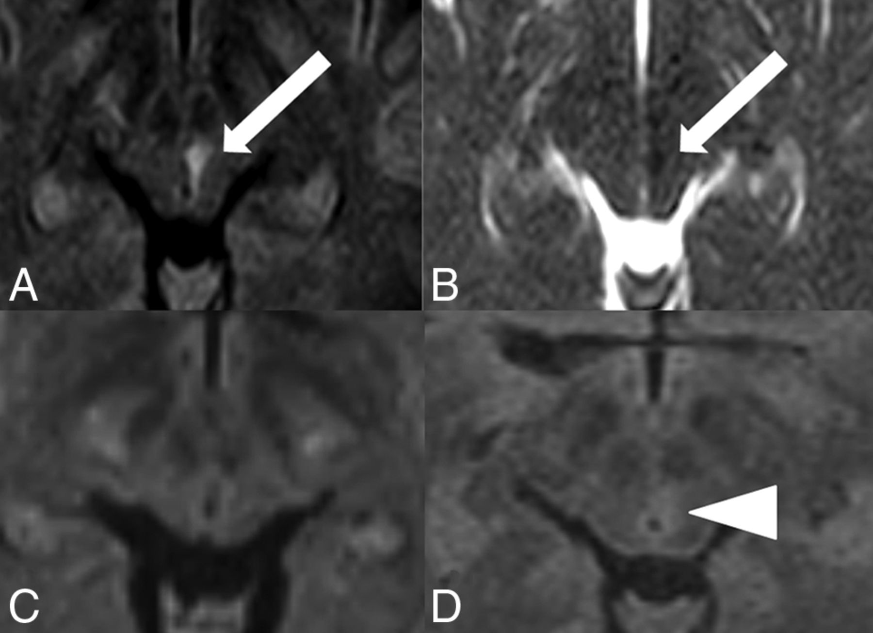
A 57-year-old woman presented with severe intractable vertigo, nystagmus, and vomiting due to isolated vestibular syndrome lasting 3 days. A punctate diffusion abnormality on DW ZOOM-EPI and ADC maps (Fig 5) in the left inferior cerebellar peduncle was present, not visible on standard DWI or FLAIR.

Fig 5.
  • Download figure
  • Open in new tab
  • Download powerpoint
Fig 5.

Acute ischemic infarct in the left inferior cerebellar peduncle. MR imaging was performed on a 3T scanner. Note a punctate area with hyperintensity in the inferior cerebellar peduncle on the left side on DW ZOOM-EPI (white arrow in A) and as a slight hypointensity on DW ZOOM-EPI ADC with an ADC of 0.562 × 10⁻³ mm2/second (white arrow in B) compared with the ADC of 0.841 × 10⁻³ mm2/second in the normal right inferior cerebellar peduncle. This diffusion abnormality was not visible on transverse standard DWI (C) or the transverse FLAIR image (D). Severe chronic microangiopathy was present in the supra- and infratentorial areas.

Acute Ischemic Infarct in the Right Nucleus Prepositus Hypoglossi.

A 40-year-old female patient presented with acute forward and backward vertigo and horizontal nystagmus. A punctate diffusion abnormality was visible on DW ZOOM-EPI and ADC maps (Fig 6) in the right nucleus prepositus hypoglossi; the lesion was not depicted on standard DWI and on FLAIR.

Fig 6.
  • Download figure
  • Open in new tab
  • Download powerpoint
Fig 6.

Acute ischemic infarct in the nucleus prepositus hypoglossi on the right side. MR imaging was performed on a 1.5T scanner. A punctate area with diffusion restriction was visible in the right nucleus prepositus hypoglossi. This lesion was hyperintense on DW ZOOM-EPI (white arrow in A) and slightly hypointense on DW ZOOM-EPI ADC with an ADC of 0.701 × 10⁻³ mm2/second (white arrow in B) compared with an ADC of 0.822 × 10⁻³ mm2/second in the normal left nucleus prepositus hypoglossi. The lesion was not visible on transverse standard DWI (C) and FLAIR (D) and was thought to be of local microangiopathic origin.

Acute Ischemic Infarct in the Right Flocculus.

A 51-year-old woman presented with vertigo and saccadic eye movements during the last 5 days. A punctate diffusion abnormality representing her infarction was present in the right flocculus on DW ZOOM-EPI and ADC maps (Fig 7). Standard DWI and FLAIR findings were normal.

Fig 7.
  • Download figure
  • Open in new tab
  • Download powerpoint
Fig 7.

Subacute ischemic infarct in the right flocculus. MR imaging was performed on a 1.5T scanner. On DW ZOOM-EPI, a punctate hyperintensity (white arrow, A) with isointensity on DW ZOOM-EPI ADC and an ADC of 0.878 × 10⁻³ mm2/second (white arrow, B) represents a subacute ischemic infarct in the right flocculus. In the normal left flocculus, the ADC was 0.926 × 10⁻³ mm2/second. Standard DWI (C) and FLAIR (D) findings were normal. The lesion was thought to be of local microangiopathic origin.

Discussion

Standard sshot SE EPI DWI is prone to geometric distortions, mainly due to the long readout time and low bandwidth in the phase-encoding direction. Reduced-FOV imaging allows acquisition of a small FOV with either reduced geometric distortion (shorter readout time) or higher spatial resolution (same readout time) compared with standard EPI DWI. An FOV smaller than the object typically induces foldover artifacts. Numerous different techniques have been introduced in the past to prevent these, especially selective excitation methods that excite only the ROI such as “inner volume imaging” and suppression-based methods that apply bands to saturate signal external to the target FOV such as “outer volume suppression” or a combination of both.3⇓⇓⇓–7

Reduced-FOV imaging or ZOOM imaging or small-FOV imaging is a commonly used technique in the context of data acquisition of small FOVs within larger objects. In this work, a noncoplanar excitation combined with outer volume suppression, originally presented by Wilm et al,8 was used. This technique is now referred to as DW ZOOM-EPI, and the main applications using small-FOV imaging are imaging of the prostate, spinal cord, pancreas, breast, and heart,5⇓⇓⇓⇓⇓–11 where a relatively small area of interest surrounded by tissue of less interest is depicted with high resolution. However, to date, the use of reduced-FOV imaging in the depiction of very small pathologic lesions with diffusion restriction in the brain stem and hippocampus has not yet been systematically evaluated.12⇓⇓⇓⇓⇓–18 The imaging examples presented in this clinical report stress but do not prove the importance of reduced-FOV imaging for the visualization of small lesions in the brain stem and hippocampus.

The increased detectability of small lesions with diffusion restriction in the brain stem and hippocampus on DW ZOOM-EPI in comparison with standard DWI might have been influenced by several technical factors:

  1. The registration effect: The standard DWI and the DW ZOOM-EPI slice selections may be slightly different. As a result, the comparison of visual lesion detectability may not entirely represent an effect of the sequence in these patients.

  2. The difference in scan time: The total scan time and thus the signal-to-noise ratio differed between standard DWI and DW ZOOM-EPI. The increased scan time in DW ZOOM-EPI automatically increased the signal-to-noise ratio and could also affect the visibility of lesions.

  3. The difference in the display window/level settings: Due to the differences in the imaging parameters between standard DWI and DW ZOOM-EPI, it was not possible to keep an identical display window/level. Window and level selection was at the discretion of the reviewer to optimally depict pathology.

Therefore, a next step might be a prospective study of standard DWI and DW ZOOM-EPI with constant imaging parameters to make a precise comparison of the 2 sequences. This would help clarify whether the improved visibility of small lesions originates from a higher SNR, higher resolution, reduced image distortion, or a combination of these factors.

Conclusions

Pathologies with diffusion restriction are routinely depicted by standard DWI. However, the visualization of very small lesions with diffusion restriction in the brain stem and hippocampus may be difficult on standard DWI. Tailored DW ZOOM-EPI enhances the visualization of very small lesions in the brain stem and hippocampus and thus can be the only MR image depicting the pathology. The acquisition of DW ZOOM-EPI may be recommended in selected patients presenting with specific brain stem syndromes or with clinical suspicion of hippocampal pathologies such as TGA or hippocampal ischemia and with negative standard DWI findings. The correct diagnosis of an ischemic-pathologic lesion can expedite correct subsequent therapy.

Acknowledgments

The authors thank Dr Simon Rauch, Institute of Radiology, Kantonsspital Winterthur, Winterthur, Switzerland, for his support in the preparation of the manuscript.

Footnotes

  • Disclosures: Christoph Binkert—UNRELATED: Consultancy: Merit Medical Systems, Biotronik; Grants/Grants Pending: Cook, Merit Medical Systems. David Czell—UNRELATED: Payment for Development of Educational Presentations: University of Zürich. Michael Wyss—UNRELATED: Employment: My position in the hospital is paid by Philips within the framework of a collaboration agreement, Comments: €4000 per year.* Erika Brüllmann—OTHER RELATIONSHIPS: Philips HealthSystems employee. *Money paid to the institution.

References

  1. 1.↵
    1. Moritani T,
    2. Smoker WRK,
    3. Sato Y, et al
    . Diffusion-weighted imaging of acute excitotoxic brain injury. AJNR Am J Neuroradiol 2005;26:216–28 pmid:15709116
    FREE Full Text
  2. 2.↵
    1. Schaefer PW,
    2. Grant PE,
    3. Gonzalez RG
    . Diffusion-weighted MRI imaging of the brain. Radiology 2000;217:331–45 doi:10.1148/radiology.217.2.r00nv24331 pmid:11058626
    CrossRefPubMed
  3. 3.↵
    1. Wargo CJ,
    2. Moore J,
    3. Gore JC
    . A comparison and evaluation of reduced-FOV methods for multi-slice 7T human imaging. Magn Reson Imaging 2013;31:1349–59 doi:10.1016/j.mri.2013.05.003 pmid:23891434
    CrossRefPubMed
  4. 4.↵
    1. Wheeler-Kingshott CA,
    2. Parker GJ,
    3. Symms MR, et al
    . ADC mapping of the human optic nerve: increased resolution, coverage, and reliability with CSF-suppressed ZOOM-EPI. Magn Reson Med 2002;47:24–31 doi:10.1002/mrm.10016 pmid:11754439
    CrossRefPubMed
  5. 5.↵
    1. Andre JB,
    2. Bammer R
    . Advanced diffusion-weighted magnetic resonance imaging techniques of the human spinal cord. Top Magn Reson Imaging 2010;21:367–78 doi:10.1097/RMR.0b013e31823e65a1 pmid:22158130
    CrossRefPubMed
  6. 6.↵
    1. Dowell NG,
    2. Jenkins TM,
    3. Ciccarelli O, et al
    . Contiguous-slice zonally oblique multislice (COZOOM) diffusion tensor imaging: examples of in vivo spinal cord and optic nerve applications. J Magn Reson Imaging 2009;29:454–60 doi:10.1002/jmri.21656 pmid:19161202
    CrossRefPubMed
  7. 7.↵
    1. Samson RS,
    2. Lévy S,
    3. Schneider T, et al
    . ZOOM or non-ZOOM? Assessing spinal cord diffusion tensor imaging protocols for multi-centre studies. PLoS One 2016;11:e0155557 doi:10.1371/journal.pone.0155557 pmid:27171194
    CrossRefPubMed
  8. 8.↵
    1. Wilm BJ,
    2. Svensson J,
    3. Henning A, et al
    . Reduced field-of-view MRI using outer volume suppression for spinal cord diffusion imaging. Magn Reson Med 2007;57:625–30 doi:10.1002/mrm.21167 pmid:17326167
    CrossRefPubMed
  9. 9.↵
    1. Dong H,
    2. Li Y,
    3. Li H, et al
    . Study of the reduced field-of-view diffusion-weighted imaging of the breast. Clin Breast Cancer 2014;14:265–71 doi:10.1016/j.clbc.2013.12.001 pmid:24462803
    CrossRefPubMed
  10. 10.↵
    1. Kim H,
    2. Lee JM,
    3. Yoon JH, et al
    . Reduced field-of-view diffusion-weighted magnetic resonance imaging of the pancreas: comparison with conventional single-shot echo-planar imaging. Korean J Radiol 2015;16:1216–25 doi:10.3348/kjr.2015.16.6.1216 pmid:26576110
    CrossRefPubMed
  11. 11.↵
    1. Reischauer C,
    2. Wilm BJ,
    3. Froehlich JM, et al
    . High-resolution diffusion tensor imaging of prostate cancer using a reduced FOV technique. Eur J Radiol 2011;80:34–41 doi:10.1016/j.ejrad.2010.06.038 pmid:20638208
    CrossRefPubMed
  12. 12.↵
    1. Förster A,
    2. Griebe M,
    3. Gass A, et al
    . Diffusion-weighted imaging for the differential diagnosis of disorders affecting the hippocampus. Cerebrovasc Dis 2012;33:104–15 doi:10.1159/000332036 pmid:22179485
    CrossRefPubMed
  13. 13.↵
    1. Bartsch T,
    2. Deuschl G
    . Transient global amnesia: functional anatomy and clinical implications. Lancet Neurol 2010;9:205–14 doi:10.1016/S1474-4422(09)70344-8 pmid:20129169
    CrossRefPubMed
  14. 14.↵
    1. Weon YC,
    2. Kim JH,
    3. Lee JS, et al
    . Optimal diffusion-weighted imaging protocol for lesion detection in transient global amnesia. AJNR Am J Neuroradiol 2008;29:1324–28 doi:10.3174/ajnr.A1105 pmid:18451087
    Abstract/FREE Full Text
  15. 15.↵
    1. Dekezyer S,
    2. De Kock S,
    3. Nikoubashman O, et al
    . “Unforgettable”: a pictorial essay on anatomy and pathology of the hippocampus. Insights Imaging 2017;8:199–212 doi:10.1007/s13244-016-0541-2 pmid:28108955
    CrossRefPubMed
  16. 16.↵
    1. Szabo K,
    2. Förster A,
    3. Jäger Th, et al
    . Hippocampal lesion patterns in acute posterior cerebral artery stroke: clinical and MRI findings. Stroke 2009;40:2042–45 doi:10.1161/STROKEAHA.108.536144 pmid:19359650
    Abstract/FREE Full Text
  17. 17.↵
    1. Kim HJ,
    2. Lee SH,
    3. Park JH, et al
    . Isolated vestibular nuclear infarction: report of two cases and review of the literature. J Neurol 2014;261:121–29 doi:10.1007/s00415-013-7139-0 pmid:24162036
    CrossRefPubMed
  18. 18.↵
    1. Kim SH,
    2. Park SH,
    3. Kim HJ, et al
    . Isolated central vestibular syndrome. Ann N.Y. Acad Sci 2015;1343:2015:80–89 doi:10.1111/nyas.12712 pmid:25735822
    CrossRefPubMed
  • Received October 19, 2017.
  • Accepted after revision March 5, 2018.
  • © 2018 by American Journal of Neuroradiology
View Abstract
PreviousNext
Back to top

In this issue

American Journal of Neuroradiology: 39 (7)
American Journal of Neuroradiology
Vol. 39, Issue 7
1 Jul 2018
  • Table of Contents
  • Index by author
  • Complete Issue (PDF)
Advertisement
Print
Download PDF
Email Article

Thank you for your interest in spreading the word on American Journal of Neuroradiology.

NOTE: We only request your email address so that the person you are recommending the page to knows that you wanted them to see it, and that it is not junk mail. We do not capture any email address.

Enter multiple addresses on separate lines or separate them with commas.
Diffusion-Weighted Zonal Oblique Multislice–EPI Enhances the Detection of Small Lesions with Diffusion Restriction in the Brain Stem and Hippocampus: A Clinical Report of Selected Cases
(Your Name) has sent you a message from American Journal of Neuroradiology
(Your Name) thought you would like to see the American Journal of Neuroradiology web site.
CAPTCHA
This question is for testing whether or not you are a human visitor and to prevent automated spam submissions.
Cite this article
T. Sartoretti, E. Sartoretti, C. Binkert, A. Gutzeit, C. Reischauer, D. Czell, M. Wyss, E. Brüllmann, S. Sartoretti-Schefer
Diffusion-Weighted Zonal Oblique Multislice–EPI Enhances the Detection of Small Lesions with Diffusion Restriction in the Brain Stem and Hippocampus: A Clinical Report of Selected Cases
American Journal of Neuroradiology Jul 2018, 39 (7) 1255-1259; DOI: 10.3174/ajnr.A5635

Citation Manager Formats

  • BibTeX
  • Bookends
  • EasyBib
  • EndNote (tagged)
  • EndNote 8 (xml)
  • Medlars
  • Mendeley
  • Papers
  • RefWorks Tagged
  • Ref Manager
  • RIS
  • Zotero
0 Responses
Respond to this article
Share
Bookmark this article
Diffusion-Weighted Zonal Oblique Multislice–EPI Enhances the Detection of Small Lesions with Diffusion Restriction in the Brain Stem and Hippocampus: A Clinical Report of Selected Cases
T. Sartoretti, E. Sartoretti, C. Binkert, A. Gutzeit, C. Reischauer, D. Czell, M. Wyss, E. Brüllmann, S. Sartoretti-Schefer
American Journal of Neuroradiology Jul 2018, 39 (7) 1255-1259; DOI: 10.3174/ajnr.A5635
del.icio.us logo Twitter logo Facebook logo Mendeley logo
  • Tweet Widget
  • Facebook Like
  • Google Plus One
Purchase

Jump to section

  • Article
    • Abstract
    • ABBREVIATIONS:
    • Materials and Methods
    • Discussion
    • Conclusions
    • Acknowledgments
    • Footnotes
    • References
  • Figures & Data
  • Info & Metrics
  • Responses
  • References
  • PDF

Related Articles

  • No related articles found.
  • PubMed
  • Google Scholar

Cited By...

  • Structural Connectivity and Cortical Thickness Alterations in Transient Global Amnesia
  • Crossref (7)
  • Google Scholar

This article has been cited by the following articles in journals that are participating in Crossref Cited-by Linking.

  • Prospective Comparison of Reduced Field-of-View (rFOV) and Full FOV (fFOV) Diffusion-Weighted Imaging (DWI) in the Assessment of Insulinoma: Image Quality and Lesion Detection
    Ming He, Jin Xu, Zhaoyong Sun, Xiaoqi Wang, Jiazheng Wang, Feng Feng, Huadan Xue, Zhengyu Jin
    Academic Radiology 2020 27 11
  • Structural Connectivity and Cortical Thickness Alterations in Transient Global Amnesia
    J. Hodel, X. Leclerc, M. Zuber, S. Gerber, P. Besson, V. Marcaud, V. Roubeau, H. Brasme, I. Ganzoui, D. Ducreux, J.-P. Pruvo, M. Bertoux, M. Zins, R. Lopes
    American Journal of Neuroradiology 2020 41 5
  • Single shot zonal oblique multislice SE-EPI diffusion-weighted imaging with low to ultra-high b-values for the differentiation of benign and malignant vertebral spinal fractures
    Elisabeth Sartoretti, Sabine Sartoretti-Schefer, Luuk van Smoorenburg, Barbara Eichenberger, Árpád Schwenk, David Czell, Alex Alfieri, Andreas Gutzeit, Manoj Mannil, Christoph A. Binkert, Michael Wyss, Thomas Sartoretti
    European Journal of Radiology Open 2021 8
  • The Abducens Nerve: Anatomy and Pathology
    Luciane Lucas Lucio, Tomás de Andrade Lourenção Freddi, Ana Carolina Ottaiano
    Seminars in Ultrasound, CT and MRI 2022 43 5
  • Comparison Between EPI DWI and PROPELLER DWI in Brain MR Imaging
    Panayiotis Mavroidis, Eleonora Giankou, Maria Papaioannou, Violeta Roka, Aleksandra Tsikrika, Spiros Kostopoulos, Dimitrios Glotsos, Giorgos K. Sakkas, Efthimios Dardiotis, Dimitrios Chaniotis, Eftichia Kapsalaki, Eleftherios Lavdas
    Current Problems in Diagnostic Radiology 2024 53 1
  • Isolated oculomotor nerve palsy due to mesencephalic infarction diagnosed by ZOOM DWI
    Shangpei Wang, Yajie Cai, Xiaosan Wu, Sunhong Yan, Longsheng Wang
    BMC Neurology 2025 25 1
  • Magnetic resonance imaging of the cerebellopontine angle: comparison between constructive interference steady-state and small field-of-view technique turbo spin echo sequences
    Valerio Vitale, Alessia Varotto, Ilaria Cracco, Mariam Mansour, Loris Di Clemente, Fabio Angelo Raneri, Alberto Tecchio, Giuseppe Iannucci
    Die Radiologie 2024 64 S1

More in this TOC Section

  • Diagnostic Neuroradiology of Monoclonal Antibodies
  • Clinical Outcomes After Chiari I Decompression
  • Segmentation of Brain Metastases with BLAST
Show more Adult Brain

Similar Articles

Advertisement

Indexed Content

  • Current Issue
  • Accepted Manuscripts
  • Article Preview
  • Past Issues
  • Editorials
  • Editor's Choice
  • Fellows' Journal Club
  • Letters to the Editor
  • Video Articles

Cases

  • Case Collection
  • Archive - Case of the Week
  • Archive - Case of the Month
  • Archive - Classic Case

Special Collections

  • AJNR Awards
  • ASNR Foundation Special Collection
  • Most Impactful AJNR Articles
  • Photon-Counting CT
  • Spinal CSF Leak Articles (Jan 2020-June 2024)

More from AJNR

  • Trainee Corner
  • Imaging Protocols
  • MRI Safety Corner

Multimedia

  • AJNR Podcasts
  • AJNR Scantastics

Resources

  • Turnaround Time
  • Submit a Manuscript
  • Submit a Video Article
  • Submit an eLetter to the Editor/Response
  • Manuscript Submission Guidelines
  • Statistical Tips
  • Fast Publishing of Accepted Manuscripts
  • Graphical Abstract Preparation
  • Imaging Protocol Submission
  • Evidence-Based Medicine Level Guide
  • Publishing Checklists
  • Author Policies
  • Become a Reviewer/Academy of Reviewers
  • News and Updates

About Us

  • About AJNR
  • Editorial Board
  • Editorial Board Alumni
  • Alerts
  • Permissions
  • Not an AJNR Subscriber? Join Now
  • Advertise with Us
  • Librarian Resources
  • Feedback
  • Terms and Conditions
  • AJNR Editorial Board Alumni

American Society of Neuroradiology

  • Not an ASNR Member? Join Now

© 2025 by the American Society of Neuroradiology All rights, including for text and data mining, AI training, and similar technologies, are reserved.
Print ISSN: 0195-6108 Online ISSN: 1936-959X

Powered by HighWire