
- Fig 1.
MR images obtained in a 13-year-old girl with early-onset thoracolumbar scoliosis.
A, Sagittal T1-weighted image (500/12 [TR/TE]) of the brain shows depression of the floor of the fourth ventricle (arrowhead). The pons and medulla oblongata have a reduced volume.
B, Axial T2-weighted image (4500/120) at the level of the medulla oblongata shows rectangular configuration of the medulla. The floor of the fourth ventricle is tent shaped (arrows), with missing prominence of the cuneate and gracile nuclei. The inferior olivary nuclei (IO) are prominent with respect to the pyramids (P).
C, Axial T2-weighted image (4500/120) at the level of the pons shows absence of the facial colliculi, with tent-shaped configuration of the floor of the fourth ventricle (arrows). A deep midsagittal cleft extends ventrally from the fourth ventricular floor, producing the split pons sign (arrowhead).
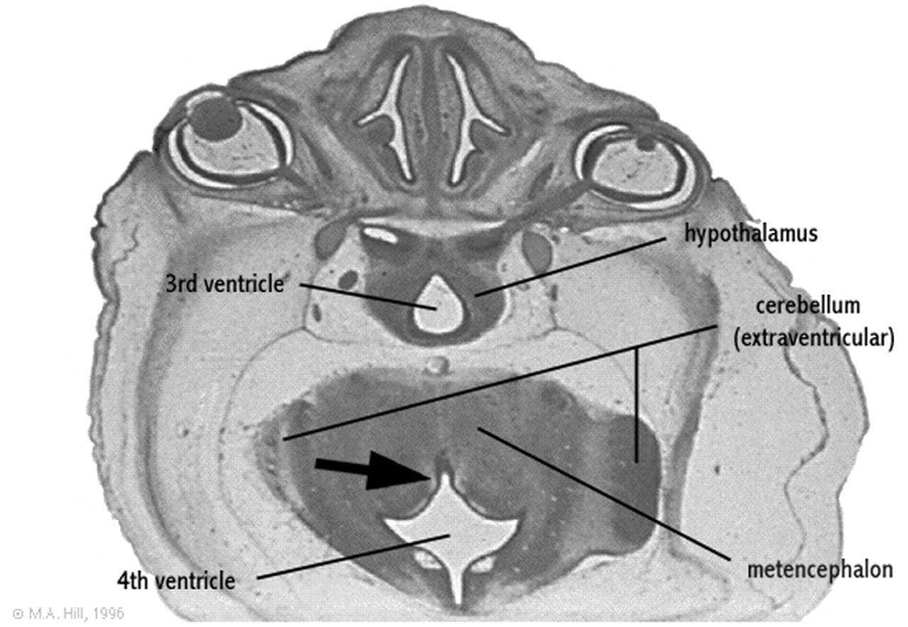
- Fig 2.
Rendering of the embryologic development of the fourth ventricle.
Axial section of a 27-mm human embryo (approximately day 56 and Carnegie stage 22) shows ventral fourth ventricular furrow (arrow) deeply indenting the posterior aspect of the developing metencephalon.
Modified from M. Hill, UNSW Embryology, version 3.0 (8), with permission.

- Copyright © American Society of Neuroradiology


