
- Fig 1.
Case 1.
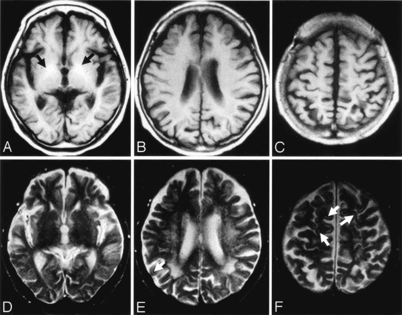
A–C, Axial T1-weighted MR images obtained 2 days after admission. T1-weighted image (A) shows hyperintensities in the globus pallidus on both sides (arrows). T1-weighted images show no definite signal intensity changes in the cerebral cortices (A–C).
D–F, T2-weighted MR images obtained 2 days after admission. Signal intensity abnormalities are difficult to discern on T2-weighted image (D). T2-weighted images (E and F) show focal, faint hyperintensities in the cerebral cortices (arrows). Confluent and diffuse T2 hyperintensities are seen in the deep and subcortical cerebral white matters (E).
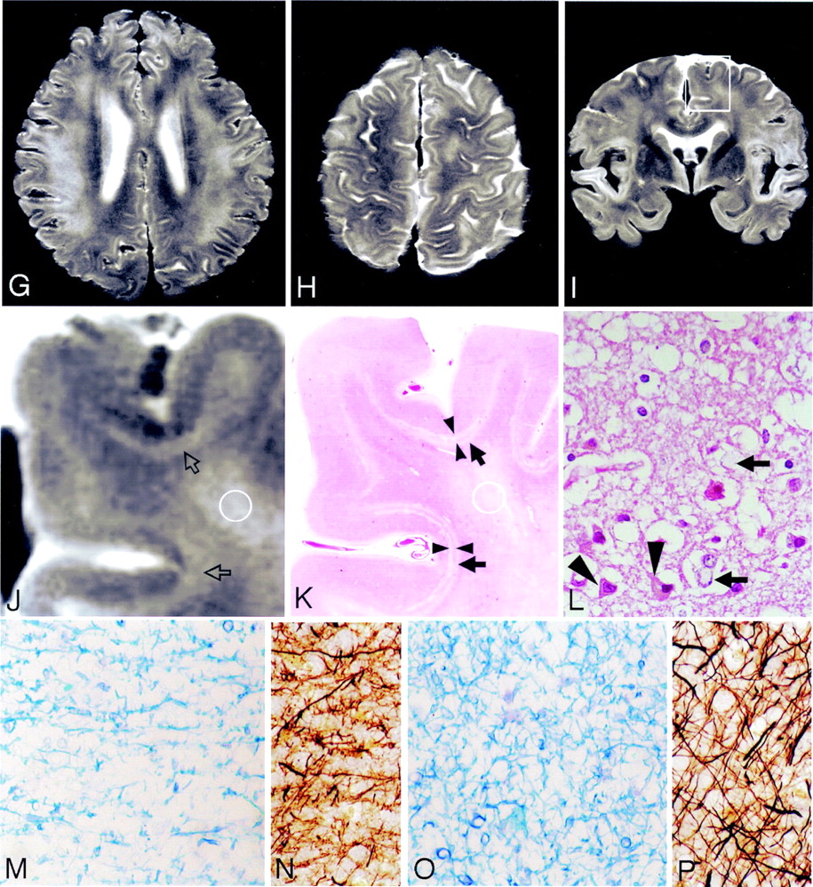
G and H, Postmortem axial T2-weighted MR images show deep cortical and subcortical laminar hyperintensities in the entire cerebral hemisphere. Diffuse T2 hypertensities are seen in the deep and subcortical cerebral white matters (G).
I and J, Coronal T2-weighted MR images show deep cortical and subcortical laminar hyperintensities in the entire cerebral hemisphere. Hyperintensities are also seen in the deep and subcortical white matters (J). Boxed area in panel I shows hyperintensities in the deep layers of the cerebral cortices and subcortical white matters (open arrows) as well as deep cerebral white matter. The boundary between deep layers of the cerebral cortex and subcortical white matter is not clear.
K, The hematoxylin and eosin stained section corresponding to panel J shows laminar pallor of the deep layers of the cerebral cortex (arrowheads) and laminar pallor of the underlying U-fibers (arrows), as well as moderate pallor of the deep cerebral white matter. (Hematoxylin and eosin stain ×100.)
L, Histologic findings of the arrowhead area in panel K shows spongy changes of the neuropil, pyknotic, and shrunken neurons (arrowheads) and Alzheimer type II astrocytes (arrows), which have large pale nuclei with marginated chromatin and scanty cytoplasms. (Hematoxylin and eosin stain ×100.)
M and N, Histologic findings of the U-fibers (K, arrows) show tissue rarefaction and severe loss of myelin (M) and axons (N). (M, Klüver-Barrera stain ×100; N, Bielschowsky stain ×100.)
O and P, Histologic findings of the circled area in panels J and K reveals moderate loss of myelin (O) and axons (P). (O, Klüver-Barrera stain ×100; P, Bielschowsky stain ×100.)
- Fig 2.
Case 2.
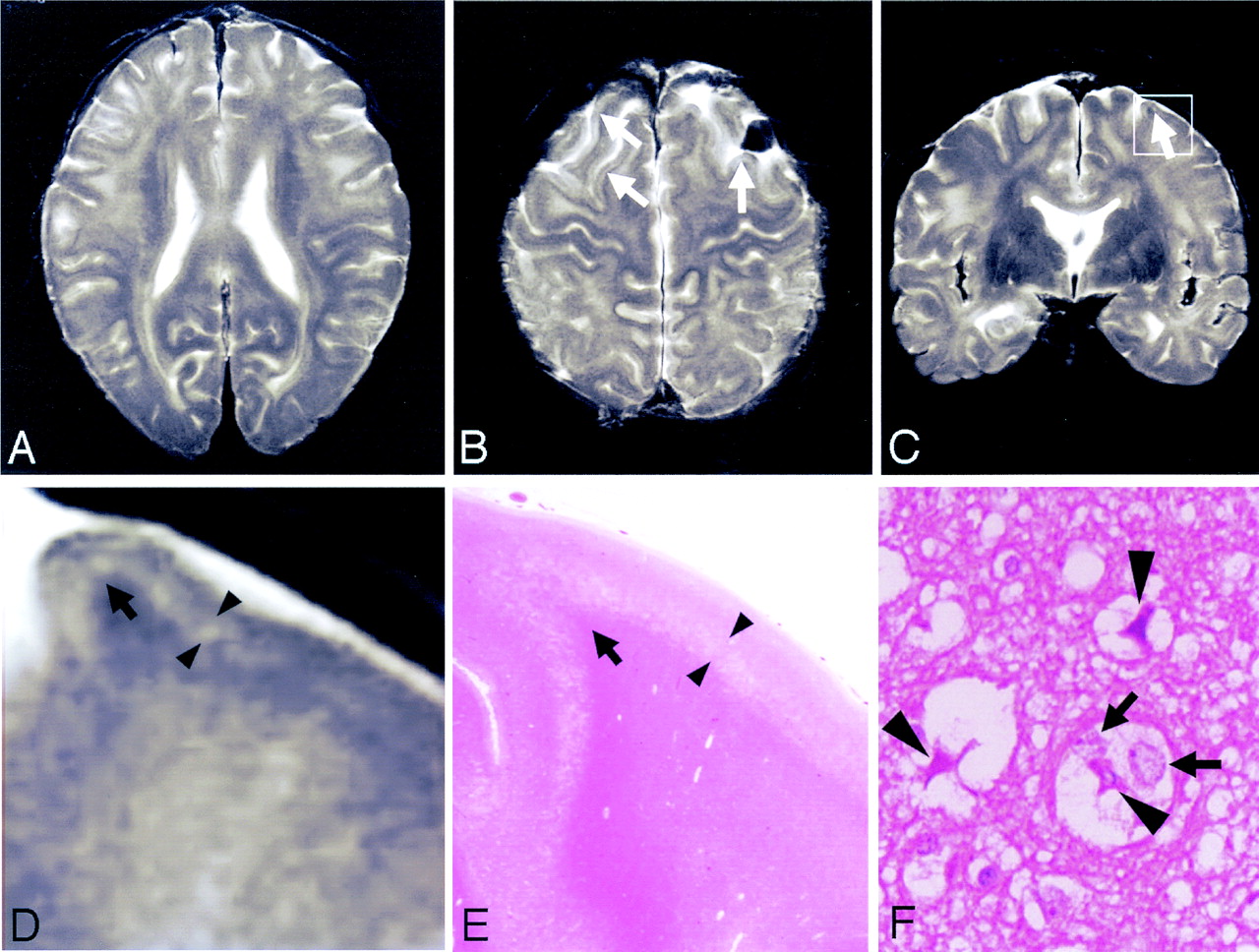
A–C, Postmortem axial (A and B) and coronal (C) T2-weighted MR images show cortical laminar hyperintensities in the frontal lobes (B and C, arrows). Hyperintensities are also seen in the deep and subcortical white matters.
D, Boxed area in panel C shows laminar hyperintensities in the deep layers of the cerebral cortex (arrowheads).
E, The hematoxylin and eosin-stained section corresponding to panel D shows laminar pallor of the deep layers of the cerebral cortex (arrowheads). U-fibers are well preserved in this case (D and E, arrows).
F, Histologic findings of the arrowhead area panel E shows spongy changes of the neuropil, pyknotic, and shrunken neurons (arrowheads) and Alzheimer type II astrocytes (arrows). (Hematoxylin and eosin ×100.)


- Copyright © American Society of Neuroradiology
In this issue


Jump to section
Related Articles
Cited By...
- Multimodality MR Imaging Findings of Low-Grade Brain Edema in Hepatic Encephalopathy
- Brain Magnetic Resonance Imaging Findings in Young Patients with Hepatosplenic Schistosomiasis Mansoni without Overt Symptoms
- Acute Hepatic Encephalopathy: Diffusion-Weighted and Fluid-Attenuated Inversion Recovery Findings, and Correlation with Plasma Ammonia Level and Clinical Outcome