
Article Figures & Data
Figures
- fig 1.
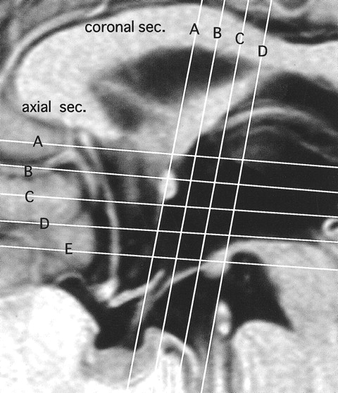
Horizontal and vertical white lines show section planes in axial and coronal views in figs 2 A–E and 3 A–D, respectively
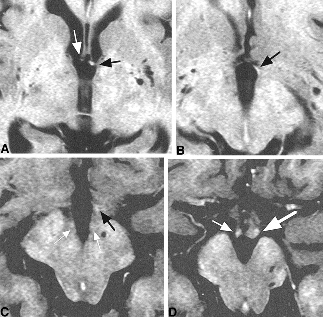
- fig 2.
Axial images of consecutive five sections in one healthy subject shown in figure 1 (A–E) and one section in another subject (F).
A, Fornical columns (arrow) were observed in one section level above the anterior commissure.
B, PF (white arrow) was observed as a high-signal-intensity spot just behind the anterior commissure (black arrow) and exposed to the cerebrospinal fluid space of the third ventricle.
C, In the next lower section, PF (arrow) was still exposed to the third ventricle.
D, In the next lower section, ovoid PF (white arrow) directing posteriorly toward the mammillary body was seen. Ill-defined high-signal-intensity spot (MT; black arrow) was visible 4 mm posterior to PF.
E, At the next level, PF (white arrow) joined to the mammillary body (black arrow) anteriorly and laterally.
F, PF (white arrow) was visible but ill-defined bilaterally. Obscure PF was seen in 6% of healthy subjects. MT (black arrow) was identifiable in this case.
- fig 3.
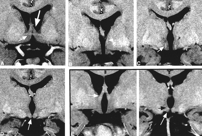
Coronal images of four consecutive sections in one healthy subject (A–D) and two sections in another subject (E and F).
A, The fornical columns (large arrow) were evident at the anterior commissure (small arrow) section.
B, In the next posterior section, the descending portion of PF (arrow) was identifiable, more clearly on the left.
C, In the next posterior section, the descending portion of PF (arrow) was evident bilaterally, above the floor of the third ventricle. It was visible in 80% of the healthy subjects.
D, In the next section, the mammillary body (arrow) was identifiable. At this level, the entry point of the PF was unidentifiable.
E, The fornical columns (arrow) were evident.
F, At the level of mammillary body (white arrow), the origin of MT (black arrow) was visible.
- fig 4.
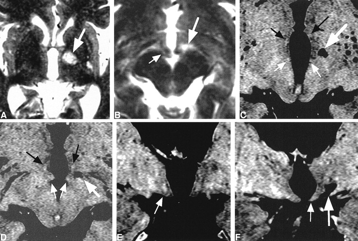
Case 1.
A, Sagittal image shows a huge left hypothalamic mass lesion.
B, Coronal image shows that the right fornical column (small arrow) and PF (large arrow) were displaced off the midline, and the left PF was not visible.
C, Right fornical column or PF (arrow) was identifiable as a slightly high-signal-intensity spot. Left one was unidentifiable because of a huge hypothalamic mass.
D and E, Right PF (small white arrow) and MT (small black arrow) were visible. Left PF (large white arrow) and MT (large black arrow) were displaced inferiorly and deformed by the mass.
F, Right PF (small white arrow) and mammillary body (small black arrow) were displaced laterally. Left mammillary body (large black arrow) was shifted inferiorly, and left PF (large white arrow) was clearly visible as a high-signal-intensity spot.
- fig 5.
Case 2.
A and B, T2-weighted axial images. A high-signal-intensity spot (large arrow), 8 mm in maximum size and compatible with the diagnosis of lacunar infarction, was visible in the left hypothalamus in A and lateral to the superior part of the mammillary body (small arrow) in B.
C and D, Axial T2R images. A lacunar infarct (large white arrow) was visible in the left hypothalamus. PFs (black arrows) and MTs (small white arrows) were visible. Both white matter tracts seemed slightly smaller or less evident in signal intensity on the left.
E, Coronal T2R image. Descending portion of PF (arrow) was more evident on the right than on the left, which is compatible with the axial images in fig 5C and D.
F, Coronal T2R image. Left mammillary body (small arrow) was decreased in size compared with the right. A lacunar infarct (large arrow) was visible a few millimeters lateral to the left third ventricle wall.
- fig 6.
Case 3.
A, T1-weighted coronal image with enhancement. A suprasellar mass with an almost homogeneous enhancement effect was noted.
B-D, T2R coronal MR images.
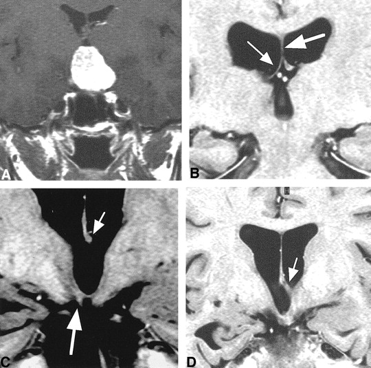
B, The right half of fornical body (small arrow), attached to the inferior surface of the septum pellucidum (large arrow), was thin and decreased in size.
C, The left half of the fornical body (small arrow) was seen. The right mammillary body was smaller than the left one (large arrow).
D, A fornical column was seen on the left only (arrow).
- fig 7.
Case 3. Axial T2R images.
A, Fornical column was visible on the left side (black arrow). The right fornical column (white arrow) was decreased in size.
B, Only the left PF (arrow) was visible.
C, PF (black arrow) was visible on the left side only. MT (white arrows) was visible bilaterally.
D, Although bilateral mammillary bodies (arrows) were visible, the right side (small arrow) was decreased in size. The size decrease was compatible with the coronal section (fig 6C).


In this issue


Jump to section
Related Articles
Cited By...
- No citing articles found.