Advertisement

Main menu

  • Home
  • Content
    • Current Issue
    • Accepted Manuscripts
    • Article Preview
    • Past Issue Archive
    • Video Articles
    • AJNR Case Collection
    • Case of the Week Archive
    • Case of the Month Archive
    • Classic Case Archive
  • Special Collections
    • AJNR Awards
    • ASNR Foundation Special Collection
    • Most Impactful AJNR Articles
    • Photon-Counting CT
    • Spinal CSF Leak Articles (Jan 2020-June 2024)
  • Multimedia
    • AJNR Podcasts
    • AJNR SCANtastic
    • Trainee Corner
    • MRI Safety Corner
    • Imaging Protocols
  • For Authors
    • Submit a Manuscript
    • Submit a Video Article
    • Submit an eLetter to the Editor/Response
    • Manuscript Submission Guidelines
    • Statistical Tips
    • Fast Publishing of Accepted Manuscripts
    • Graphical Abstract Preparation
    • Imaging Protocol Submission
    • Author Policies
  • About Us
    • About AJNR
    • Editorial Board
    • Editorial Board Alumni
  • More
    • Become a Reviewer/Academy of Reviewers
    • Subscribers
    • Permissions
    • Alerts
    • Feedback
    • Advertisers
    • ASNR Home

User menu

  • Alerts
  • Log in

Search

  • Advanced search
American Journal of Neuroradiology

American Journal of Neuroradiology

ASHNR American Society of Functional Neuroradiology ASHNR American Society of Pediatric Neuroradiology ASSR
  • Alerts
  • Log in

Advanced Search

  • Home
  • Content
    • Current Issue
    • Accepted Manuscripts
    • Article Preview
    • Past Issue Archive
    • Video Articles
    • AJNR Case Collection
    • Case of the Week Archive
    • Case of the Month Archive
    • Classic Case Archive
  • Special Collections
    • AJNR Awards
    • ASNR Foundation Special Collection
    • Most Impactful AJNR Articles
    • Photon-Counting CT
    • Spinal CSF Leak Articles (Jan 2020-June 2024)
  • Multimedia
    • AJNR Podcasts
    • AJNR SCANtastic
    • Trainee Corner
    • MRI Safety Corner
    • Imaging Protocols
  • For Authors
    • Submit a Manuscript
    • Submit a Video Article
    • Submit an eLetter to the Editor/Response
    • Manuscript Submission Guidelines
    • Statistical Tips
    • Fast Publishing of Accepted Manuscripts
    • Graphical Abstract Preparation
    • Imaging Protocol Submission
    • Author Policies
  • About Us
    • About AJNR
    • Editorial Board
    • Editorial Board Alumni
  • More
    • Become a Reviewer/Academy of Reviewers
    • Subscribers
    • Permissions
    • Alerts
    • Feedback
    • Advertisers
    • ASNR Home
  • Follow AJNR on Twitter
  • Visit AJNR on Facebook
  • Follow AJNR on Instagram
  • Join AJNR on LinkedIn
  • RSS Feeds

AJNR is seeking candidates for the AJNR Podcast Editor. Read the position description.

Research ArticleHEAD AND NECK

Foramen Tympanicum, or Foramen of Huschke: Pathologic Cases and Anatomic CT Study

Alexis Lacout, Kathlyn Marsot-Dupuch, Wendy R. K. Smoker and Pierre Lasjaunias
American Journal of Neuroradiology June 2005, 26 (6) 1317-1323;
Alexis Lacout
  • Find this author on Google Scholar
  • Find this author on PubMed
  • Search for this author on this site
Kathlyn Marsot-Dupuch
  • Find this author on Google Scholar
  • Find this author on PubMed
  • Search for this author on this site
Wendy R. K. Smoker
  • Find this author on Google Scholar
  • Find this author on PubMed
  • Search for this author on this site
Pierre Lasjaunias
  • Find this author on Google Scholar
  • Find this author on PubMed
  • Search for this author on this site
  • Article
  • Figures & Data
  • Info & Metrics
  • Responses
  • References
  • PDF
Loading

Article Figures & Data

Figures

  • Tables
  • Fig 1.
    • Download figure
    • Open in new tab
    • Download powerpoint
    Fig 1.

    HRCT sections of the temporal bone through the inferior portion of the EAC. Images show the anteroinferior location of the foramen tympanicum, posterior to the TMJ (arrowhead).

    A, Axial.

    B, Sagittal.

  • Fig 2.
    • Download figure
    • Open in new tab
    • Download powerpoint
    Fig 2.

    Axial HRCT sections of the temporal bone in two patients through the same level at the inferior portion of the EAC.

    A, Normal tympanic bone.

    B, Focal anteroinferior reduction in tympanic bone thickness 2 mm lateral to the tympanic membrane (arrowhead).

  • Fig 3.
    • Download figure
    • Open in new tab
    • Download powerpoint
    Fig 3.

    Axial HRCT sialograms (soft tissue window) of the left parotid gland through the inferior portion of the EAC show (arrow) submucosal nodular enhancement in the foramen tympanicum, suggesting ectopic salivary-gland tissue.

  • Fig 4.
    • Download figure
    • Open in new tab
    • Download powerpoint
    Fig 4.

    Axial HRCT sections (bone window) of the temporal bone through the inferior portion of the EAC.

    A, Closed-mouth view shows herniation of soft tissue herniation into the EAC 2 mm lateral to the tympanic membrane (arrowhead).

    B, Open-mouth view show subcutaneous air pockets spreading along the TMJ (arrow). Slight retraction of the soft tissue material is noted when the mouth is open.

    C, Contrast-enhanced T1-weighted MR image (TE/TR, 500 /15) through the inferior portion of the EAC shows enhancement of the herniating soft tissue (arrowhead).
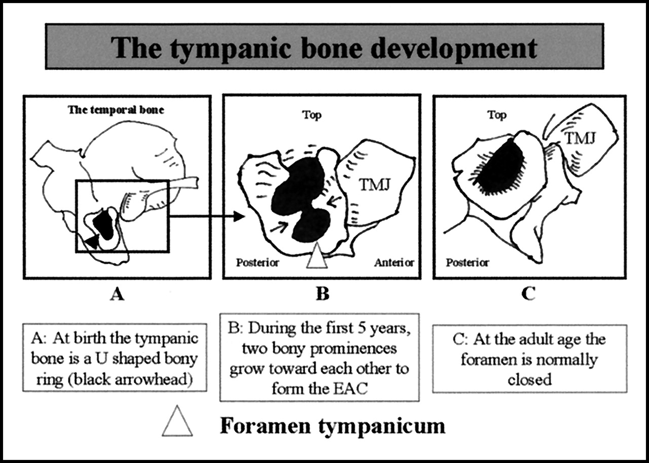
  • Fig 5.
    • Download figure
    • Open in new tab
    • Download powerpoint
    Fig 5.

    Schema illustrates normal postnatal development of the tympanic bone (used with permission from Anson BJ, Donaldson JA. Surgical Anatomy of the Temporal Bone. 3rd ed. Philadelphia: W. B. Saunders; 1981:122).

Tables

  • Figures
  • Foramen tympanicum reported in the literature

    Patient Age (y)/SexSide and HistoryPhysical FindingImaging FindingDiagnosis
    Heffez et al. 19893
        67/FR, painSwelling/OMRCT: bony defect, MRI: herniationTMJ herniation
        53/FR, painSwelling/OMRPolystomograms: tympanic plate defectTMJ herniation
    Weissman et al, 19914
        15/FLSwelling/OMRNATMJ herniation
    Sharma et al, 19845
        58/FR, otorrheaAnterior punctum, flow while sucking on a sweetNASalivary fistula
    Chilla, 20026
        62/FL, otorrheaAnterior polypNASalivary fistula
    Dingle, 19927
        75/MR, painSwelling/OMRNAEAC fistula
    Tahir and Rubinstein 200017
        64/MR, HLClear fluid on anterior wallCT: bony defectArthritis
    Hawke et al, 198718
        68/FR/L, HLSwelling/OMRCT: anterior bony defect, soft tissue herniation/OM anterior retractionMeniscus herniation
    Cecire and Austin, 199119
        60/ML, otorrheaPolypCT: anterior defect, soft tissue herniationPolyp
    Hawke et al, 198820
        58/FR, otorrheaSwelling/OMRCT: R/L dehiscence of anterior wallEAC fistula
    Our study
        52/FL, otorrheaNormalSialo-CT: subcutaneous nodular enhancementSalivary fistula
        90/FR, painSwellingCT: soft tissue herniationTMJ herniation
    Our series
        58/FLNACT: foramen tympanicumNA
        38/FR/LNACT: foramen tympanicumNA
        92/FRNACT: foramen tympanicumNA
        39/MR/LNACT: foramen tympanicumNA
    • Note.—NA = not applicable, OMR = retraction upon opening of mouth, HL = hearing loss.

PreviousNext
Back to top

In this issue

American Journal of Neuroradiology: 26 (6)
American Journal of Neuroradiology
Vol. 26, Issue 6
1 Jun 2005
  • Table of Contents
  • Index by author
Advertisement
Print
Download PDF
Email Article

Thank you for your interest in spreading the word on American Journal of Neuroradiology.

NOTE: We only request your email address so that the person you are recommending the page to knows that you wanted them to see it, and that it is not junk mail. We do not capture any email address.

Enter multiple addresses on separate lines or separate them with commas.
Foramen Tympanicum, or Foramen of Huschke: Pathologic Cases and Anatomic CT Study
(Your Name) has sent you a message from American Journal of Neuroradiology
(Your Name) thought you would like to see the American Journal of Neuroradiology web site.
CAPTCHA
This question is for testing whether or not you are a human visitor and to prevent automated spam submissions.
Cite this article
Alexis Lacout, Kathlyn Marsot-Dupuch, Wendy R. K. Smoker, Pierre Lasjaunias
Foramen Tympanicum, or Foramen of Huschke: Pathologic Cases and Anatomic CT Study
American Journal of Neuroradiology Jun 2005, 26 (6) 1317-1323;

Citation Manager Formats

  • BibTeX
  • Bookends
  • EasyBib
  • EndNote (tagged)
  • EndNote 8 (xml)
  • Medlars
  • Mendeley
  • Papers
  • RefWorks Tagged
  • Ref Manager
  • RIS
  • Zotero
0 Responses
Respond to this article
Share
Bookmark this article
Foramen Tympanicum, or Foramen of Huschke: Pathologic Cases and Anatomic CT Study
Alexis Lacout, Kathlyn Marsot-Dupuch, Wendy R. K. Smoker, Pierre Lasjaunias
American Journal of Neuroradiology Jun 2005, 26 (6) 1317-1323;
del.icio.us logo Twitter logo Facebook logo Mendeley logo
  • Tweet Widget
  • Facebook Like
  • Google Plus One

Jump to section

  • Article
    • Abstract
    • Methodsm
    • Results
    • Discussion
    • Conclusion
    • Acknowledgments
    • Footnotes
    • References
  • Figures & Data
  • Info & Metrics
  • Responses
  • References
  • PDF

Related Articles

  • No related articles found.
  • PubMed
  • Google Scholar

Cited By...

  • A Persistent Foramen of Huschke: A Small Road to Misery in Necrotizing External Otitis
  • Cone beam CT detection of foramen tympanicum or foramen of huschke
  • Foramen tympanicum or foramen of Huschke: anatomical cone beam CT study
  • Crossref
  • Google Scholar

This article has not yet been cited by articles in journals that are participating in Crossref Cited-by Linking.

More in this TOC Section

  • Correlation of Apparent Diffusion Coefficient at 3T with Prognostic Parameters of Retinoblastoma
  • Parathyroid Lesions: Characterization with Dual-Phase Arterial and Venous Enhanced CT of the Neck
  • MR Diagnosis of Facial Neuritis: Diagnostic Performance of Contrast-Enhanced 3D-FLAIR Technique Compared with Contrast-Enhanced 3D-T1-Fast-Field Echo with Fat Suppression
Show more HEAD AND NECK

Similar Articles

Advertisement

Indexed Content

  • Current Issue
  • Accepted Manuscripts
  • Article Preview
  • Past Issues
  • Editorials
  • Editor's Choice
  • Fellows' Journal Club
  • Letters to the Editor
  • Video Articles

Cases

  • Case Collection
  • Archive - Case of the Week
  • Archive - Case of the Month
  • Archive - Classic Case

Special Collections

  • AJNR Awards
  • ASNR Foundation Special Collection
  • Most Impactful AJNR Articles
  • Photon-Counting CT
  • Spinal CSF Leak Articles (Jan 2020-June 2024)

More from AJNR

  • Trainee Corner
  • Imaging Protocols
  • MRI Safety Corner

Multimedia

  • AJNR Podcasts
  • AJNR Scantastics

Resources

  • Turnaround Time
  • Submit a Manuscript
  • Submit a Video Article
  • Submit an eLetter to the Editor/Response
  • Manuscript Submission Guidelines
  • Statistical Tips
  • Fast Publishing of Accepted Manuscripts
  • Graphical Abstract Preparation
  • Imaging Protocol Submission
  • Evidence-Based Medicine Level Guide
  • Publishing Checklists
  • Author Policies
  • Become a Reviewer/Academy of Reviewers
  • News and Updates

About Us

  • About AJNR
  • Editorial Board
  • Editorial Board Alumni
  • Alerts
  • Permissions
  • Not an AJNR Subscriber? Join Now
  • Advertise with Us
  • Librarian Resources
  • Feedback
  • Terms and Conditions
  • AJNR Editorial Board Alumni

American Society of Neuroradiology

  • Not an ASNR Member? Join Now

© 2025 by the American Society of Neuroradiology All rights, including for text and data mining, AI training, and similar technologies, are reserved.
Print ISSN: 0195-6108 Online ISSN: 1936-959X

Powered by HighWire