Advertisement

Main menu

  • Home
  • Content
    • Current Issue
    • Accepted Manuscripts
    • Article Preview
    • Past Issue Archive
    • Video Articles
    • AJNR Case Collection
    • Case of the Week Archive
    • Case of the Month Archive
    • Classic Case Archive
  • Special Collections
    • AJNR Awards
    • ASNR Foundation Special Collection
    • Most Impactful AJNR Articles
    • Photon-Counting CT
    • Spinal CSF Leak Articles (Jan 2020-June 2024)
  • Multimedia
    • AJNR Podcasts
    • AJNR SCANtastic
    • Trainee Corner
    • MRI Safety Corner
    • Imaging Protocols
  • For Authors
    • Submit a Manuscript
    • Submit a Video Article
    • Submit an eLetter to the Editor/Response
    • Manuscript Submission Guidelines
    • Statistical Tips
    • Fast Publishing of Accepted Manuscripts
    • Graphical Abstract Preparation
    • Imaging Protocol Submission
    • Author Policies
  • About Us
    • About AJNR
    • Editorial Board
    • Editorial Board Alumni
  • More
    • Become a Reviewer/Academy of Reviewers
    • Subscribers
    • Permissions
    • Alerts
    • Feedback
    • Advertisers
    • ASNR Home

User menu

  • Alerts
  • Log in

Search

  • Advanced search
American Journal of Neuroradiology

American Journal of Neuroradiology

ASHNR American Society of Functional Neuroradiology ASHNR American Society of Pediatric Neuroradiology ASSR
  • Alerts
  • Log in

Advanced Search

  • Home
  • Content
    • Current Issue
    • Accepted Manuscripts
    • Article Preview
    • Past Issue Archive
    • Video Articles
    • AJNR Case Collection
    • Case of the Week Archive
    • Case of the Month Archive
    • Classic Case Archive
  • Special Collections
    • AJNR Awards
    • ASNR Foundation Special Collection
    • Most Impactful AJNR Articles
    • Photon-Counting CT
    • Spinal CSF Leak Articles (Jan 2020-June 2024)
  • Multimedia
    • AJNR Podcasts
    • AJNR SCANtastic
    • Trainee Corner
    • MRI Safety Corner
    • Imaging Protocols
  • For Authors
    • Submit a Manuscript
    • Submit a Video Article
    • Submit an eLetter to the Editor/Response
    • Manuscript Submission Guidelines
    • Statistical Tips
    • Fast Publishing of Accepted Manuscripts
    • Graphical Abstract Preparation
    • Imaging Protocol Submission
    • Author Policies
  • About Us
    • About AJNR
    • Editorial Board
    • Editorial Board Alumni
  • More
    • Become a Reviewer/Academy of Reviewers
    • Subscribers
    • Permissions
    • Alerts
    • Feedback
    • Advertisers
    • ASNR Home
  • Follow AJNR on Twitter
  • Visit AJNR on Facebook
  • Follow AJNR on Instagram
  • Join AJNR on LinkedIn
  • RSS Feeds

AJNR is seeking candidates for the AJNR Podcast Editor. Read the position description.

Research ArticleBRAIN

Diffusion Tensor Imaging in Glioblastoma Multiforme and Brain Metastases: The Role of p, q, L, and Fractional Anisotropy

W. Wang, C.E. Steward and P.M. Desmond
American Journal of Neuroradiology January 2009, 30 (1) 203-208; DOI: https://doi.org/10.3174/ajnr.A1303
W. Wang
  • Find this author on Google Scholar
  • Find this author on PubMed
  • Search for this author on this site
C.E. Steward
  • Find this author on Google Scholar
  • Find this author on PubMed
  • Search for this author on this site
P.M. Desmond
  • Find this author on Google Scholar
  • Find this author on PubMed
  • Search for this author on this site
  • Article
  • Figures & Data
  • Supplemental
  • Info & Metrics
  • Responses
  • References
  • PDF
Loading

Article Figures & Data

Figures

  • Fig 1.
    • Download figure
    • Open in new tab
    • Download powerpoint
    Fig 1.

    Axial images of a patient with GBM. A–C, T1-weighted CE, T2-weighted SE EPI, and FA map, respectively. D, T1-weighted CE image with 4 regions of interest: tumor (necrotic/hemorrhagic core excluded) and corresponding contralateral region of interest in black and peritumoral margin and corresponding contralateral region of interest in white. E, SE EPI image with 4 regions of interest: peritumoral edema and corresponding contralateral region of interest in black and adjacent NAWM and corresponding contralateral region of interest in white. F, FA map with the region of interest of tumor in black and of peritumoral margin in white. Arrows point to areas of abnormality: gross tumor, necrotic core, peritumoral edema, and FA abnormality.
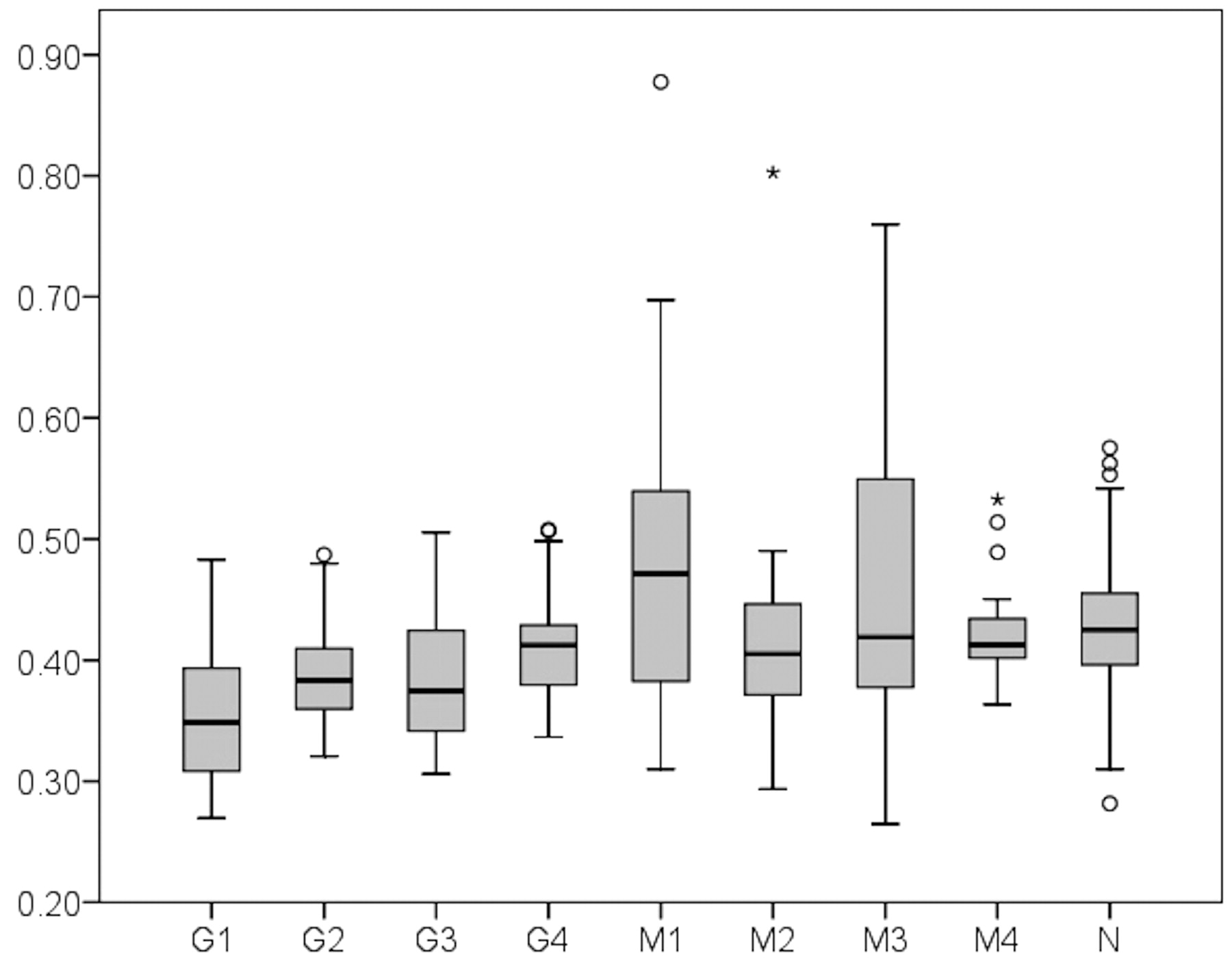
  • Fig 2.
    • Download figure
    • Open in new tab
    • Download powerpoint
    Fig 2.

    Boxplot of mean q value shows from left to right: GBM gross tumor (G1), peritumoral edema (G2) and margin (G3), and adjacent NAWM (G4); metastases gross tumor (M1), peritumoral edema (M2) and margin (M3), and adjacent NAWM (M4); and normal brain compiled from all regions (N). Also shown are outliers: mild outliers represented by a circle and extreme outliers, by an asterisk.

  • Fig 3.
    • Download figure
    • Open in new tab
    • Download powerpoint
    Fig 3.

    Boxplot of mean FA value shows from left to right: GBM gross tumor (G1), peritumoral edema (G2) and margin (G3), and adjacent NAWM (G4); metastases gross tumor (M1), peritumoral edema (M2) and margin (M3), and adjacent NAWM (M4); and normal brain compiled from all regions (N). Also shown are outliers: mild outliers represented by a circle and extreme outliers, by an asterisk.

PreviousNext
Back to top

In this issue

American Journal of Neuroradiology: 30 (1)
American Journal of Neuroradiology
Vol. 30, Issue 1
January 2009
  • Table of Contents
  • Index by author
Advertisement
Print
Download PDF
Email Article

Thank you for your interest in spreading the word on American Journal of Neuroradiology.

NOTE: We only request your email address so that the person you are recommending the page to knows that you wanted them to see it, and that it is not junk mail. We do not capture any email address.

Enter multiple addresses on separate lines or separate them with commas.
Diffusion Tensor Imaging in Glioblastoma Multiforme and Brain Metastases: The Role of p, q, L, and Fractional Anisotropy
(Your Name) has sent you a message from American Journal of Neuroradiology
(Your Name) thought you would like to see the American Journal of Neuroradiology web site.
CAPTCHA
This question is for testing whether or not you are a human visitor and to prevent automated spam submissions.
Cite this article
W. Wang, C.E. Steward, P.M. Desmond
Diffusion Tensor Imaging in Glioblastoma Multiforme and Brain Metastases: The Role of p, q, L, and Fractional Anisotropy
American Journal of Neuroradiology Jan 2009, 30 (1) 203-208; DOI: 10.3174/ajnr.A1303

Citation Manager Formats

  • BibTeX
  • Bookends
  • EasyBib
  • EndNote (tagged)
  • EndNote 8 (xml)
  • Medlars
  • Mendeley
  • Papers
  • RefWorks Tagged
  • Ref Manager
  • RIS
  • Zotero
0 Responses
Respond to this article
Share
Bookmark this article
Diffusion Tensor Imaging in Glioblastoma Multiforme and Brain Metastases: The Role of p, q, L, and Fractional Anisotropy
W. Wang, C.E. Steward, P.M. Desmond
American Journal of Neuroradiology Jan 2009, 30 (1) 203-208; DOI: 10.3174/ajnr.A1303
del.icio.us logo Twitter logo Facebook logo Mendeley logo
  • Tweet Widget
  • Facebook Like
  • Google Plus One
Purchase

Jump to section

  • Article
    • Abstract
    • Materials and Methods
    • Results
    • Discussion
    • Conclusions
    • Footnotes
    • References
  • Figures & Data
  • Supplemental
  • Info & Metrics
  • Responses
  • References
  • PDF

Related Articles

  • PubMed
  • Google Scholar

Cited By...

  • Mesoscopic Assessment of Microstructure in Glioblastomas and Metastases by Merging Advanced Diffusion Imaging with Immunohistopathology
  • Diffusion-Weighted Imaging and Diffusion Tensor Imaging for Differentiating High-Grade Glioma from Solitary Brain Metastasis: A Systematic Review and Meta-Analysis
  • Combining Diffusion Tensor Metrics and DSC Perfusion Imaging: Can It Improve the Diagnostic Accuracy in Differentiating Tumefactive Demyelination from High-Grade Glioma?
  • MR Fingerprinting of Adult Brain Tumors: Initial Experience
  • Multimodal Imaging in Malignant Brain Tumors: Enhancing the Preoperative Risk Evaluation for Motor Deficits with a Combined Hybrid MRI-PET and Navigated Transcranial Magnetic Stimulation Approach
  • Diffusion-Weighted Imaging in Cancer: Physical Foundations and Applications of Restriction Spectrum Imaging
  • Diagnostic Utility of Diffusion Tensor Imaging in Differentiating Glioblastomas from Brain Metastases
  • Utility of Diffusion Tensor Imaging in Evaluation of the Peritumoral Region in Patients with Primary and Metastatic Brain Tumors
  • Differentiation of Brain Abscesses from Necrotic Glioblastomas and Cystic Metastatic Brain Tumors with Diffusion Tensor Imaging
  • Crossref (106)
  • Google Scholar

This article has been cited by the following articles in journals that are participating in Crossref Cited-by Linking.

  • Diffusion-Weighted Imaging in Cancer: Physical Foundations and Applications of Restriction Spectrum Imaging
    Nathan S. White, Carrie R. McDonald, Niky Farid, Josh Kuperman, David Karow, Natalie M. Schenker-Ahmed, Hauke Bartsch, Rebecca Rakow-Penner, Dominic Holland, Ahmed Shabaik, Atle Bjørnerud, Tuva Hope, Jona Hattangadi-Gluth, Michael Liss, J. Kellogg Parsons, Clark C. Chen, Steve Raman, Daniel Margolis, Robert E. Reiter, Leonard Marks, Santosh Kesari, Arno J. Mundt, Christopher J. Kane, Bob S. Carter, William G. Bradley, Anders M. Dale
    Cancer Research 2014 74 17
  • MR Fingerprinting of Adult Brain Tumors: Initial Experience
    C. Badve, A. Yu, S. Dastmalchian, M. Rogers, D. Ma, Y. Jiang, S. Margevicius, S. Pahwa, Z. Lu, M. Schluchter, J. Sunshine, M. Griswold, A. Sloan, V. Gulani
    American Journal of Neuroradiology 2017 38 3
  • Assessment of diffusion tensor imaging metrics in differentiating low-grade from high-grade gliomas
    Lamiaa El-Serougy, Ahmed Abdel Khalek Abdel Razek, Amani Ezzat, Hany Eldawoody, Ahmad El-Morsy
    The Neuroradiology Journal 2016 29 5
  • Diffusion tensor imaging discriminates between glioblastoma and cerebral metastases in vivo
    Tiernan J. D. Byrnes, Thomas R. Barrick, B. Anthony Bell, Chris A. Clark
    NMR in Biomedicine 2011 24 1
  • Differentiation of Brain Abscesses from Necrotic Glioblastomas and Cystic Metastatic Brain Tumors with Diffusion Tensor Imaging
    C.H. Toh, K.-C. Wei, S.-H. Ng, Y.-L. Wan, C.-P. Lin, M. Castillo
    American Journal of Neuroradiology 2011 32 9
  • Potential role of advanced MRI techniques for the peritumoural region in differentiating glioblastoma multiforme and solitary metastatic lesions
    E.J. Lee, K.J. Ahn, E.K. Lee, Y.S. Lee, D.B. Kim
    Clinical Radiology 2013 68 12
  • Utility of Diffusion Tensor Imaging in Evaluation of the Peritumoral Region in Patients with Primary and Metastatic Brain Tumors
    E.J. Sternberg, M.L. Lipton, J. Burns
    American Journal of Neuroradiology 2014 35 3
  • Diagnostic Utility of Diffusion Tensor Imaging in Differentiating Glioblastomas from Brain Metastases
    S. Wang, S. J. Kim, H. Poptani, J. H. Woo, S. Mohan, R. Jin, M. R. Voluck, D. M. O'Rourke, R. L. Wolf, E. R. Melhem, S. Kim
    American Journal of Neuroradiology 2014 35 5
  • Human Brain and Spinal Cord Tumors: From Bench to Bedside. Volume 2
    Linda M. Wang, Zachary K. Englander, Michael L. Miller, Jeffrey N. Bruce
    2023 1405
  • Chemical exchange saturation transfer for predicting response to stereotactic radiosurgery in human brain metastasis
    Kimberly L. Desmond, Hatef Mehrabian, Sofia Chavez, Arjun Sahgal, Hany Soliman, Radoslaw Rola, Greg J. Stanisz
    Magnetic Resonance in Medicine 2017 78 3

More in this TOC Section

  • Usefulness of Quantitative Susceptibility Mapping for the Diagnosis of Parkinson Disease
  • White Matter Alterations in the Brains of Patients with Active, Remitted, and Cured Cushing Syndrome: A DTI Study
  • Qualitative and Quantitative Analysis of MR Imaging Findings in Patients with Middle Cerebral Artery Stroke Implanted with Mesenchymal Stem Cells
Show more BRAIN

Similar Articles

Advertisement

Indexed Content

  • Current Issue
  • Accepted Manuscripts
  • Article Preview
  • Past Issues
  • Editorials
  • Editor's Choice
  • Fellows' Journal Club
  • Letters to the Editor
  • Video Articles

Cases

  • Case Collection
  • Archive - Case of the Week
  • Archive - Case of the Month
  • Archive - Classic Case

Special Collections

  • AJNR Awards
  • ASNR Foundation Special Collection
  • Most Impactful AJNR Articles
  • Photon-Counting CT
  • Spinal CSF Leak Articles (Jan 2020-June 2024)

More from AJNR

  • Trainee Corner
  • Imaging Protocols
  • MRI Safety Corner

Multimedia

  • AJNR Podcasts
  • AJNR Scantastics

Resources

  • Turnaround Time
  • Submit a Manuscript
  • Submit a Video Article
  • Submit an eLetter to the Editor/Response
  • Manuscript Submission Guidelines
  • Statistical Tips
  • Fast Publishing of Accepted Manuscripts
  • Graphical Abstract Preparation
  • Imaging Protocol Submission
  • Evidence-Based Medicine Level Guide
  • Publishing Checklists
  • Author Policies
  • Become a Reviewer/Academy of Reviewers
  • News and Updates

About Us

  • About AJNR
  • Editorial Board
  • Editorial Board Alumni
  • Alerts
  • Permissions
  • Not an AJNR Subscriber? Join Now
  • Advertise with Us
  • Librarian Resources
  • Feedback
  • Terms and Conditions
  • AJNR Editorial Board Alumni

American Society of Neuroradiology

  • Not an ASNR Member? Join Now

© 2025 by the American Society of Neuroradiology All rights, including for text and data mining, AI training, and similar technologies, are reserved.
Print ISSN: 0195-6108 Online ISSN: 1936-959X

Powered by HighWire