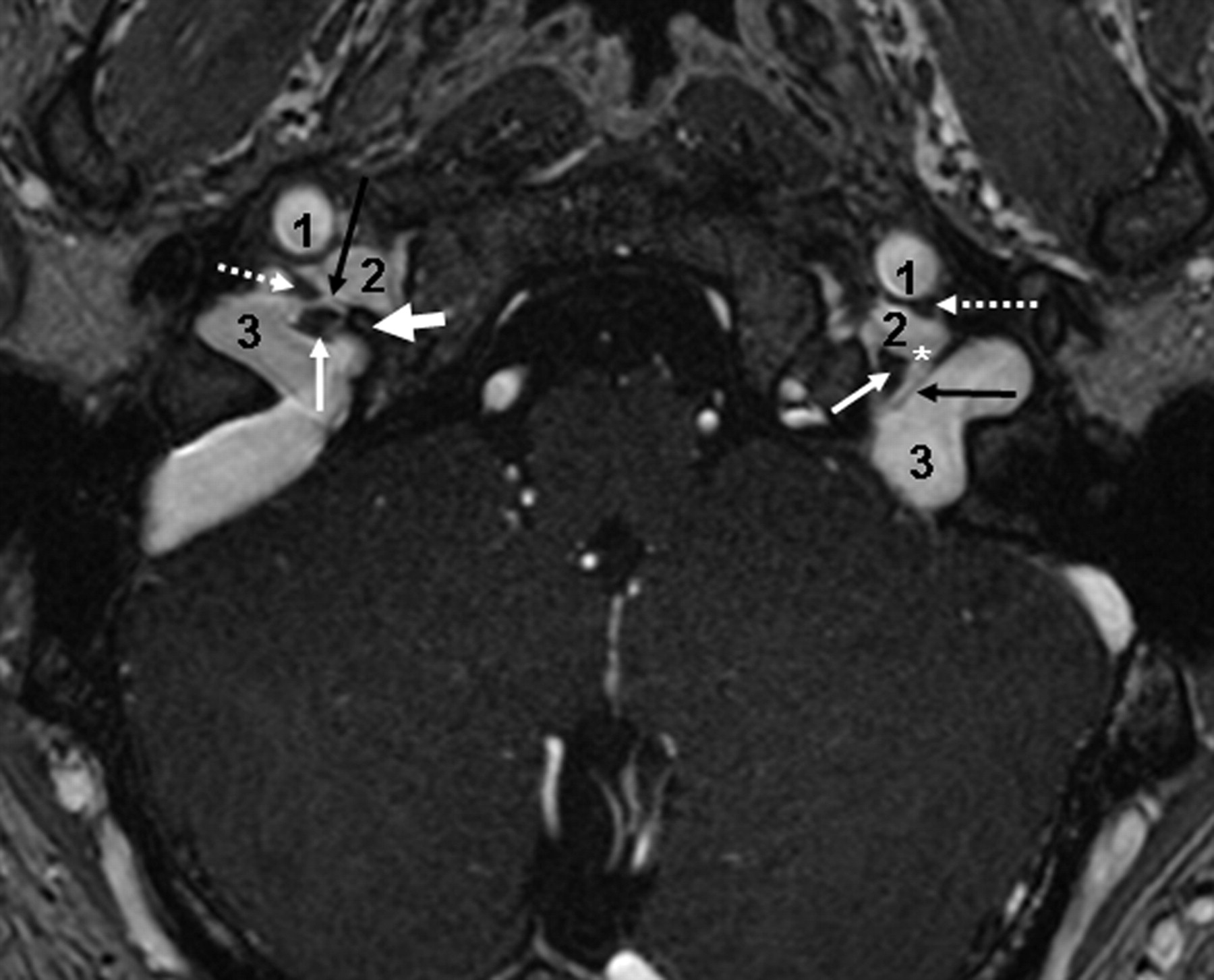
Advertisement

Main menu

  • Home
  • Content
    • Current Issue
    • Accepted Manuscripts
    • Article Preview
    • Past Issue Archive
    • Video Articles
    • AJNR Case Collection
    • Case of the Week Archive
    • Case of the Month Archive
    • Classic Case Archive
  • Special Collections
    • AJNR Awards
    • ASNR Foundation Special Collection
    • Most Impactful AJNR Articles
    • Photon-Counting CT
    • Spinal CSF Leak Articles (Jan 2020-June 2024)
  • Multimedia
    • AJNR Podcasts
    • AJNR SCANtastic
    • Trainee Corner
    • MRI Safety Corner
    • Imaging Protocols
  • For Authors
    • Submit a Manuscript
    • Submit a Video Article
    • Submit an eLetter to the Editor/Response
    • Manuscript Submission Guidelines
    • Statistical Tips
    • Fast Publishing of Accepted Manuscripts
    • Graphical Abstract Preparation
    • Imaging Protocol Submission
    • Author Policies
  • About Us
    • About AJNR
    • Editorial Board
    • Editorial Board Alumni
  • More
    • Become a Reviewer/Academy of Reviewers
    • Subscribers
    • Permissions
    • Alerts
    • Feedback
    • Advertisers
    • ASNR Home

User menu

  • Alerts
  • Log in

Search

  • Advanced search
American Journal of Neuroradiology

American Journal of Neuroradiology

ASHNR American Society of Functional Neuroradiology ASHNR American Society of Pediatric Neuroradiology ASSR
  • Alerts
  • Log in

Advanced Search

  • Home
  • Content
    • Current Issue
    • Accepted Manuscripts
    • Article Preview
    • Past Issue Archive
    • Video Articles
    • AJNR Case Collection
    • Case of the Week Archive
    • Case of the Month Archive
    • Classic Case Archive
  • Special Collections
    • AJNR Awards
    • ASNR Foundation Special Collection
    • Most Impactful AJNR Articles
    • Photon-Counting CT
    • Spinal CSF Leak Articles (Jan 2020-June 2024)
  • Multimedia
    • AJNR Podcasts
    • AJNR SCANtastic
    • Trainee Corner
    • MRI Safety Corner
    • Imaging Protocols
  • For Authors
    • Submit a Manuscript
    • Submit a Video Article
    • Submit an eLetter to the Editor/Response
    • Manuscript Submission Guidelines
    • Statistical Tips
    • Fast Publishing of Accepted Manuscripts
    • Graphical Abstract Preparation
    • Imaging Protocol Submission
    • Author Policies
  • About Us
    • About AJNR
    • Editorial Board
    • Editorial Board Alumni
  • More
    • Become a Reviewer/Academy of Reviewers
    • Subscribers
    • Permissions
    • Alerts
    • Feedback
    • Advertisers
    • ASNR Home
  • Follow AJNR on Twitter
  • Visit AJNR on Facebook
  • Follow AJNR on Instagram
  • Join AJNR on LinkedIn
  • RSS Feeds

AJNR is seeking candidates for the AJNR Podcast Editor. Read the position description.

Research ArticleHEAD & NECK

The Jugular Foramen: Imaging Strategy and Detailed Anatomy at 3T

J. Linn, F. Peters, B. Moriggl, T.P. Naidich, H. Brückmann and I. Yousry
American Journal of Neuroradiology January 2009, 30 (1) 34-41; DOI: https://doi.org/10.3174/ajnr.A1281
J. Linn
  • Find this author on Google Scholar
  • Find this author on PubMed
  • Search for this author on this site
F. Peters
  • Find this author on Google Scholar
  • Find this author on PubMed
  • Search for this author on this site
B. Moriggl
  • Find this author on Google Scholar
  • Find this author on PubMed
  • Search for this author on this site
T.P. Naidich
  • Find this author on Google Scholar
  • Find this author on PubMed
  • Search for this author on this site
H. Brückmann
  • Find this author on Google Scholar
  • Find this author on PubMed
  • Search for this author on this site
I. Yousry
  • Find this author on Google Scholar
  • Find this author on PubMed
  • Search for this author on this site

Fig 1.

Fig 1.
  • Download figure
  • Open in new tab
  • Download powerpoint
Fig 1.

Petrosal and sigmoid parts of the JF. Axial CE-MRA image demonstrates the petrosal and sigmoid parts of the JF as well as the cranial nerves within the intrajugular compartment. The thick white arrow marks the right interjugular process of the occipital bone; the black arrow depicts the dural septum between the petrosal and sigmoid part of the JF. Asterisks mark the drainage of the inferior petrosal sinus into the jugular bulb between CNIX anterolaterally (dotted arrows) and the CNX/XI complex at the level of the supCNX posteromedially (thin white arrow). 1 indicates the ICA; 2, the inferior petrosal sinus; 3, the sigmoid sinus.

  View article

In this issue

American Journal of Neuroradiology: 46 (5)
American Journal of Neuroradiology
Vol. 46, Issue 5
1 May 2025
  • Table of Contents
  • Index by author
  • Complete Issue (PDF)
Print
Download PDF
Email Article

Thank you for your interest in spreading the word on American Journal of Neuroradiology.

NOTE: We only request your email address so that the person you are recommending the page to knows that you wanted them to see it, and that it is not junk mail. We do not capture any email address.

Enter multiple addresses on separate lines or separate them with commas.
Fig 1.
(Your Name) has sent you a message from American Journal of Neuroradiology
(Your Name) thought you would like to see the American Journal of Neuroradiology web site.
CAPTCHA
This question is for testing whether or not you are a human visitor and to prevent automated spam submissions.
Cite this article
J. Linn, F. Peters, B. Moriggl, T.P. Naidich, H. Brückmann, I. Yousry
The Jugular Foramen: Imaging Strategy and Detailed Anatomy at 3T
American Journal of Neuroradiology Jan 2009, 30 (1) 34-41; DOI: 10.3174/ajnr.A1281

Citation Manager Formats

  • BibTeX
  • Bookends
  • EasyBib
  • EndNote (tagged)
  • EndNote 8 (xml)
  • Medlars
  • Mendeley
  • Papers
  • RefWorks Tagged
  • Ref Manager
  • RIS
  • Zotero
0 Responses
Respond to this article
Share
Bookmark this article
The Jugular Foramen: Imaging Strategy and Detailed Anatomy at 3T
J. Linn, F. Peters, B. Moriggl, T.P. Naidich, H. Brückmann, I. Yousry
American Journal of Neuroradiology Jan 2009, 30 (1) 34-41; DOI: 10.3174/ajnr.A1281
del.icio.us logo Twitter logo Facebook logo Mendeley logo
  • Tweet Widget
  • Facebook Like
  • Google Plus One

Related Articles

  • No related articles found.
  • PubMed
  • Google Scholar

Cited By...

  • 3D Double-Echo Steady-State with Water Excitation MR Imaging of the Intraparotid Facial Nerve at 1.5T: A Pilot Study
  • Detailed MR Imaging Anatomy of the Cisternal Segments of the Glossopharyngeal, Vagus, and Spinal Accessory Nerves in the Posterior Fossa: The Use of 3D Balanced Fast-Field Echo MR Imaging
  • Crossref (36)
  • Google Scholar

This article has been cited by the following articles in journals that are participating in Crossref Cited-by Linking.

  • Trigeminal Neuralgia due to Neurovascular Compression: High-Spatial-Resolution Diffusion-Tensor Imaging Reveals Microstructural Neural Changes
    Juergen Lutz, Jennifer Linn, Jan H. Mehrkens, Niklas Thon, Robert Stahl, Klaus Seelos, Hartmut Brückmann, Markus Holtmannspötter
    Radiology 2011 258 2
  • Visualization of Cranial Nerves Using High-Definition Fiber Tractography
    Masanori Yoshino, Kumar Abhinav, Fang-Cheng Yeh, Sandip Panesar, David Fernandes, Sudhir Pathak, Paul A. Gardner, Juan C. Fernandez-Miranda
    Neurosurgery 2016 79 1
  • 3D Double-Echo Steady-State with Water Excitation MR Imaging of the Intraparotid Facial Nerve at 1.5T: A Pilot Study
    Y. Qin, J. Zhang, P. Li, Y. Wang
    American Journal of Neuroradiology 2011 32 7
  • Anatomic Considerations, Nomenclature, and Advanced Cross-sectional Imaging Techniques for Visualization of the Cranial Nerve Segments by MR Imaging
    Ari M. Blitz, Asim F. Choudhri, Zachary D. Chonka, Ahmet T. Ilica, Leonardo L. Macedo, Avneesh Chhabra, Gary L. Gallia, Nafi Aygun
    Neuroimaging Clinics of North America 2014 24 1
  • Detailed MR Imaging Anatomy of the Cisternal Segments of the Glossopharyngeal, Vagus, and Spinal Accessory Nerves in the Posterior Fossa: The Use of 3D Balanced Fast-Field Echo MR Imaging
    W.-J. Moon, H.G. Roh, E.C. Chung
    American Journal of Neuroradiology 2009 30 6
  • Fast imaging employing steady-state acquisition (FIESTA) MRI to investigate cerebrospinal fluid (CSF) within dural reflections of posterior fossa cranial nerves
    David J Noble, Daniel Scoffings, Thankamma Ajithkumar, Michael V Williams, Sarah J Jefferies
    The British Journal of Radiology 2016 89 1067
  • Magnetic Resonance Imaging of Cranial Nerves at 7 Tesla
    A. E. Grams, O. Kraff, J. Kalkmann, S. Orzada, S. Maderwald, M. E. Ladd, M. Forsting, E. R. Gizewski
    Clinical Neuroradiology 2013 23 1
  • Tracking the glossopharyngeal nerve pathway through anatomical references in cross-sectional imaging techniques: a pictorial review
    José María García Santos, Sandra Sánchez Jiménez, Marta Tovar Pérez, Matilde Moreno Cascales, Javier Lailhacar Marty, Miguel A. Fernández-Villacañas Marín
    Insights into Imaging 2018 9 4
  • Detailed imaging of the normal anatomy and pathologic conditions of the cavernous region at 3 Tesla using a contrast-enhanced MR angiography
    Jennifer Linn, Friederike Peters, Nina Lummel, Christoph Schankin, Walter Rachinger, Hartmut Brueckmann, Indra Yousry
    Neuroradiology 2011 53 12
  • Compartmental Subdivisions of the Jugular Foramen: A Review of the Current Models
    Jacob D. Bond, Ming Zhang
    World Neurosurgery 2020 136

More in this TOC Section

  • Chondrosarcoma vs Synovial Chondromatosis: Imaging
  • WHO Classification Update: Nasal&Skull Base Tumors
  • Peritumoral Signal in Vestibular Schwannomas
Show more HEAD & NECK

Similar Articles

Advertisement

Indexed Content

  • Current Issue
  • Accepted Manuscripts
  • Article Preview
  • Past Issues
  • Editorials
  • Editor's Choice
  • Fellows' Journal Club
  • Letters to the Editor
  • Video Articles

Cases

  • Case Collection
  • Archive - Case of the Week
  • Archive - Case of the Month
  • Archive - Classic Case

Special Collections

  • AJNR Awards
  • ASNR Foundation Special Collection
  • Most Impactful AJNR Articles
  • Photon-Counting CT
  • Spinal CSF Leak Articles (Jan 2020-June 2024)

More from AJNR

  • Trainee Corner
  • Imaging Protocols
  • MRI Safety Corner

Multimedia

  • AJNR Podcasts
  • AJNR Scantastics

Resources

  • Turnaround Time
  • Submit a Manuscript
  • Submit a Video Article
  • Submit an eLetter to the Editor/Response
  • Manuscript Submission Guidelines
  • Statistical Tips
  • Fast Publishing of Accepted Manuscripts
  • Graphical Abstract Preparation
  • Imaging Protocol Submission
  • Evidence-Based Medicine Level Guide
  • Publishing Checklists
  • Author Policies
  • Become a Reviewer/Academy of Reviewers
  • News and Updates

About Us

  • About AJNR
  • Editorial Board
  • Editorial Board Alumni
  • Alerts
  • Permissions
  • Not an AJNR Subscriber? Join Now
  • Advertise with Us
  • Librarian Resources
  • Feedback
  • Terms and Conditions
  • AJNR Editorial Board Alumni

American Society of Neuroradiology

  • Not an ASNR Member? Join Now

© 2025 by the American Society of Neuroradiology All rights, including for text and data mining, AI training, and similar technologies, are reserved.
Print ISSN: 0195-6108 Online ISSN: 1936-959X

Powered by HighWire