
Article Figures & Data
Figures
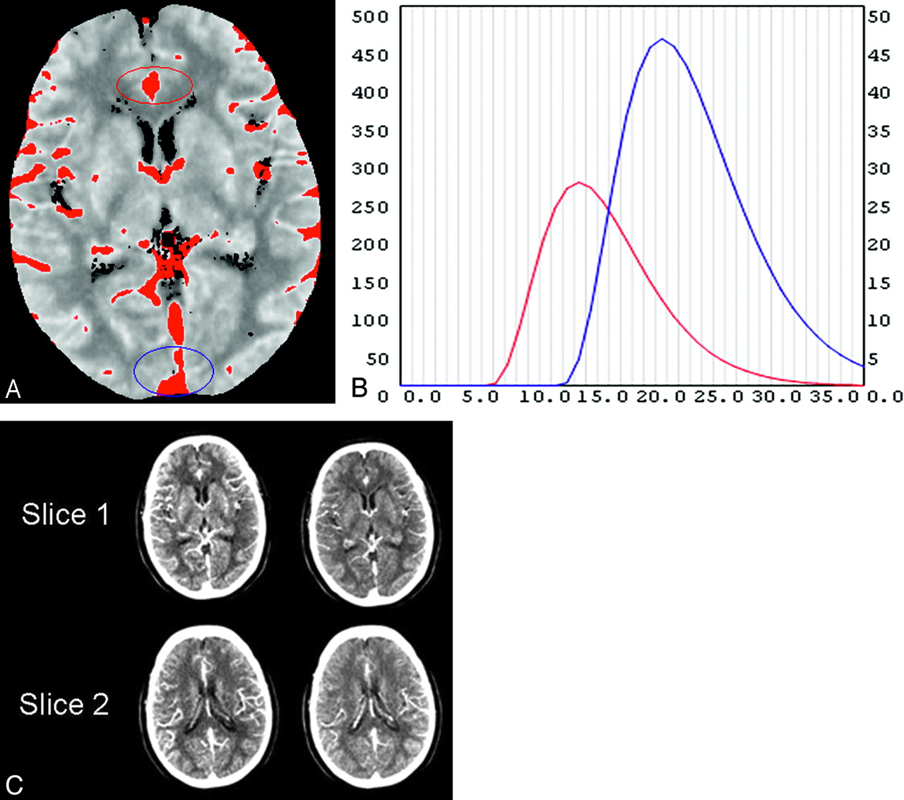
- Fig 1.
The arterial phase CT perfusion source image (CTP-SI) and the venous phase CTP-SI are respectively obtained at the peak points of the arterial (B, red curve) and venous time attenuation curves (TACs) (B, blue curve). On the maximum-intensity-projection image (A), the region of interest is on the area of the anterior cerebral artery (A, red circle); for the arterial TAC curve (B, red curve), the region of interest is on the area of the venous sinus (A, blue circle) for the venous TAC (B, blue curve). On the TAC (B), the horizontal axis is time (seconds) and the vertical axis is attenuation (Hounsfield units). There are 40 seconds in the horizontal axis, every second corresponding to 1 image in each section. With this patient as an example, the peak point of the arterial phase is 15 seconds after scanning begins (at the peak point of the red curve, B).The arterial phase CTP-SI is the fifteenth image in each section (C, left side); the peak point of the venous phase is 23 seconds after scanning begins at the peak point of blue curve (B). The venous phase CTP-SI is the twenty-third image in each section (C, right side).
- Fig 2.
Bland-Altman plots of the differences against the mean value for the arterial phase CTP-SI (ACTP-SI) Alberta Stroke Program Early CT Score (ASPECTS) and cerebral blood flow (CBF) ASPECTS (A); and venous phase CTP-SI (VCTP-SI) ASPECTS and cerebral blood volume (CBV) ASPECTS (B). The solid line represents the mean bias, and the dotted lines indicate the upper and lower limits of agreement.
- Fig 3.
A 74-year-old women imaged 6 hours 40 minutes after sudden onset of left hemiparesis. Non-contrast CT (NCCT) demonstrates sulcal effacement in the right M5 territory, yielding an ASPECTS of 9. ACTP-SI reveals a hypoattenuation in the right middle cerebral artery territory; ACTP-SI ASPECTS is consistent with CBF (white arrows), yielding an ASPECTS of 3. VCTP-SI and CBV ASPECTS is 7 (black arrows), which was confirmed by follow-up NCCT.
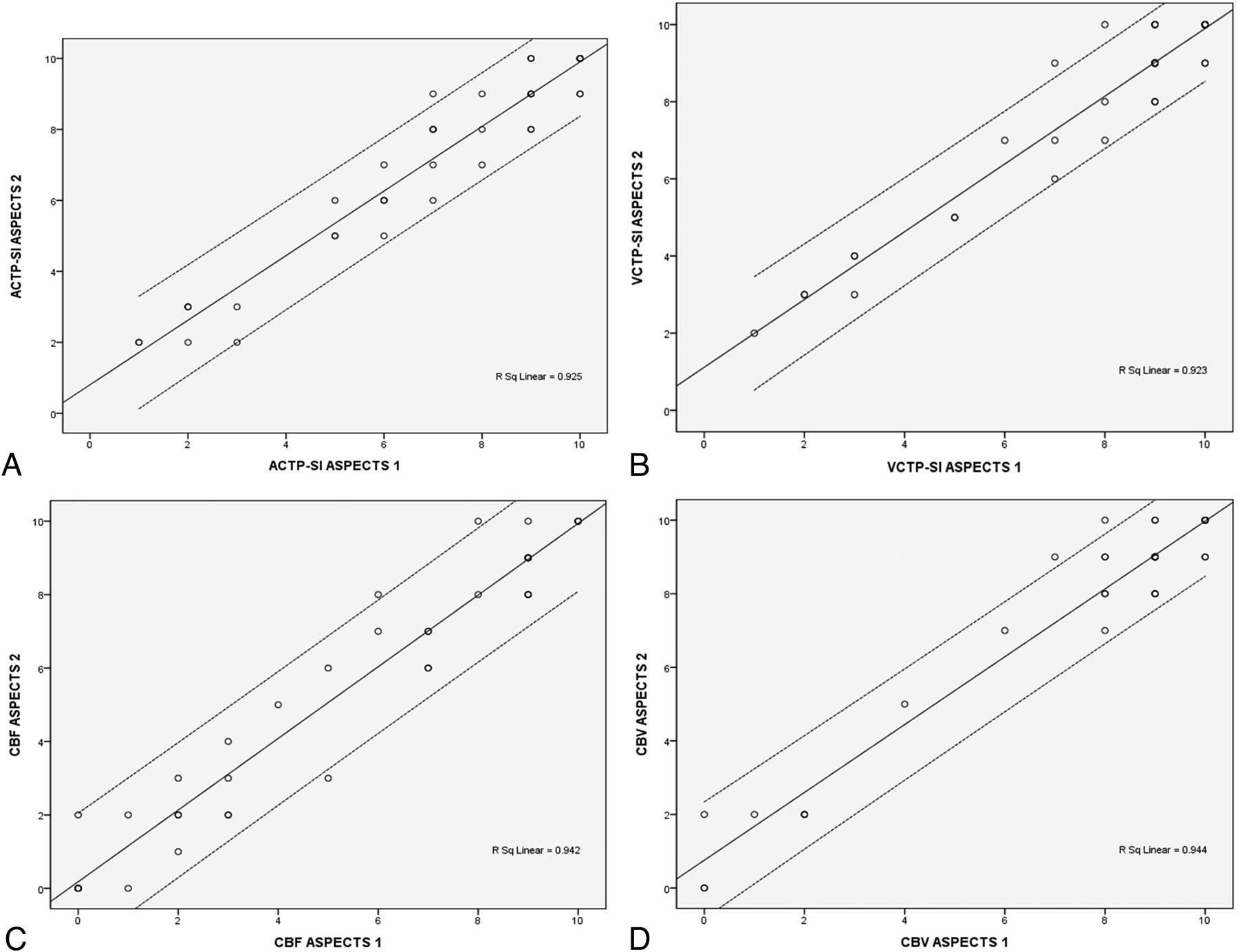
- Fig 4.
Scatterplot demonstrating the good agreement between raters for ACTP-SI ASPECTS (A), VCTP-SI ASPECTS (B), CBF ASPECTS (C), and CBV ASPECTS (D) in patients with acute stroke. The best-fit linear regression line (single solid line) and 95% confidence intervals for the data points (paired dotted lines) are shown.


Tables
Comparison of lesion ASPECTS at baseline CTP-SI and color maps
ASPECTS Mean ± SD 95% CI for Mean ASPECTS Compared Wilcoxon Signed Rank Test (P value) Lower Bound Upper Bound ACTP-SI 6.869 ± 2.734 6.017 7.721 ACTP-SI−VCTP-SI <.001 VCTP-SI 7.869 ± 2.465 7.101 8.637 VCTP-SI−CBV .393 CBF 5.405 ± 3.633 4.273 6.537 CBF−ACTP-SI <.001 CBV 7.595 ± 3.079 6.636 8.555 CBV-CBF <.001 -
Note:—ASPECTS indicates Alberta Stroke Program Early CT Score; CBF, cerebral blood flow; CBV, cerebral blood volume; CTP-SI, CT perfusion source image; ACTP-SI, arterial phase CTP-SI; VCTP-SI, venous phase CTP-SI; CI, confidence interval.
-
In this issue


Jump to section
Related Articles
Cited By...
- Optimal Perfusion Computed Tomographic Thresholds for Ischemic Core and Penumbra Are Not Time Dependent in the Clinically Relevant Time Window
- Pre-intervention triage incorporating perfusion imaging improves outcomes in patients undergoing endovascular stroke therapy: a comparison with the device trials
- CT Angiographic Source Images Predict Outcome and Final Infarct Volume Better Than Noncontrast CT in Proximal Vascular Occlusions