Advertisement

Main menu

  • Home
  • Content
    • Current Issue
    • Accepted Manuscripts
    • Article Preview
    • Past Issue Archive
    • Video Articles
    • AJNR Case Collection
    • Case of the Week Archive
    • Case of the Month Archive
    • Classic Case Archive
  • Special Collections
    • AJNR Awards
    • ASNR Foundation Special Collection
    • Most Impactful AJNR Articles
    • Photon-Counting CT
    • Spinal CSF Leak Articles (Jan 2020-June 2024)
  • Multimedia
    • AJNR Podcasts
    • AJNR SCANtastic
    • Trainee Corner
    • MRI Safety Corner
    • Imaging Protocols
  • For Authors
    • Submit a Manuscript
    • Submit a Video Article
    • Submit an eLetter to the Editor/Response
    • Manuscript Submission Guidelines
    • Statistical Tips
    • Fast Publishing of Accepted Manuscripts
    • Graphical Abstract Preparation
    • Imaging Protocol Submission
    • Author Policies
  • About Us
    • About AJNR
    • Editorial Board
    • Editorial Board Alumni
  • More
    • Become a Reviewer/Academy of Reviewers
    • Subscribers
    • Permissions
    • Alerts
    • Feedback
    • Advertisers
    • ASNR Home

User menu

  • Alerts
  • Log in

Search

  • Advanced search
American Journal of Neuroradiology

American Journal of Neuroradiology

ASHNR American Society of Functional Neuroradiology ASHNR American Society of Pediatric Neuroradiology ASSR
  • Alerts
  • Log in

Advanced Search

  • Home
  • Content
    • Current Issue
    • Accepted Manuscripts
    • Article Preview
    • Past Issue Archive
    • Video Articles
    • AJNR Case Collection
    • Case of the Week Archive
    • Case of the Month Archive
    • Classic Case Archive
  • Special Collections
    • AJNR Awards
    • ASNR Foundation Special Collection
    • Most Impactful AJNR Articles
    • Photon-Counting CT
    • Spinal CSF Leak Articles (Jan 2020-June 2024)
  • Multimedia
    • AJNR Podcasts
    • AJNR SCANtastic
    • Trainee Corner
    • MRI Safety Corner
    • Imaging Protocols
  • For Authors
    • Submit a Manuscript
    • Submit a Video Article
    • Submit an eLetter to the Editor/Response
    • Manuscript Submission Guidelines
    • Statistical Tips
    • Fast Publishing of Accepted Manuscripts
    • Graphical Abstract Preparation
    • Imaging Protocol Submission
    • Author Policies
  • About Us
    • About AJNR
    • Editorial Board
    • Editorial Board Alumni
  • More
    • Become a Reviewer/Academy of Reviewers
    • Subscribers
    • Permissions
    • Alerts
    • Feedback
    • Advertisers
    • ASNR Home
  • Follow AJNR on Twitter
  • Visit AJNR on Facebook
  • Follow AJNR on Instagram
  • Join AJNR on LinkedIn
  • RSS Feeds

AJNR is seeking candidates for the AJNR Podcast Editor. Read the position description.

Research ArticlePediatrics

Spectrum of Temporal Bone Abnormalities in Patients with Waardenburg Syndrome and SOX10 Mutations

M. Elmaleh-Bergès, C. Baumann, N. Noël-Pétroff, A. Sekkal, V. Couloigner, K. Devriendt, M. Wilson, S. Marlin, G. Sebag and V. Pingault
American Journal of Neuroradiology June 2013, 34 (6) 1257-1263; DOI: https://doi.org/10.3174/ajnr.A3367
M. Elmaleh-Bergès
aFrom the Departments of Pediatric Imaging (M.E.-B., A.S., G.S.)
  • Find this author on Google Scholar
  • Find this author on PubMed
  • Search for this author on this site
C. Baumann
bClinical Genetics (C.B.)
  • Find this author on Google Scholar
  • Find this author on PubMed
  • Search for this author on this site
N. Noël-Pétroff
cOtorhinolaryngology (N.N.-P.), Hôpital Robert Debré, Paris, France
  • Find this author on Google Scholar
  • Find this author on PubMed
  • Search for this author on this site
A. Sekkal
aFrom the Departments of Pediatric Imaging (M.E.-B., A.S., G.S.)
  • Find this author on Google Scholar
  • Find this author on PubMed
  • Search for this author on this site
V. Couloigner
eDepartment of Otorhinolaryngology (V.C.), Hôpital Necker-Enfants Malades, Paris, France
  • Find this author on Google Scholar
  • Find this author on PubMed
  • Search for this author on this site
K. Devriendt
dCenter for Human Genetics (K.D.), University Hospitals Leuven, Leuven, Belgium
  • Find this author on Google Scholar
  • Find this author on PubMed
  • Search for this author on this site
M. Wilson
fDepartment of Clinical Genetics (M.W.), The Children's Hospital at Westmead, Sydney, Australia
  • Find this author on Google Scholar
  • Find this author on PubMed
  • Search for this author on this site
S. Marlin
gCentre de Référence des Surdités Génétiques (S.M.), Hôpital Armand-Trousseau, Paris, France
  • Find this author on Google Scholar
  • Find this author on PubMed
  • Search for this author on this site
G. Sebag
aFrom the Departments of Pediatric Imaging (M.E.-B., A.S., G.S.)
  • Find this author on Google Scholar
  • Find this author on PubMed
  • Search for this author on this site
V. Pingault
hLaboratoire de Biochimie et Génétique (V.P.), AP-HP, Hôpital Henri Mondor–Albert Chenevier, Créteil, France.
  • Find this author on Google Scholar
  • Find this author on PubMed
  • Search for this author on this site
  • Article
  • Figures & Data
  • Supplemental
  • Info & Metrics
  • Responses
  • References
  • PDF
Loading

Article Figures & Data

Figures

  • Tables
  • Fig 1.
    • Download figure
    • Open in new tab
    • Download powerpoint
    Fig 1.

    Patient H. High-resolution 3D T2 MR imaging. Axial views through the cochlea and lateral SCC show a cochlea reduced in size with a flattened aspect of the midturn and apex; an enlarged vestibule; and a lateral SCC with a small diameter, a thin arch, and a small bone island. The basal turn was normal in this patient (not shown). The left cochlea is thinner than the right; this finding was confirmed by oblique sagittal views perpendicular to the nerves (not shown).

  • Fig 2.
    • Download figure
    • Open in new tab
    • Download powerpoint
    Fig 2.

    Patient J. CT. Axial views (A and B) through the cochlea and the vestibule show the flattened apex and midturn of the cochlea. The vestibular cavity is large and shows evaginations that could represent SCC anlages. The vestibular aqueduct is visible and is not dilated. Coronal view (C) favors the hypothesis of a lateral SCC anlagen. The superior SCCs are absent, but the superior margin of the vestibule is convex and a small anlagen cannot be excluded.

  • Fig 3.
    • Download figure
    • Open in new tab
    • Download powerpoint
    Fig 3.

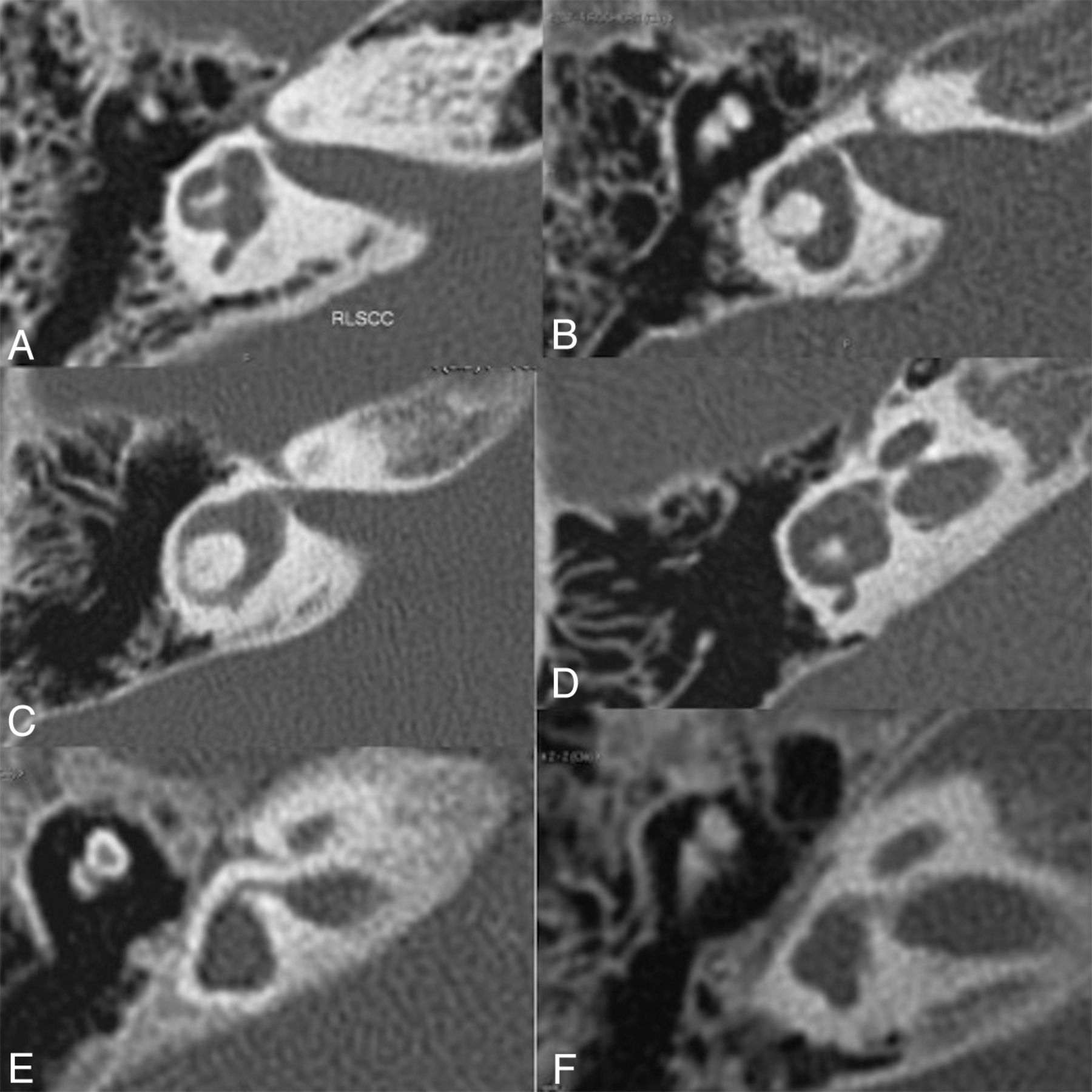
    Different patterns of semicircular canal abnormalities shown on high-resolution axial CT (right ear), confirmed by multiplanar reconstructions for the posterior and superior SCCs: posterior SCC with a thick arch of small diameter (A and D); posterior SCC agenesis or potential SCC anlage (B, C, E, and F); superior SCC agenesis or potential SCC anlage (E and F); lateral SCC agenesis or potential SCC anlage (E and F); lateral SCC with a thick arch of small diameter (A and D); lateral SCC with a thin arch of small diameter (B); and lateral SCC with a thin arch of large diameter (C).

  • Fig 4.
    • Download figure
    • Open in new tab
    • Download powerpoint
    Fig 4.

    Patient M. CT. Axial views (A and B) through the cochlea and the vestibule show a small cochlea, flattened with a partition hardly visible and atresia of cochlear nerve canals, an enlarged vestibular cavity, and agenesis of all of the semicircular canals. Coronal view (C) confirms the absence of the superior and lateral SCC. Posterior deformity of the vestibule cannot exclude an anlage.

  • Fig 5.
    • Download figure
    • Open in new tab
    • Download powerpoint
    Fig 5.

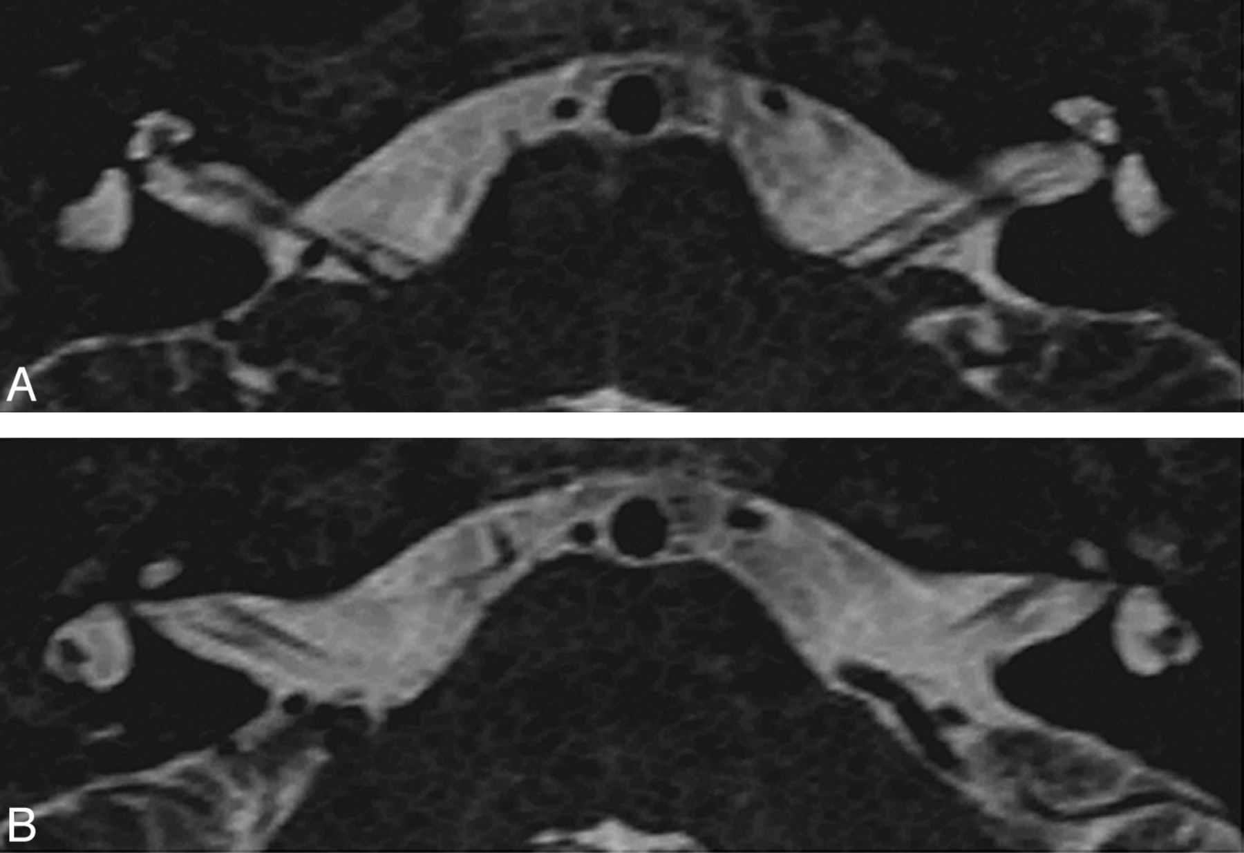
    Patient B. 3D T2 TSE MR image driven equilibrium (Philips). Reformatted axial view in the lateral SCC plane (A) and maximum intensity projection of the superior and posterior SCC (B and C). Same patient as in Fig 2C. Axial view shows a short linear structure along the posterior aspect of the lateral SCC, but multiplanar reformations and maximum intensity projection demonstrate a deformity of the posterior aspect of the vestibule and no posterior SCC. The superior SCC has a thin arch with a large diameter. Hypersignal of the inferior cerebellar peduncles is noticeable due to hypomyelination.

Tables

  • Figures
    • View popup
    Table 1:

    Inner ear imaging findings in patients included in this studya

    PatientAge at MRI/CTCochleaCochlear NerveVestibulePosterior SCCSuperior SCCLateral SCC
    A?/?FlattenedPresentEnlargedAgenesisNLarge arch, thin
    B32 mo/32 moSmallPresentEnlargedSmall arch, thickLarge arch, thinLarge arch, thin
    C32 mo/32 moSmallPresentEnlargedSmall arch, thickNSmall arch, thick
    D1b5 wk/–Small, flattenedPresentEnlargedSmall arch, thickSmall arch, thickSmall arch, thick
    D2b2 mo/2 moSmall, flattenedPresentEnlargedNR: small arch, thick L: NSmall arch, thick
    E3 yr/3 yrFlattenedPresentEnlargedAgenesisAgenesisR: agenesis; L: small arch, thick
    F2 mo/–Small, flattenedAbsentEnlargedAgenesisAgenesisR: agenesis; L: small, thin
    G5 yr/5 yrFlattenedPresentEnlargedAgenesisAgenesisAgenesis
    H18 yr/–Small, flattenedR: present; L: present, thinEnlargedAgenesisAgenesisSmall, thin
    I20 mo/20 moSmall, flattenedPresentEnlargedAgenesisSmall arch, thickR: small, thin, incomplete; L: small, thin
    J4 yr/4 yrSmall, flattenedR: present; L: absentEnlargedAgenesisAgenesisAgenesis
    K18 mo/18 moNPresentEnlargedAgenesisAgenesisSmall, thin
    L–/16 yrSmallN/AR: enlarged; L: NSmall arch, thickAgenesisSmall arch, thick
    M8 days/ 8 daysSmall, flattenedAbsentEnlargedAgenesisAgenesisAgenesis
    N31 yr/31 yrFlattenedPresentEnlargedAgenesisSmall arch, thickSmall arch, thick
    Agenesis or absence (%)21675333
    Defect (%)93291009387100
    • Note:—N indicates normal; N/A, could not be analyzed; R, right; and L, left; –, not performed; ?, age unknown.

    • ↵a When right or left is not specified, the same findings were observed in both sides. In the case of an asymmetric defect, the percentage of absence and/or defects was calculated on the basis of the more severe side.

    • ↵b Brothers.

    • View popup
    Table 2:

    Summary of other imaging findings in patients included in this study

    PatientAge at MRI/CTFacial NervePosterior FossaWhite MatterOlfactory BulbsLacrimal GlandsParotid Glands
    A?/?PresentNNN/AHypoplasticN/A
    B32 mo/32 moPresentICPAbnormalAgenesis ×2HypoplasticHypoplastic
    C32 mo/32 moPresentNNAgenesis ×2NN
    D1a5 wk/–PresentNAbnormalN/AHypoplasticHypoplastic
    D2a2 mo/2 moPresentNNN/AHypoplasticHypoplastic
    E3 yr/3 yrPresentNNAgenesis ×2HypoplasticHypoplastic
    F2 mo/–Right agenesisHypoplastic brain stemAbnormalAgenesis ×2AbsentHypoplastic
    G5 yr/5 yrPresentNNN/ANHypoplastic
    H18 yr/–PresentNLarge VRSAgenesis ×2AbsentHypoplastic
    I20 mo/20 moPresentNAbnormalAgenesis ×2NN
    J4 yr/4 yrPresentNNN/AHypoplasticHypoplastic
    K18 mo/18 moPresentNAbnormalN/AHypoplasticHypoplastic
    L–/16 yrN/AN/AN/AN/AN/AHypoplastic
    M8 days/ 8 daysPresentNAbnormalPresentsHypoplasticHypoplastic
    N31 yr/31 yrPresentNN/AAgenesis ×2HypoplasticHypoplastic
    Agenesis or absence (%)78814
    Defect (%)14507985
    • Note:—N indicates normal; N/A, could not be analyzed; R, right; and L, left; –, not performed; ?, age unknown; ICP, inferior cerebellar peduncles; VRS, Virchow Robin spaces; ×2, similar findings on both sides.

    • ↵a Brothers.

PreviousNext
Back to top

In this issue

American Journal of Neuroradiology: 34 (6)
American Journal of Neuroradiology
Vol. 34, Issue 6
1 Jun 2013
  • Table of Contents
  • Index by author
Advertisement
Print
Download PDF
Email Article

Thank you for your interest in spreading the word on American Journal of Neuroradiology.

NOTE: We only request your email address so that the person you are recommending the page to knows that you wanted them to see it, and that it is not junk mail. We do not capture any email address.

Enter multiple addresses on separate lines or separate them with commas.
Spectrum of Temporal Bone Abnormalities in Patients with Waardenburg Syndrome and SOX10 Mutations
(Your Name) has sent you a message from American Journal of Neuroradiology
(Your Name) thought you would like to see the American Journal of Neuroradiology web site.
CAPTCHA
This question is for testing whether or not you are a human visitor and to prevent automated spam submissions.
Cite this article
M. Elmaleh-Bergès, C. Baumann, N. Noël-Pétroff, A. Sekkal, V. Couloigner, K. Devriendt, M. Wilson, S. Marlin, G. Sebag, V. Pingault
Spectrum of Temporal Bone Abnormalities in Patients with Waardenburg Syndrome and SOX10 Mutations
American Journal of Neuroradiology Jun 2013, 34 (6) 1257-1263; DOI: 10.3174/ajnr.A3367

Citation Manager Formats

  • BibTeX
  • Bookends
  • EasyBib
  • EndNote (tagged)
  • EndNote 8 (xml)
  • Medlars
  • Mendeley
  • Papers
  • RefWorks Tagged
  • Ref Manager
  • RIS
  • Zotero
0 Responses
Respond to this article
Share
Bookmark this article
Spectrum of Temporal Bone Abnormalities in Patients with Waardenburg Syndrome and SOX10 Mutations
M. Elmaleh-Bergès, C. Baumann, N. Noël-Pétroff, A. Sekkal, V. Couloigner, K. Devriendt, M. Wilson, S. Marlin, G. Sebag, V. Pingault
American Journal of Neuroradiology Jun 2013, 34 (6) 1257-1263; DOI: 10.3174/ajnr.A3367
del.icio.us logo Twitter logo Facebook logo Mendeley logo
  • Tweet Widget
  • Facebook Like
  • Google Plus One
Purchase

Jump to section

  • Article
    • Abstract
    • ABBREVIATIONS:
    • Materials and Methods
    • Results
    • Discussion
    • Conclusions
    • Acknowledgments
    • Footnotes
    • REFERENCES
  • Figures & Data
  • Supplemental
  • Info & Metrics
  • Responses
  • References
  • PDF

Related Articles

  • PubMed
  • Google Scholar

Cited By...

  • SOX10: 20 years of phenotypic plurality and current understanding of its developmental function
  • GENETICS IN ENDOCRINOLOGY: Genetic counseling for congenital hypogonadotropic hypogonadism and Kallmann syndrome: new challenges in the era of oligogenism and next-generation sequencing
  • Olfactory bulb agenesis with normal sexual hormones
  • Spectrum of Clinical and Associated MR Imaging Findings in Children with Olfactory Anomalies
  • Crossref (94)
  • Google Scholar

This article has been cited by the following articles in journals that are participating in Crossref Cited-by Linking.

  • GENETICS IN ENDOCRINOLOGY: Genetic counseling for congenital hypogonadotropic hypogonadism and Kallmann syndrome: new challenges in the era of oligogenism and next-generation sequencing
    Luigi Maione, Andrew A Dwyer, Bruno Francou, Anne Guiochon-Mantel, Nadine Binart, Jérôme Bouligand, Jacques Young
    European Journal of Endocrinology 2018 178 3
  • Hypomyelinating leukodystrophies — unravelling myelin biology
    Nicole I. Wolf, Charles ffrench-Constant, Marjo S. van der Knaap
    Nature Reviews Neurology 2021 17 2
  • Hearing loss in Waardenburg syndrome: a systematic review
    J. Song, Y. Feng, F.R. Acke, P. Coucke, K. Vleminckx, I.J. Dhooge
    Clinical Genetics 2016 89 4
  • Neurosensory development and cell fate determination in the human cochlea
    Heiko Locher, Johan HM Frijns, Liesbeth van Iperen, John CMJ de Groot, Margriet A Huisman, Susana M Chuva de Sousa Lopes
    Neural Development 2013 8 1
  • SOX10: 20 years of phenotypic plurality and current understanding of its developmental function
    Veronique Pingault, Lisa Zerad, William Bertani-Torres, Nadege Bondurand
    Journal of Medical Genetics 2022 59 2
  • Lacrimal gland development: From signaling interactions to regenerative medicine
    Ankur Garg, Xin Zhang
    Developmental Dynamics 2017 246 12
  • Efficient Generation of Gene-Modified Pigs Harboring Precise Orthologous Human Mutation via CRISPR/Cas9-Induced Homology-Directed Repair in Zygotes
    Xiaoyang Zhou, Lulu Wang, Yinan Du, Fei Xie, Liang Li, Yu Liu, Chuanhong Liu, Shiqiang Wang, Shibing Zhang, Xingxu Huang, Yong Wang, Hong Wei
    Human Mutation 2016 37 1
  • The Zebrafish - Disease Models and Chemical Screens
    B. Blanco-Sánchez, A. Clément, J.B. Phillips, M. Westerfield
    2017 138
  • Biology of human melanocyte development, Piebaldism, and Waardenburg syndrome
    Mohammed D. Saleem
    Pediatric Dermatology 2019 36 1
  • CT findings of the temporal bone in CHARGE syndrome: aspects of importance in cochlear implant surgery
    A. C. Vesseur, B. M. Verbist, H. E. Westerlaan, F. J. J. Kloostra, R. J. C. Admiraal, C. M. A. van Ravenswaaij-Arts, R. H. Free, E. A. M. Mylanus
    European Archives of Oto-Rhino-Laryngology 2016 273 12

More in this TOC Section

  • SyMRI & MR Fingerprinting in Brainstem Myelination
  • Comparison of Image Quality and Radiation Dose in Pediatric Temporal Bone CT Using Photon-Counting Detector CT and Energy-Integrating Detector CT
  • Pons&Vermis Localization on Fetal MRI Using U-Net
Show more Pediatrics

Similar Articles

Advertisement

Indexed Content

  • Current Issue
  • Accepted Manuscripts
  • Article Preview
  • Past Issues
  • Editorials
  • Editor's Choice
  • Fellows' Journal Club
  • Letters to the Editor
  • Video Articles

Cases

  • Case Collection
  • Archive - Case of the Week
  • Archive - Case of the Month
  • Archive - Classic Case

Special Collections

  • AJNR Awards
  • ASNR Foundation Special Collection
  • Most Impactful AJNR Articles
  • Photon-Counting CT
  • Spinal CSF Leak Articles (Jan 2020-June 2024)

More from AJNR

  • Trainee Corner
  • Imaging Protocols
  • MRI Safety Corner

Multimedia

  • AJNR Podcasts
  • AJNR Scantastics

Resources

  • Turnaround Time
  • Submit a Manuscript
  • Submit a Video Article
  • Submit an eLetter to the Editor/Response
  • Manuscript Submission Guidelines
  • Statistical Tips
  • Fast Publishing of Accepted Manuscripts
  • Graphical Abstract Preparation
  • Imaging Protocol Submission
  • Evidence-Based Medicine Level Guide
  • Publishing Checklists
  • Author Policies
  • Become a Reviewer/Academy of Reviewers
  • News and Updates

About Us

  • About AJNR
  • Editorial Board
  • Editorial Board Alumni
  • Alerts
  • Permissions
  • Not an AJNR Subscriber? Join Now
  • Advertise with Us
  • Librarian Resources
  • Feedback
  • Terms and Conditions
  • AJNR Editorial Board Alumni

American Society of Neuroradiology

  • Not an ASNR Member? Join Now

© 2025 by the American Society of Neuroradiology All rights, including for text and data mining, AI training, and similar technologies, are reserved.
Print ISSN: 0195-6108 Online ISSN: 1936-959X

Powered by HighWire