
Article Figures & Data
Figures
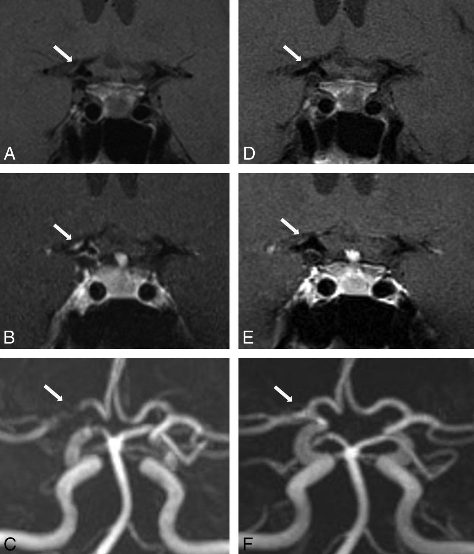
- Fig 1.
A 41-year-old woman with CNS vasculitis. 3T-HRMRI pre- and postgadolinium (A and B) T1-weighted arterial wall coronal images of terminal supraclinoid ICA and proximal M1 with strong smooth, concentric wall enhancement and thickening. C, MRA shows stenosis of the lumen and narrowing of M1 and the terminal supraclinoid ICA. Follow-up at 12 months. D and E, Pre- and post-gadolinium T1WI with near resolution of enhancement on coronal view. F, MRA with interval resolution of luminal narrowing and stenosis.
- Fig 2.
A 61-year-old woman with RCVS. 3T-HRMRI postgadolinium T1-weighted arterial wall with fat suppression and saturation band. A and B, Axial images in the same planes show bilateral A1, M1, and P2 with diffuse uniform wall thickening and wall narrowing with mild enhancement. C, MRA shows corresponding bilateral A1, M1, posterior communicating artery, and P2 narrowing. Follow-up vessel wall imaging at 3 months. D and E, Postgadolinium axial imaging with interval near resolution of uniform wall thickening and wall narrowing in the A1 and M1 segments and resolution of uniform wall thickening and wall narrowing in the P2 segment. F, Follow-up MRA with resolution of luminal narrowing with patent bilateral A1, M1, posterior communicating artery, and P2.


Jump to section
Related Articles
Cited By...
- CT-Based Intrathrombus and Perithrombus Radiomics for Prediction of Prognosis after Endovascular Thrombectomy: A Retrospective Study across 2 Centers
- Imaging Features of Symptomatic MCA Stenosis in Patients of Different Ages: A Vessel Wall MR Imaging Study
- Consensus disease definitions for neurologic immune-related adverse events of immune checkpoint inhibitors
- Black blood imaging of intracranial vessel walls
- The diagnosis of primary central nervous system vasculitis
- Yield of diagnostic imaging in atraumatic convexity subarachnoid hemorrhage
- HIV vasculopathy versus VZV vasculitis in an HIV patient with multiple brain ischaemic infarcts
- High-Resolution Vessel Wall MR Imaging as an Alternative to Brain Biopsy
- Comparison of 3T Intracranial Vessel Wall MRI Sequences
- Cryptogenic stroke as initial manifestation of CNS vasculitis: demonstration of vessel wall enhancement on 1.5T MRI using volumetric T1 TSE sequence
- Added Value of Vessel Wall Magnetic Resonance Imaging for Differentiation of Nonocclusive Intracranial Vasculopathies
- Concordance of Time-of-Flight MRA and Digital Subtraction Angiography in Adult Primary Central Nervous System Vasculitis
- Subtypes of primary angiitis of the CNS identified by MRI patterns reflect the size of affected vessels
- Predicting Progression of Intracranial Arteriopathies in Childhood Stroke With Vessel Wall Imaging
- Primary Angiitis of the Central Nervous System: Magnetic Resonance Imaging Spectrum of Parenchymal, Meningeal, and Vascular Lesions at Baseline
- Intracranial Vessel Wall MRI: Principles and Expert Consensus Recommendations of the American Society of Neuroradiology
- Comparison of High-Resolution MR Imaging and Digital Subtraction Angiography for the Characterization and Diagnosis of Intracranial Artery Disease
- Vessel wall imaging for intracranial vascular disease evaluation
- Clinical Images: Vessel Wall Imaging in the Management of Subarachnoid Hemorrhage and Multiple Intracranial Aneurysms
- High-resolution intracranial vessel wall imaging: imaging beyond the lumen
- Imaging Inflammation in Cerebrovascular Disease
- Reversible Cerebral Vasoconstriction Syndrome, Part 2: Diagnostic Work-Up, Imaging Evaluation, and Differential Diagnosis
- Multicontrast High-Resolution Vessel Wall Magnetic Resonance Imaging and Its Value in Differentiating Intracranial Vasculopathic Processes
- Challenge of Identifying the Cause of Intracranial Artery Stenosis in Patients With Ischemic Stroke
- Isolated MCA Disease in Patients Without Significant Atherosclerotic Risk Factors: A High-Resolution Magnetic Resonance Imaging Study
- Multimodal 3 Tesla MRI Confirms Intact Arterial Wall in Acute Stroke Patients After Stent-Retriever Thrombectomy