Advertisement

Main menu

  • Home
  • Content
    • Current Issue
    • Accepted Manuscripts
    • Article Preview
    • Past Issue Archive
    • Video Articles
    • AJNR Case Collection
    • Case of the Week Archive
    • Case of the Month Archive
    • Classic Case Archive
  • Special Collections
    • AJNR Awards
    • ASNR Foundation Special Collection
    • Most Impactful AJNR Articles
    • Photon-Counting CT
    • Spinal CSF Leak Articles (Jan 2020-June 2024)
  • Multimedia
    • AJNR Podcasts
    • AJNR SCANtastic
    • Trainee Corner
    • MRI Safety Corner
    • Imaging Protocols
  • For Authors
    • Submit a Manuscript
    • Submit a Video Article
    • Submit an eLetter to the Editor/Response
    • Manuscript Submission Guidelines
    • Statistical Tips
    • Fast Publishing of Accepted Manuscripts
    • Graphical Abstract Preparation
    • Imaging Protocol Submission
    • Author Policies
  • About Us
    • About AJNR
    • Editorial Board
    • Editorial Board Alumni
  • More
    • Become a Reviewer/Academy of Reviewers
    • Subscribers
    • Permissions
    • Alerts
    • Feedback
    • Advertisers
    • ASNR Home

User menu

  • Alerts
  • Log in

Search

  • Advanced search
American Journal of Neuroradiology

American Journal of Neuroradiology

ASHNR American Society of Functional Neuroradiology ASHNR American Society of Pediatric Neuroradiology ASSR
  • Alerts
  • Log in

Advanced Search

  • Home
  • Content
    • Current Issue
    • Accepted Manuscripts
    • Article Preview
    • Past Issue Archive
    • Video Articles
    • AJNR Case Collection
    • Case of the Week Archive
    • Case of the Month Archive
    • Classic Case Archive
  • Special Collections
    • AJNR Awards
    • ASNR Foundation Special Collection
    • Most Impactful AJNR Articles
    • Photon-Counting CT
    • Spinal CSF Leak Articles (Jan 2020-June 2024)
  • Multimedia
    • AJNR Podcasts
    • AJNR SCANtastic
    • Trainee Corner
    • MRI Safety Corner
    • Imaging Protocols
  • For Authors
    • Submit a Manuscript
    • Submit a Video Article
    • Submit an eLetter to the Editor/Response
    • Manuscript Submission Guidelines
    • Statistical Tips
    • Fast Publishing of Accepted Manuscripts
    • Graphical Abstract Preparation
    • Imaging Protocol Submission
    • Author Policies
  • About Us
    • About AJNR
    • Editorial Board
    • Editorial Board Alumni
  • More
    • Become a Reviewer/Academy of Reviewers
    • Subscribers
    • Permissions
    • Alerts
    • Feedback
    • Advertisers
    • ASNR Home
  • Follow AJNR on Twitter
  • Visit AJNR on Facebook
  • Follow AJNR on Instagram
  • Join AJNR on LinkedIn
  • RSS Feeds

AJNR is seeking candidates for the AJNR Podcast Editor. Read the position description.

Research ArticleHEAD & NECK

MRI-Based Texture Analysis to Differentiate Sinonasal Squamous Cell Carcinoma from Inverted Papilloma

S. Ramkumar, S. Ranjbar, S. Ning, D. Lal, C.M. Zwart, C.P. Wood, S.M. Weindling, T. Wu, J.R. Mitchell, J. Li and J.M. Hoxworth
American Journal of Neuroradiology May 2017, 38 (5) 1019-1025; DOI: https://doi.org/10.3174/ajnr.A5106
S. Ramkumar
aFrom the School of Computing, Informatics, and Decision Systems Engineering (S.Ramkumar, S.N., T.W., J.L.)
  • Find this author on Google Scholar
  • Find this author on PubMed
  • Search for this author on this site
  • ORCID record for S. Ramkumar
S. Ranjbar
bDepartment of Biomedical Informatics (S.Ranjbar), Arizona State University, Tempe, Arizona
  • Find this author on Google Scholar
  • Find this author on PubMed
  • Search for this author on this site
  • ORCID record for S. Ranjbar
S. Ning
aFrom the School of Computing, Informatics, and Decision Systems Engineering (S.Ramkumar, S.N., T.W., J.L.)
  • Find this author on Google Scholar
  • Find this author on PubMed
  • Search for this author on this site
  • ORCID record for S. Ning
D. Lal
cDepartments of Otolaryngology (D.L.)
  • Find this author on Google Scholar
  • Find this author on PubMed
  • Search for this author on this site
  • ORCID record for D. Lal
C.M. Zwart
dRadiology (C.M.Z., J.M.H.), Mayo Clinic, Phoenix, Arizona
  • Find this author on Google Scholar
  • Find this author on PubMed
  • Search for this author on this site
  • ORCID record for C.M. Zwart
C.P. Wood
eDepartment of Radiology (C.P.W.), Mayo Clinic, Rochester, Minnesota
  • Find this author on Google Scholar
  • Find this author on PubMed
  • Search for this author on this site
  • ORCID record for C.P. Wood
S.M. Weindling
fDepartment of Radiology (S.M.W.), Mayo Clinic, Jacksonville, Florida
  • Find this author on Google Scholar
  • Find this author on PubMed
  • Search for this author on this site
  • ORCID record for S.M. Weindling
T. Wu
aFrom the School of Computing, Informatics, and Decision Systems Engineering (S.Ramkumar, S.N., T.W., J.L.)
  • Find this author on Google Scholar
  • Find this author on PubMed
  • Search for this author on this site
  • ORCID record for T. Wu
J.R. Mitchell
gDepartment of Research (J.R.M.), Mayo Clinic, Scottsdale, Arizona.
  • Find this author on Google Scholar
  • Find this author on PubMed
  • Search for this author on this site
  • ORCID record for J.R. Mitchell
J. Li
aFrom the School of Computing, Informatics, and Decision Systems Engineering (S.Ramkumar, S.N., T.W., J.L.)
  • Find this author on Google Scholar
  • Find this author on PubMed
  • Search for this author on this site
  • ORCID record for J. Li
J.M. Hoxworth
dRadiology (C.M.Z., J.M.H.), Mayo Clinic, Phoenix, Arizona
  • Find this author on Google Scholar
  • Find this author on PubMed
  • Search for this author on this site
  • ORCID record for J.M. Hoxworth
  • Article
  • Figures & Data
  • Info & Metrics
  • Responses
  • References
  • PDF
Loading

Article Figures & Data

Figures

  • Tables
  • Fig 1.
    • Download figure
    • Open in new tab
    • Download powerpoint
    Fig 1.

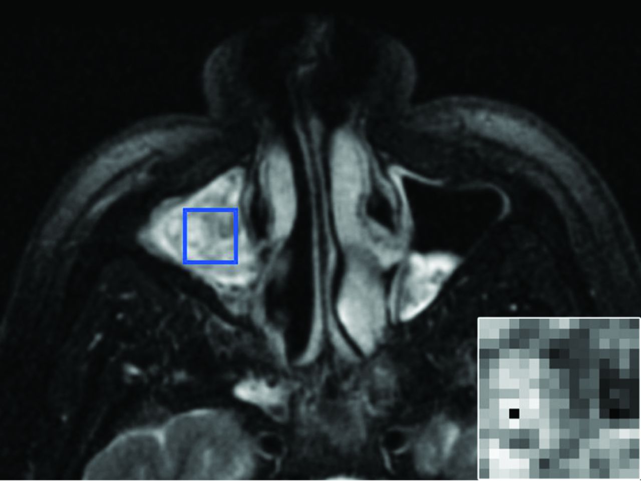
    ROI placement. A 51-year-old man with an IP involving the right maxillary sinus. Axial T2-weighted fat-suppressed MR imaging pulse sequence demonstrates the manual placement of the largest rectangular ROI that would fit within the tumor margins on the axial image with the greatest tumor cross-sectional area. The inset image in the lower right corner is representative of the final 16 × 16 matrix that was derived from the ROI isocenter and served as the input for texture analysis.

  • Fig 2.
    • Download figure
    • Open in new tab
    • Download powerpoint
    Fig 2.

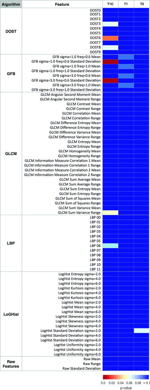
    Heat map showing MR imaging texture feature significance in distinguishing tumor type. Univariate analysis compared the pathology status (SCC versus IP) with MR imaging–texture features. Color maps show the false discovery rate–adjusted P values of a 2-sample t test. MR imaging contrasts (pulse sequences) are listed above the columns, and MR imaging–based texture features are listed in rows. DOST features 0–9 correspond with low-to-high frequency patterns. LBP 0–11 are the normalized bin counts in the LBP histogram. The reader is referred to the “Materials and Methods” section for additional details about the features.

  • Fig 3.
    • Download figure
    • Open in new tab
    • Download powerpoint
    Fig 3.

    Relative contributions to model accuracy. Of the 90.9% overall model accuracy for the training dataset, the bar graph demonstrates the accuracy attributable to PCs derived from T1C-GFB, T1-DOST, and T1-GLCM (upper panel). Across all texture algorithms, the contribution to total model accuracy was derived predominantly from T1C, with minor contributions from T1 and no input from T2 (lower panel).

  • Fig 4.
    • Download figure
    • Open in new tab
    • Download powerpoint
    Fig 4.

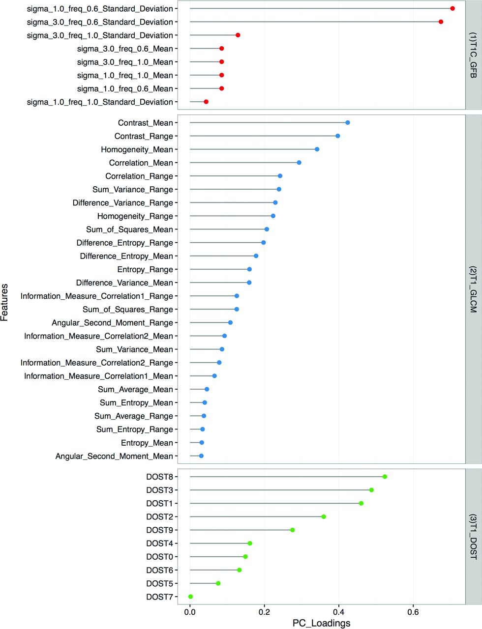
    PC loading. The model with the greatest accuracy for discriminating SCC from IP was derived from T1C-GFB, T1-GLCM, and T1-DOST texture features (right). For the individually specified texture features (left), PC loadings are graphically represented, and larger values in the PC loading indicate greater significance in the final model.

Tables

  • Figures
    • View popup
    Table 1:

    Patient demographic characteristics and tumor featuresa

    Study GroupSample SizeSex (Female/Male)Age (yr)Tumor Volume (cm3)Tumor Stageb
    T1T2T3T4
    IP training164:1258.0 ± 12.121.2 ± 17.713102
    IP validation61:558.2 ± 15.322.0 ± 6.91131
    IP combined225:17c58.1 ± 13.1d21.4 ± 15.5e24133
    SCC training174:1354.0 ± 13.555.8 ± 40.501412
    SCC validation71:654.6 ± 9.443.5 ± 27.90124
    SCC combined245:19c54.2 ± 12.5d52.2 ± 37.7e02616
    • ↵a Data are presented separately for the training and validation sets and also as a single combined cohort for each tumor type. Age and tumor volume are presented as means.

    • ↵b Tumor stage represents the Krouse staging system39 for IP and the American Joint Committee on Cancer staging40 for SCC.

    • ↵c Fisher exact test, P = .578.

    • ↵d Two-sample t test, P = .317.

    • ↵e Two-sample t test, P = .001.

    • View popup
    Table 2:

    Diagnostic performance of machine-learning classification in training and validation datasets

    Tumor Type (Pathologic Diagnosis)Diagnostic Performance
    SCCIP
    Model prediction for training dataset
        SCC162Accuracy90.9%a
    Sensitivity94.1%
        IP114Specificity87.5%
    PPV88.9%
        Total1716NPV93.3%
    Model prediction for validation dataset
        SCC61Accuracy84.6%a
    Sensitivity85.7%
        IP15Specificity83.3%
    PPV85.7%
        Total76NPV83.3%
    Model prediction for entire cohort
        SCC223Accuracy89.1%
    Sensitivity91.7%
        IP219Specificity86.4%
    PPV88.0%
        Total2422NPV90.5%
    • Note:—NPV indicates negative predictive value; PPV, positive predictive value.

    • ↵a With a 2-tailed test of population proportion, the accuracies for the training and validation datasets were not significantly different (P = .537).

    • View popup
    Table 3:

    Diagnostic performance of texture analysis with machine learning compared with neuroradiologists' review for the differentiation of SCC from IPa

    Analysis MethodAccuracybSensitivitySpecificityPPVNPV
    Texture analysis with machine learning89.1%91.7%86.4%88.0%90.5%
    Neuroradiologists' review, ROI56.5% (P = .0004)54.2%59.1%59.1%54.2%
    Neuroradiologists' review, tumor73.9% (P = .060)75.0%72.7%75.0%72.7%
    Neuroradiologists' review, image87.0% (P = .748)91.7%81.8%84.6%90.0%
    • Note:—NPV indicates negative predictive value; PPV, positive predictive value.

    • ↵a Results are shown for the entire cohort (22 IPs, 24 SCCs) and reflect the best classification model. The labels for the neuroradiologists' assessment indicate whether they reviewed the 16 × 16 ROI (ROI), tumor alone (tumor), or entire images (image).

    • ↵b P values represent comparison of texture analysis with machine learning against each neuroradiologist's review using a 2-tailed test of population proportion.

PreviousNext
Back to top

In this issue

American Journal of Neuroradiology: 38 (5)
American Journal of Neuroradiology
Vol. 38, Issue 5
1 May 2017
  • Table of Contents
  • Index by author
  • Complete Issue (PDF)
Advertisement
Print
Download PDF
Email Article

Thank you for your interest in spreading the word on American Journal of Neuroradiology.

NOTE: We only request your email address so that the person you are recommending the page to knows that you wanted them to see it, and that it is not junk mail. We do not capture any email address.

Enter multiple addresses on separate lines or separate them with commas.
MRI-Based Texture Analysis to Differentiate Sinonasal Squamous Cell Carcinoma from Inverted Papilloma
(Your Name) has sent you a message from American Journal of Neuroradiology
(Your Name) thought you would like to see the American Journal of Neuroradiology web site.
CAPTCHA
This question is for testing whether or not you are a human visitor and to prevent automated spam submissions.
Cite this article
S. Ramkumar, S. Ranjbar, S. Ning, D. Lal, C.M. Zwart, C.P. Wood, S.M. Weindling, T. Wu, J.R. Mitchell, J. Li, J.M. Hoxworth
MRI-Based Texture Analysis to Differentiate Sinonasal Squamous Cell Carcinoma from Inverted Papilloma
American Journal of Neuroradiology May 2017, 38 (5) 1019-1025; DOI: 10.3174/ajnr.A5106

Citation Manager Formats

  • BibTeX
  • Bookends
  • EasyBib
  • EndNote (tagged)
  • EndNote 8 (xml)
  • Medlars
  • Mendeley
  • Papers
  • RefWorks Tagged
  • Ref Manager
  • RIS
  • Zotero
0 Responses
Respond to this article
Share
Bookmark this article
MRI-Based Texture Analysis to Differentiate Sinonasal Squamous Cell Carcinoma from Inverted Papilloma
S. Ramkumar, S. Ranjbar, S. Ning, D. Lal, C.M. Zwart, C.P. Wood, S.M. Weindling, T. Wu, J.R. Mitchell, J. Li, J.M. Hoxworth
American Journal of Neuroradiology May 2017, 38 (5) 1019-1025; DOI: 10.3174/ajnr.A5106
del.icio.us logo Twitter logo Facebook logo Mendeley logo
  • Tweet Widget
  • Facebook Like
  • Google Plus One
Purchase

Jump to section

  • Article
    • Abstract
    • ABBREVIATIONS:
    • Materials and Methods
    • Results
    • Discussion
    • Conclusions
    • Footnotes
    • REFERENCES
  • Figures & Data
  • Info & Metrics
  • Responses
  • References
  • PDF

Related Articles

  • No related articles found.
  • PubMed
  • Google Scholar

Cited By...

  • No citing articles found.
  • Crossref (51)
  • Google Scholar

This article has been cited by the following articles in journals that are participating in Crossref Cited-by Linking.

  • Artificial Intelligence-based methods in head and neck cancer diagnosis: an overview
    Hanya Mahmood, Muhammad Shaban, Nasir Rajpoot, Syed A. Khurram
    British Journal of Cancer 2021 124 12
  • Exploring Applications of Radiomics in Magnetic Resonance Imaging of Head and Neck Cancer: A Systematic Review
    Amit Jethanandani, Timothy A. Lin, Stefania Volpe, Hesham Elhalawani, Abdallah S. R. Mohamed, Pei Yang, Clifton D. Fuller
    Frontiers in Oncology 2018 8
  • Quantitative variations in texture analysis features dependent on MRI scanning parameters: A phantom model
    Karen Buch, Hirofumi Kuno, Muhammad M. Qureshi, Baojun Li, Osamu Sakai
    Journal of Applied Clinical Medical Physics 2018 19 6
  • The utility of MRI histogram and texture analysis for the prediction of histological diagnosis in head and neck malignancies
    Noriyuki Fujima, Akihiro Homma, Taisuke Harada, Yukie Shimizu, Khin Khin Tha, Satoshi Kano, Takatsugu Mizumachi, Ruijiang Li, Kohsuke Kudo, Hiroki Shirato
    Cancer Imaging 2019 19 1
  • Textural analysis on contrast-enhanced CT in pancreatic neuroendocrine neoplasms: association with WHO grade
    Chuangen Guo, Xiaoling Zhuge, Zhongqiu Wang, Qidong Wang, Ke Sun, Zhan Feng, Xiao Chen
    Abdominal Radiology 2019 44 2
  • Predicting early recurrence of hepatocellular carcinoma with texture analysis of preoperative MRI: a radiomics study
    T.C.H. Hui, T.K. Chuah, H.M. Low, C.H. Tan
    Clinical Radiology 2018 73 12
  • Big Data in Head and Neck Cancer
    Carlo Resteghini, Annalisa Trama, Elio Borgonovi, Hykel Hosni, Giovanni Corrao, Ester Orlandi, Giuseppina Calareso, Loris De Cecco, Cesare Piazza, Luca Mainardi, Lisa Licitra
    Current Treatment Options in Oncology 2018 19 12
  • Enhancing head and neck tumor management with artificial intelligence: Integration and perspectives
    Nian-Nian Zhong, Han-Qi Wang, Xin-Yue Huang, Zi-Zhan Li, Lei-Ming Cao, Fang-Yi Huo, Bing Liu, Lin-Lin Bu
    Seminars in Cancer Biology 2023 95
  • Recent Advances in the Application of Artificial Intelligence in Otorhinolaryngology-Head and Neck Surgery
    Bayu Adhi Tama, Do Hyun Kim, Gyuwon Kim, Soo Whan Kim, Seungchul Lee
    Clinical and Experimental Otorhinolaryngology 2020 13 4
  • Radiomic biomarkers for head and neck squamous cell carcinoma
    Stephanie Tanadini-Lang, Panagiotis Balermpas, Matthias Guckenberger, Matea Pavic, Oliver Riesterer, Diem Vuong, Marta Bogowicz
    Strahlentherapie und Onkologie 2020 196 10

More in this TOC Section

  • Chondrosarcoma vs Synovial Chondromatosis: Imaging
  • WHO Classification Update: Nasal&Skull Base Tumors
  • Peritumoral Signal in Vestibular Schwannomas
Show more HEAD & NECK

Similar Articles

Advertisement

Indexed Content

  • Current Issue
  • Accepted Manuscripts
  • Article Preview
  • Past Issues
  • Editorials
  • Editor's Choice
  • Fellows' Journal Club
  • Letters to the Editor
  • Video Articles

Cases

  • Case Collection
  • Archive - Case of the Week
  • Archive - Case of the Month
  • Archive - Classic Case

Special Collections

  • AJNR Awards
  • ASNR Foundation Special Collection
  • Most Impactful AJNR Articles
  • Photon-Counting CT
  • Spinal CSF Leak Articles (Jan 2020-June 2024)

More from AJNR

  • Trainee Corner
  • Imaging Protocols
  • MRI Safety Corner

Multimedia

  • AJNR Podcasts
  • AJNR Scantastics

Resources

  • Turnaround Time
  • Submit a Manuscript
  • Submit a Video Article
  • Submit an eLetter to the Editor/Response
  • Manuscript Submission Guidelines
  • Statistical Tips
  • Fast Publishing of Accepted Manuscripts
  • Graphical Abstract Preparation
  • Imaging Protocol Submission
  • Evidence-Based Medicine Level Guide
  • Publishing Checklists
  • Author Policies
  • Become a Reviewer/Academy of Reviewers
  • News and Updates

About Us

  • About AJNR
  • Editorial Board
  • Editorial Board Alumni
  • Alerts
  • Permissions
  • Not an AJNR Subscriber? Join Now
  • Advertise with Us
  • Librarian Resources
  • Feedback
  • Terms and Conditions
  • AJNR Editorial Board Alumni

American Society of Neuroradiology

  • Not an ASNR Member? Join Now

© 2025 by the American Society of Neuroradiology All rights, including for text and data mining, AI training, and similar technologies, are reserved.
Print ISSN: 0195-6108 Online ISSN: 1936-959X

Powered by HighWire