
Article Figures & Data
Figures
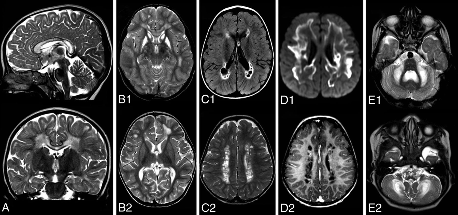
- FIG 1.
MR imaging features suggestive of mitochondrial leukodystrophy. A, Sagittal and coronal T2-weighted images of a patient with a complex 2 deficiency show longitudinal T2 hyperintensity affecting the middle blade of the corpus callosum while sparing the inner and outer blades. B, Axial T2-weigthed images of 2 patients. B1, Symmetric involvement of the striatum in a patient with NDUFV1 variants. B2, Symmetric hyperintensity in the thalami in a patient with NDUFAF5 variants. C1, FLAIR image of a patient with LYRM7 variants shows small cysts in the periventricular and deep cerebral white matter. C2, T2-weighted image shows the well-defined border of cysts in a different patient with LYRM7 variants. D1, Axial diffusion trace images show extensive diffusion restriction at the edge of the lesions in a patient with BOLA3 variants, D2, Postcontrast T1-weighted image shows subtle enhancement at the edge of cysts in the same patient as in C2. E1 and E2, Axial T2-weighted images. E1, Symmetric hyperintensity of the middle cerebellar peduncles and cerebellar white matter in a patient with ISCA2 variants. E2, Hyperintensity of the medulla oblongata, middle cerebellar peduncles, and cerebellar white matter in a patient with IBA57 variants.
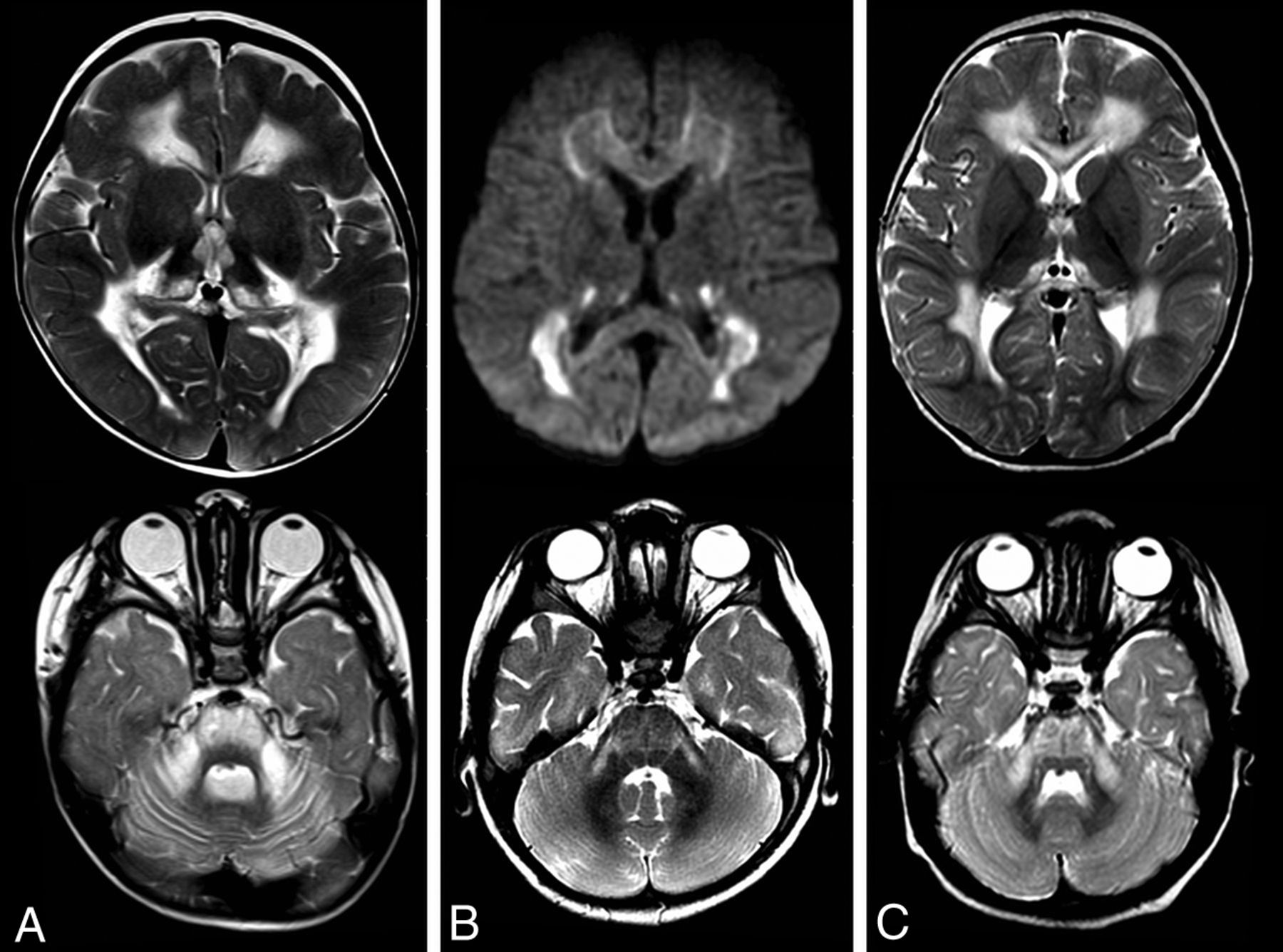
- FIG 2.
Features characteristic of SDH deficiency in 3 patients (A–C) showing symmetric thalamic involvement and pontine and middle cerebellar hyperintensities on T2-weighted images. B, Note the diffusion restriction.
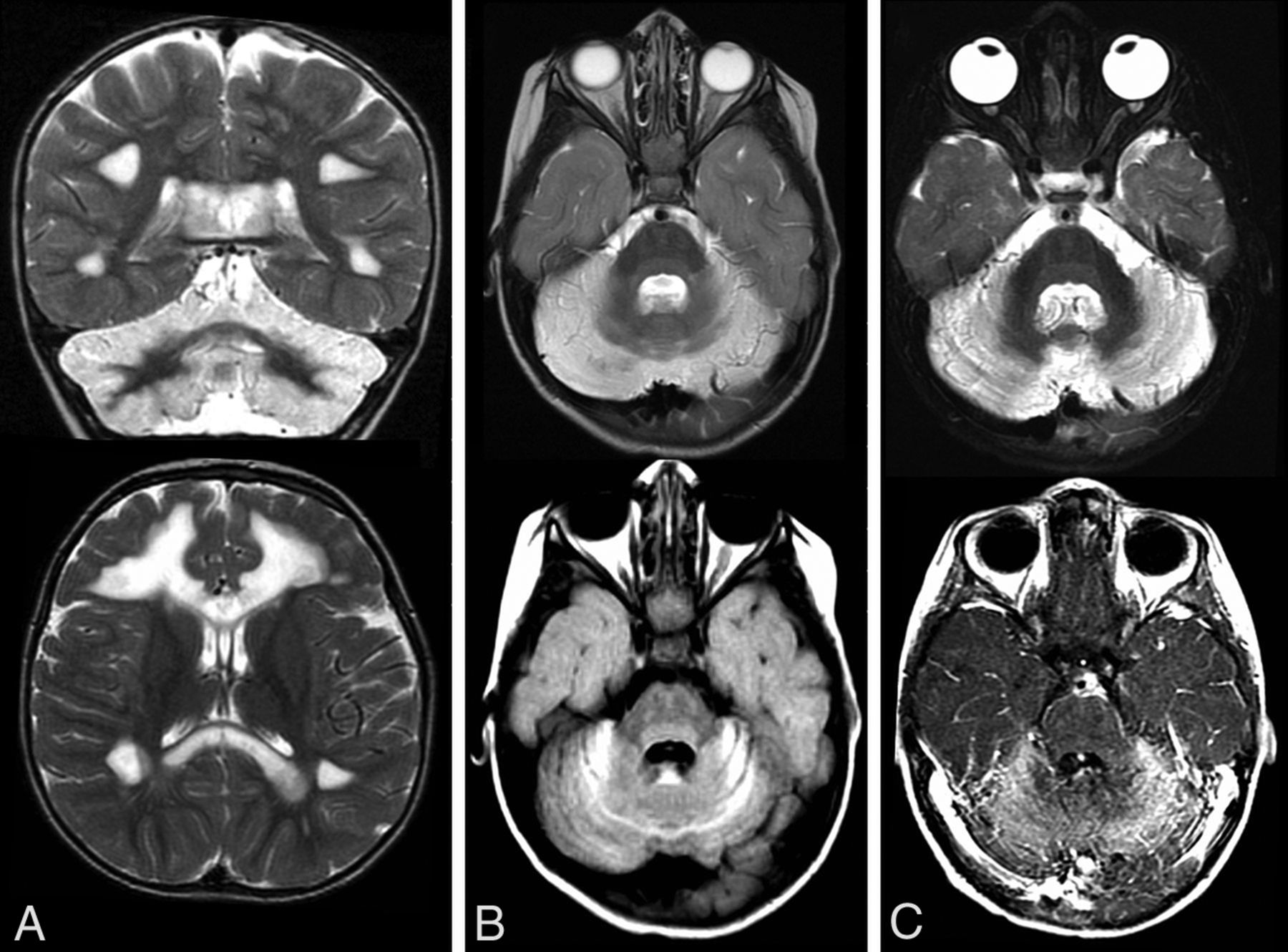
- FIG 3.
Characteristic features of defects in complexes 3 (A) and 4 (B and C). Upper rows are T2-weighted images; lower rows show FLAIR images and a diffusion trace image. Predominantly periventricular T2 hyperintensities, well-defined cystic lesions with a distinct border. Diffusion restriction at the edge of the lesions.
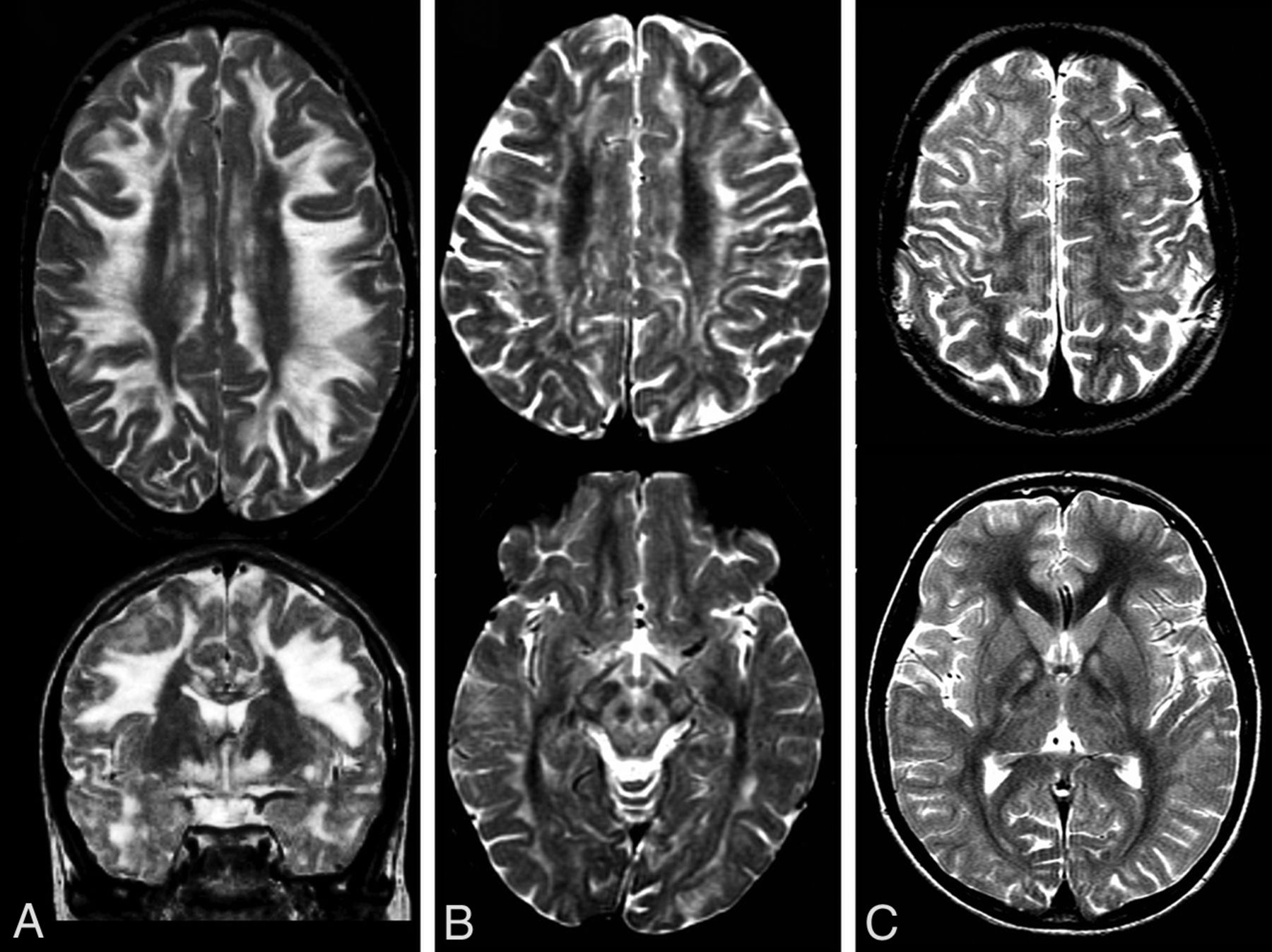
- FIG 4.
Characteristic features of NUBPL variants shown in 3 patients (A–C). The T2-weighted images show hyperintensity of the cerebellum, which is also hyperintense on FLAIR (B) and enhances (C). Involvement of supratentorial white matter can be extensive (A).
- FIG 5.
T2-weighted images of 3 patients with KSS (A–C) showing hyperintensity affecting (predominantly the subcortical and also deep) white matter. A and C, In the lower panel, hyperintensities in the globus pallidus exist. B, Involvement of the substantia nigra and, in this case, larger areas of the midbrain are shown.


Tables
Imaging Features Selective longitudinal involvement of the middle blade of the corpus callosum
Cerebral white matter rarefaction and cysts with a well-delineated rim, which may show contrast enhancement and diffusion restriction
Symmetric deep gray matter abnormalities
Brain stem abnormalities
Symmetric abnormalities in the middle cerebellar pedunclesMR Imaging Patterns Succinate dehydrogenase (complex 2) deficiency
Abnormalities in the brain stem, more specifically in the transverse pontine fibers
Abnormalities in the middle cerebellar peduncles
Abnormalities in the thalami
Succinate in MR spectroscopyComplex 3 and 4 defects
Numerous small cysts with well-defined borders in the periventricular and deep cerebral white matterKearns-Sayre syndrome
Abnormalities predominantly affecting the directly subcortical cerebral white matter plus abnormalities in the globus pallidus and substantia nigraNUBPL defects
T2 hyperintensity of the cerebellar cortex plus cerebral white matter abnormalities


Jump to section
Related Articles
Cited By...
- Biallelic mutation of SUPV3L1 causes an inherited leukodystrophy-associated neurodevelopmental disorder due to aberrant mitochondrial double stranded RNA processing
- Teaching NeuroImage: IBA57 Mutation-Associated Infantile Cavitating Leukoencephalopathy
- Pattern Recognition in Mitochondrial Leukodystrophies is Hampered by the Peculiarities of Mitochondrial Genetics