Advertisement

Main menu

  • Home
  • Content
    • Current Issue
    • Accepted Manuscripts
    • Article Preview
    • Past Issue Archive
    • Video Articles
    • AJNR Case Collection
    • Case of the Week Archive
    • Case of the Month Archive
    • Classic Case Archive
  • Special Collections
    • AJNR Awards
    • ASNR Foundation Special Collection
    • Most Impactful AJNR Articles
    • Photon-Counting CT
    • Spinal CSF Leak Articles (Jan 2020-June 2024)
  • Multimedia
    • AJNR Podcasts
    • AJNR SCANtastic
    • Trainee Corner
    • MRI Safety Corner
    • Imaging Protocols
  • For Authors
    • Submit a Manuscript
    • Submit a Video Article
    • Submit an eLetter to the Editor/Response
    • Manuscript Submission Guidelines
    • Statistical Tips
    • Fast Publishing of Accepted Manuscripts
    • Graphical Abstract Preparation
    • Imaging Protocol Submission
    • Author Policies
  • About Us
    • About AJNR
    • Editorial Board
    • Editorial Board Alumni
  • More
    • Become a Reviewer/Academy of Reviewers
    • Subscribers
    • Permissions
    • Alerts
    • Feedback
    • Advertisers
    • ASNR Home

User menu

  • Alerts
  • Log in

Search

  • Advanced search
American Journal of Neuroradiology

American Journal of Neuroradiology

ASHNR American Society of Functional Neuroradiology ASHNR American Society of Pediatric Neuroradiology ASSR
  • Alerts
  • Log in

Advanced Search

  • Home
  • Content
    • Current Issue
    • Accepted Manuscripts
    • Article Preview
    • Past Issue Archive
    • Video Articles
    • AJNR Case Collection
    • Case of the Week Archive
    • Case of the Month Archive
    • Classic Case Archive
  • Special Collections
    • AJNR Awards
    • ASNR Foundation Special Collection
    • Most Impactful AJNR Articles
    • Photon-Counting CT
    • Spinal CSF Leak Articles (Jan 2020-June 2024)
  • Multimedia
    • AJNR Podcasts
    • AJNR SCANtastic
    • Trainee Corner
    • MRI Safety Corner
    • Imaging Protocols
  • For Authors
    • Submit a Manuscript
    • Submit a Video Article
    • Submit an eLetter to the Editor/Response
    • Manuscript Submission Guidelines
    • Statistical Tips
    • Fast Publishing of Accepted Manuscripts
    • Graphical Abstract Preparation
    • Imaging Protocol Submission
    • Author Policies
  • About Us
    • About AJNR
    • Editorial Board
    • Editorial Board Alumni
  • More
    • Become a Reviewer/Academy of Reviewers
    • Subscribers
    • Permissions
    • Alerts
    • Feedback
    • Advertisers
    • ASNR Home
  • Follow AJNR on Twitter
  • Visit AJNR on Facebook
  • Follow AJNR on Instagram
  • Join AJNR on LinkedIn
  • RSS Feeds

AJNR is seeking candidates for the AJNR Podcast Editor. Read the position description.

Review ArticleREVIEW ARTICLE
Open Access

Spontaneous Intracranial Hypotension: Atypical Radiologic Appearances, Imaging Mimickers, and Clinical Look-Alikes

K.M. Bond, J.C. Benson, J.K. Cutsforth-Gregory, D.K. Kim, F.E. Diehn and C.M. Carr
American Journal of Neuroradiology July 2020, DOI: https://doi.org/10.3174/ajnr.A6637
K.M. Bond
aFrom the Mayo Clinic School of Medicine (K.M.B.), Rochester, Minnesota
  • Find this author on Google Scholar
  • Find this author on PubMed
  • Search for this author on this site
  • ORCID record for K.M. Bond
J.C. Benson
band Departments of Radiology (J.C.B., D.K.K., F.E.D., C.M.C.)
  • Find this author on Google Scholar
  • Find this author on PubMed
  • Search for this author on this site
  • ORCID record for J.C. Benson
J.K. Cutsforth-Gregory
cNeurology (J.K.C.-G.), Mayo Clinic, Rochester, Minnesota.
  • Find this author on Google Scholar
  • Find this author on PubMed
  • Search for this author on this site
  • ORCID record for J.K. Cutsforth-Gregory
D.K. Kim
band Departments of Radiology (J.C.B., D.K.K., F.E.D., C.M.C.)
  • Find this author on Google Scholar
  • Find this author on PubMed
  • Search for this author on this site
  • ORCID record for D.K. Kim
F.E. Diehn
band Departments of Radiology (J.C.B., D.K.K., F.E.D., C.M.C.)
  • Find this author on Google Scholar
  • Find this author on PubMed
  • Search for this author on this site
  • ORCID record for F.E. Diehn
C.M. Carr
band Departments of Radiology (J.C.B., D.K.K., F.E.D., C.M.C.)
  • Find this author on Google Scholar
  • Find this author on PubMed
  • Search for this author on this site
  • ORCID record for C.M. Carr
  • Article
  • Figures & Data
  • Info & Metrics
  • Responses
  • References
  • PDF
Loading

Abstract

SUMMARY: Spontaneous intracranial hypotension is a condition characterized by low CSF volume secondary to leakage through a dural defect with no identifiable cause. Patients classically present with orthostatic headaches, but this symptom is not specific to spontaneous intracranial hypotension, and initial misdiagnosis is common. The most prominent features of spontaneous intracranial hypotension on intracranial MR imaging include “brain sag” and diffuse pachymeningeal enhancement, but these characteristics can be seen in several other conditions. Understanding the clinical and imaging features of spontaneous intracranial hypotension and its mimickers will lead to more prompt and accurate diagnoses. Here we discuss conditions that mimic the radiologic and clinical presentation of spontaneous intracranial hypotension as well as other disorders that CSF leaks can imitate.

ABBREVIATIONS:

IgG4
immunoglobulin G4
POTS
postural orthostatic tachycardia syndrome
SIH
spontaneous intracranial hypotension
SS
superficial siderosis

The clinical syndrome of orthostatic headache that resolves with recumbency or worsens with Valsalva maneuvers raises suspicion of low CSF pressure, most frequently due to a spinal CSF leak. These leaks may be from iatrogenic dural defects, such as lumbar punctures or drains (secondary intracranial hypotension). In contrast, spontaneous intracranial hypotension (SIH) or CSF hypovolemia refers to leaks that arise spontaneously. These terms can be misleading, however, because most patients have normal opening pressure.1 The term “spontaneous spinal CSF leak” may be a more accurate description of this condition, though we will continue to refer to it as SIH throughout this review.

SIH is estimated to affect 5 per 100,000 people per year, with predominance for women.2 It is thought to arise when mechanical stressors cause or exacerbate underlying weakness in the spinal dura, which may be exacerbated by connective tissue disease. Spiculated osteophytes, herniated discs, and nerve root diverticula can incite small dural tears that allow CSF to leak out of the thecal sac into the extradural space.2,3 CSF-venous fistulas can also allow leakage into the epidural space in the absence of a dural tear.4,5

The most characteristic features of SIH on intracranial MR imaging include diffuse pachymeningeal enhancement and “brain sag,” which, in a retrospective review of 99 cases, were seen in 83% and 61% of cases, resepectively.6 While some patients experience spontaneous resolution of symptoms,2 many will require more definitive treatment, including patching with blood or fibrin and surgical closure of the defect.7,8

As SIH has gained recognition within the medical community, particularly after the widespread adoption of MR imaging as a noninvasive diagnostic tool, patients with classic signs and symptoms have benefitted from earlier diagnosis and treatment.2,9 Many patients, however, present with vague neurologic symptoms such as nonpositional daily headache, cranial nerve palsy, gait disturbance, and cognitive dysfunction. Moreover, the source of the leak may remain occult despite advanced imaging in an estimated 46%–55% of patients.10 Further complicating matters, several conditions have imaging features that overlap SIH.

Understanding the clinical and imaging features of SIH and its mimickers will lead to more prompt and accurate diagnoses. Here we discuss conditions that mimic the radiologic and clinical presentation of SIH (Table 1), as well as other disorders that CSF leaks can imitate (Table 2). Nonspecific radiographic findings to be discussed include bilateral subdural fluid collections or hematomas and conditions with dural thickening. Conditions with some shared imaging features include Chiari type I malformations and diencephalic-mesencephalic junction dysplasia. Nonspecific clinical mimics include orthostatic headaches, cervicogenic headache, and vestibular migraine. Postural orthostatic tachycardia syndrome (POTS) is an alternative diagnosis with shared symptoms. SIH may present as an alternate clinical entity with the leak ultimately found as the cause for the condition. These secondary conditions include cerebral venous sinus thrombosis, frontotemporal dementia, and superficial siderosis. Other entities that are associated with SIH include pituitary tumors and apoplexy.

View this table:
  • View inline
  • View popup
Table 1:

Distinguishing features of alternative diagnoses that occasionally mimic spontaneous intracranial hypotension

View this table:
  • View inline
  • View popup
Table 2:

Conditions that occasionally coexist with spontaneous intracranial hypotension

Radiologic Findings of SIH and Its Mimickers

If SIH is suspected, brain MR imaging with and without gadolinium contrast enhancement is the initial imaging examination of choice. Brain sag, or the downward displacement of the cerebellar tonsils and brain stem, is a classic finding.9 Flattening of the ventral pons, effacement of the subarachnoid spaces including the prepontine and perichiasmatic cisterns, and descent of the iter (superior opening of the cerebral aqueduct) below the incisural line are additional manifestations of brain sag. With or without brain sag, many patients’ scans will demonstrate diffuse, smooth dural thickening and enhancement.11 Other possible imaging features include ventricular collapse, dural venous sinus engorgement, atraumatic subdural hematomas or hygromas, and pituitary enlargement. Due to the effacement of the perichiasmatic cistern and pituitary engorgement, the optic chiasm can appear to be directly draped over the pituitary gland. Note that there is wide variability as to how many signs may or may not be present in a given patient. Dobrocky et al12 have proposed a scoring method to determine the likelihood of SIH (low, intermediate, or high) based on the presence of 6 of these MR imaging findings. Additionally, superficial siderosis may be present in situations in which there is chronic bleeding into the thecal sac, typically related to recurrent trauma from a spiculated osteophyte or disk protrusion.13 Even when brain MR imaging findings are normal, a few case reports have described radioisotope cisternography showing reduced tracer activity over the cerebral convexities 24 hours after tracer injection, thereby supporting the diagnosis of SIH.14⇓-16

Approaches to spinal imaging in patients with suspected SIH vary widely among institutions. Either a spinal MR imaging or CT myelography may be performed to evaluate the presence of extradural fluid collections. A “fast” leak is suspected if such a collection is identified, often leading to the use of either a dynamic CT myelogram or a digital subtraction myelogram to localize the leak.9,16 Spine MR imaging may also demonstrate secondary/supportive findings of SIH such as dural thickening and enhancement or engorgement of the epidural venous plexus.17 Nerve root sleeve diverticula are commonly seen, though these are nonspecific and of unclear significance unless they are very large and irregular.18

Chiari Type I Malformation.

A Chiari type I malformation is an abnormal morphology at the craniocervical junction, classically defined as descent of the cerebellar tonsils at least 5 mm through the foramen magnum. The prevalence is estimated to be <1% in the general population, and most of these patients are asymptomatic.19,20 Tonsillar descent can obstruct CSF outflow and thereby cause symptoms of cough-induced headache, paresthesia, lower cranial neuropathies, and cerebellar signs.21 Disruption of normal flow of the CSF can also result in syringomyelia. Treatment of symptomatic Chiari is by posterior fossa decompression.

SIH can be mistaken for a Chiari type I malformation on imaging (Fig 1) due to the superficially similar radiologic appearance of cerebellar tonsillar descent.9 Although in some cases the differentiation can be challenging, there are several key imaging differences. In SIH, the tonsils usually maintain their normal shape rather than being inferiorly pointed and often do not descend more than 5 mm below the foramen magnum.22,23 In addition, the midbrain also descends in SIH, whereas it will maintain a normal position in Chiari type I. The superior aspect of the cerebral aqueduct (iter) may fall beneath the incisural line, which extends from the anterior tuberculum sellae to the inferior point of the venous confluence of the straight sinus, whereas it does not change position in Chiari type I.24,25 The presence of a syrinx will generally indicate a Chiari type I malformation, though there are rare reports of this developing in SIH.26 The subarachnoid spaces in the posterior fossa, foramen magnum, and normal ventricular caliber will be preserved in Chiari I malformations, whereas they will be effaced in SIH. Flattening of the pons is commonly seen with pronounced SIH-associated brain sag but will not be present with Chiari type I malformations, in which the prepontine cistern will be maintained. Finally, patients with a Chiari malformation will not exhibit diffuse dural enhancement.

FIG 1.
  • Download figure
  • Open in new tab
  • Download powerpoint
FIG 1.

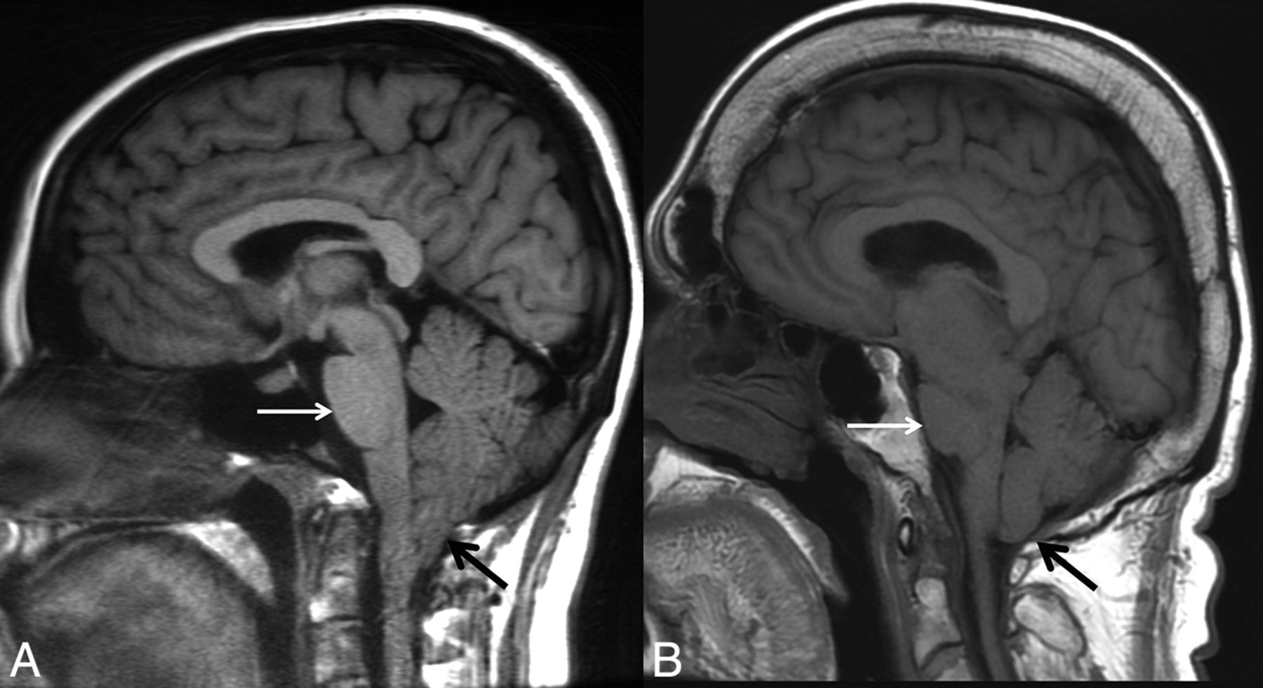
Chiari type I malformation versus SIH. A, Sagittal T1WI demonstrates a Chiari type I malformation with descent of cerebellar tonsils 1.4 cm below the foramen magnum (black arrow). Note the typical “peglike” morphology. The prepontine cistern is preserved, and the pons maintains a rounded ventral morphology (white arrow). B, Sagittal T1WI in a patient with severe brain sag due to SIH shows mild descent of the cerebellar tonsils at the foramen magnum (black arrow), but with normal morphology. The pons is flattened (white arrow) with effacement of the prepontine cistern. There is also profound effacement of the subarachnoid spaces in the posterior fossa.

The literature describes multiple reports of “acquired” Chiari I malformations in the setting of spinal CSF drainage (eg, ventriculoperitoneal shunts). Images from these reports do show cerebellar descent, but many also show findings of intracranial hypotension. Overshunting, in which CSF diverted at a rate greater than production, results in an iatrogenic cause of intracranial hypotension or hypovolemia. The underlying physiology is similar to that of SIH (though iatrogenic in origin) and shares similar findings of slit-like ventricles, effacement of the subarachnoid spaces in the posterior fossa, flattening of the ventral pons, and descent of the brain stem and tonsils (Fig 2).

FIG 2.
  • Download figure
  • Open in new tab
  • Download powerpoint
FIG 2.

“Acquired” Chiari malformation showing findings of CSF hypotension. A, Sagittal T1WI demonstrates a normal appearance of the posterior fossa of a 6-year-old patient with a ventriculoperitoneal shunt. B, Six years later, imaging demonstrates findings of overshunting resulting in CSF hypotension. This has previously (mistakenly) been called “acquired Chiari malformation.” In addition to descent of the tonsils, there is also effacement of the subarachnoid spaces, flattening of the pons, descent of the brain stem, and a decrease in the volume of the lateral and fourth ventricles, all of which indicate reduced CSF volume.

Subdural Fluid Collections or Hematomas.

Subdural hematoma is a condition that disproportionately affects elderly patients and is often associated with minor head trauma. Bridging veins are predisposed to tear in this population because they cross the subarachnoid spaces, which expand as a function of age due to brain parenchymal atrophy. Anticoagulation is a factor in 5%–20% of patients with chronic subdural hematomas.27,28 Subdural hygromas occur due to compensatory enlargement of the subdural/subarachnoid space secondary to the loss of the CSF volume. A subdural hematoma can also occur if there is tearing of abnormally engorged cortical veins. In a series of 98 patients with chronic subdural hematomas, 26% were found to be bilateral.29 Bilateral subdural collections have been known to occur in the setting of SIH (Fig 3).29⇓-31 These bilateral subdural collections can occur either with or without the characteristic intracranial stigmata of SIH (eg, brain sag and focal dural enhancement). The fluid collections may occur as only a hygroma or a subacute/chronic hematoma. Unilateral occurrence is rare. It is imperative that the appropriate etiology be identified because subdural fluid collections will not resolve until there is successful treatment of the underlying SIH. If the collections are drained, they will recur due to the underlying abnormal CSF dynamics.32 Therapy should be focused on finding and treating the leak rather than drainage of the collections. With appropriate treatment, the collections will gradually resolve.33

FIG 3.
  • Download figure
  • Open in new tab
  • Download powerpoint
FIG 3.

Bilateral subdural hematomas as an initial presentation in SIH. A, Sagittal T1WI shows a subtle decrease in the mamillopontine distance (white arrow). Otherwise, the typical stigmata of SIH are not present. B, Coronal postcontrast T1WI demonstrates large bilateral subdural hematomas with mass effect on the cerebral hemispheres. A hyperdynamic CT myelogram showed a leak in the thoracic spine, and a targeted blood patch was performed. The subdural hematomas subsequently required evacuation due to cerebral compression. All of the patient’s symptoms resolved, and the hematomas did not recur.

Conditions with Dural Thickening.

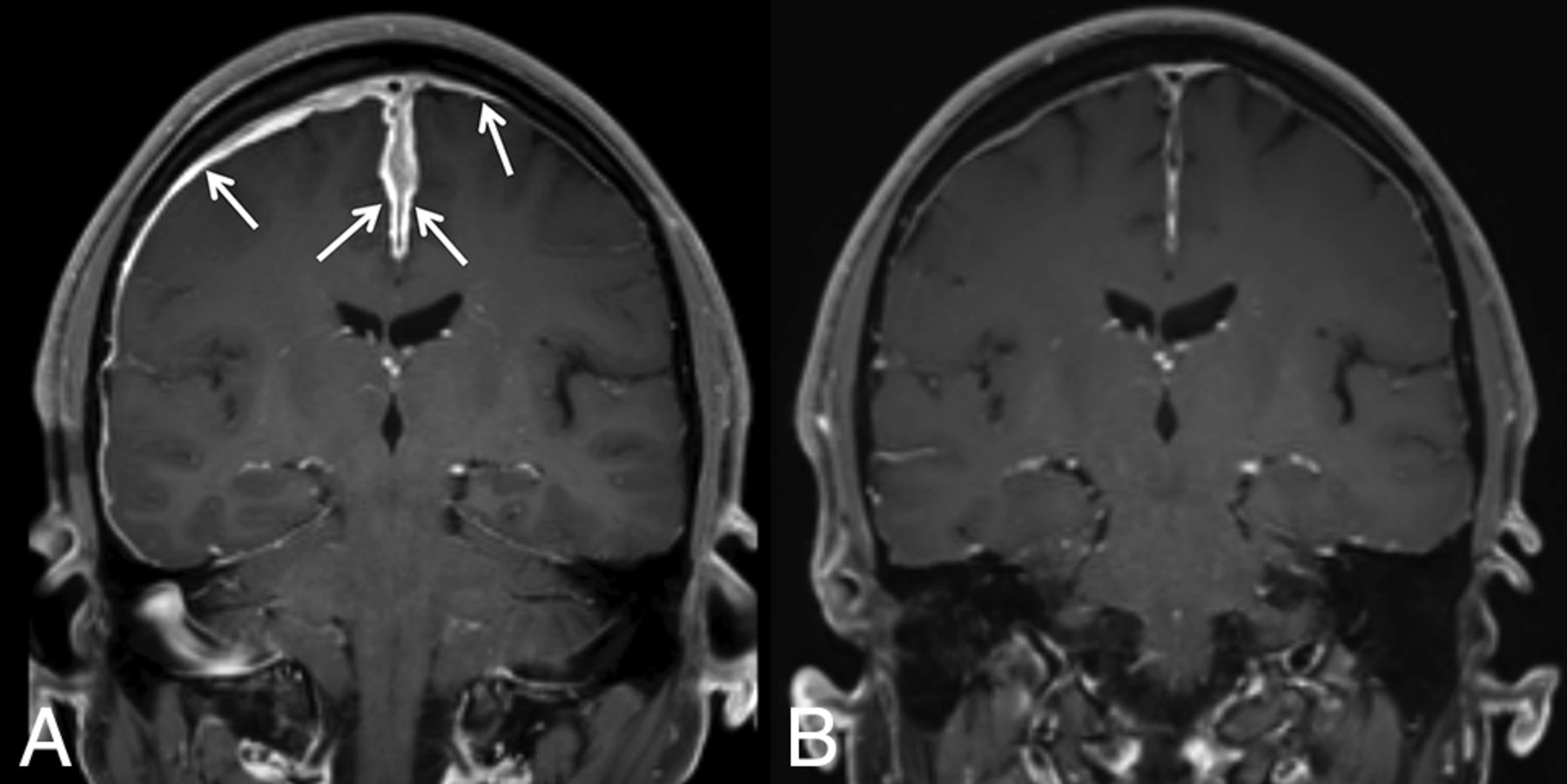
A key radiologic finding of SIH is dural thickening and enhancement, which are usually diffuse and non-nodular. Several rare conditions can have a similar appearance on MR imaging. Such hypertrophic pachymeningitis can have a specific etiology or be idiopathic. Immunoglobulin 4 (IgG4)-related disease, a known cause of pachymeningitis, is a fibroinflammatory condition caused by the infiltration of lymphoplasmacytic cells into tissues and subsequent fibrosis.34,35 It can manifest in nearly every organ system and, therefore, has a wide variety of presentations. The hypertrophic pachymeningitis due to IgG4-related disease can affect the dura intracranially or in the spinal canal.35 On MR imaging, the dura is thickened, enhances, and has a shortened T2 signal due to fibrotic changes (Fig 4). Neurosarcoidosis, another idiopathic systemic disease with variable clinical presentation, can cause pachymeningeal and/or leptomeningeal thickening and enhancement, which are generally most prominent along the skull base.36 In addition, lesions of the brain parenchyma, cranial and peripheral nerves, and bones are not unusual.37,38 Dural thickening can also occur secondary to a variety of noninfectious, non-neoplastic conditions (broadly including immune-mediated, histiocytic, granulomatous, and idiopathic), such as rheumatoid arthritis,39,40 temporal arteritis,41 Rosai-Dorfman disease,42,43 Erdheim-Chester disease,44 and polyangiitis with granulomatosis.45

FIG 4.
  • Download figure
  • Open in new tab
  • Download powerpoint
FIG 4.

IgG4-related pachymeningitis. A, Coronal contrast-enhanced T1WI from a 53-year-old man with IgG4-related disease with Castleman-like features shows abnormal pachymeningeal enhancement (arrows). B, Follow-up imaging after steroid and rituximab (Rituxan) treatment demonstrates interval near resolution of the pachymeningeal enhancement.

Tuberculous meningitis is a severe manifestation of extrapulmonary tuberculosis with high associated mortality.46,47 Focal or diffuse dural thickening may be present on MR imaging, and the diagnosis is confirmed with biopsy.47 Tuberculosis can also affect the leptomeninges, form intracranial tuberculomas, and cause spinal tuberculous arachnoiditis.48,49 Other infectious causes of hypertrophic pachymeningitis include syphilis, Cryptococcus, and Lyme disease.50⇓-52 Each of the conditions discussed here has the potential to affect multiple organ systems, and patients’ clinical presentations often reflect involvement beyond the nervous system.

SIH should not be included in the differential if focal or nodular dural thickening is present because neoplastic or infectious etiologies would be the primary differential consideration. An operation or lumbar punctures may also show diffuse smooth dural enhancement similar to what is seen in SIH; however, correlation with relevant history will distinguish these entities.

Clinical Mimics of SIH

As the recognized clinical phenotype of spontaneous spinal CSF leak has broadened, so too has the differential diagnosis of the many signs and symptoms associated with this entity broadened. Several atypical presentations of bona fide SIH are discussed in the next section. Here we will focus on mimics of the most common symptom of SIH, orthostatic headache. Indeed, we have encountered patients with orthostatic headache due to a variety of causes other than spinal CSF leak, most often an autonomic disorder or an alternative secondary headache disorder.

POTS.

POTS is perhaps the most common nonleak cause of headache that is triggered or worsened with upright posture. At least 25% of patients with POTS report headaches, and by definition, all have orthostatic worsening of symptoms.53 POTS is a diverse syndrome due to various etiologies, among which may occasionally be spinal CSF leakage.54 Some patients likely develop POTS secondary to the prolonged supine deconditioning that often occurs in the setting of a spinal CSF leak. There are also patients with POTS and orthostatic headaches in the absence of any evidence of CSF leakage.55 Complicating matters further, POTS shares risk factors with spinal CSF leak, including joint hypermobility and underlying disorders of the connective tissue matrix such as Ehlers-Danlos syndrome.53 POTS with or without spinal CSF leak should be considered when evaluating a patient with orthostatic headache. Measurement of the sitting and standing heart rate and blood pressure is, in our opinion, a reasonable screen for this diagnosis; patients with POTS demonstrate a rapid increase in heart rate and relatively stable blood pressure on standing.56

Orthostatic Hypotension.

Orthostatic hypotension, which can be caused by autonomic failure (Fig 5), medication effect, or serious hypovolemia but not spinal CSF leakage, is another reasonably common cause of orthostatic headache. Whereas spinal CSF leak–associated headache is typically centered in the head, its focus in the occipital region and the frequent accompanying neck and back pain may be confused with the “coat hanger” pain in the neck and shoulders that results from paraspinal muscle ischemia in patients with orthostatic hypotension.57 Other symptoms that may occur with either spinal CSF leak or orthostatic hypotension include tinnitus, distorted hearing, fatigue, and worsening headache with Valsalva maneuvers. Similar to POTS, measurement of orthostatic vital signs should distinguish patients with orthostatic hypotension from those with suspected spinal CSF leak. Patients with orthostatic hypotension have a fall in systolic and/or diastolic blood pressure of 20 mm Hg and 10 mm Hg, respectively, during a lying-to-standing, lying-to-sitting, or the head-up tilt test.58

FIG 5.
  • Download figure
  • Open in new tab
  • Download powerpoint
FIG 5.

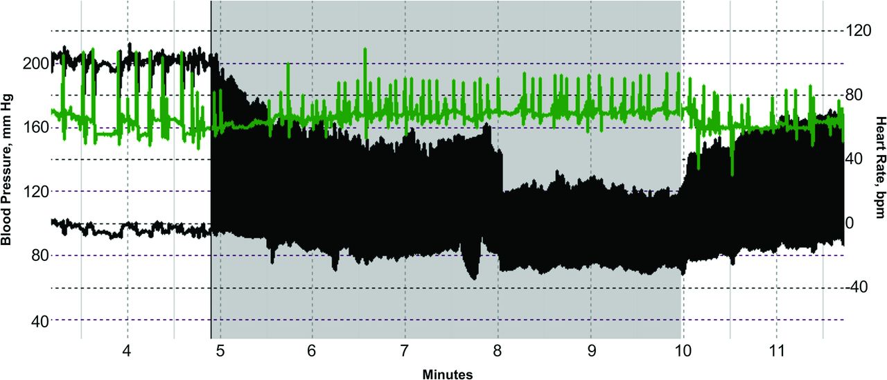
Autonomic failure presenting as orthostatic headache. Beat-to-beat blood pressure (black) and heart rate (green) of an 80-year-old woman during a tilt-table test show a sustained drop in blood pressure during head-up tilt (gray shading). The lack of heart rate acceleration suggests neurogenic orthostatic hypotension. When her mean blood pressure fell below 85 mm Hg, she developed posterior head and neck tightness (coat hanger pain) and lightheadedness. Thus, pure autonomic failure was initially misdiagnosed as orthostatic headache.

Other Headache Disorders.

Patients with cervicogenic headache or vestibular migraine will typically have worsening headache when attaining an upright position, which may raise suspicion of a spinal CSF leak. Cervicogenic headache results from a disorder or lesion of the bony cervical spine or soft tissues of the neck and is usually accompanied by neck pain.59 Most commonly, the offending pathology is osteoarthritis of the upper cervical spine or the atlanto-occipital junction.59 The clinical symptoms are typically distinct from those of SIH: Cervicogenic headaches are usually unilateral and are accompanied by neck pain that is worsened during neck movement.59 Most important, they are also relieved by pharmaceutical blockade (ie, steroid injection) of the inciting cervical structure.59

Vestibular migraine shares many features with classic migraine (eg, unilateral location, pulsating quality, photophobia, phonophobia, visual aura) but is distinguished by the presence of head motion–induced, visually-induced, spontaneous, or positional vertigo.59 It is this postural worsening that introduces possible confusion with spinal CSF leak. Often, the migrainous features of the headache, as well as a history of documented migraines, may help distinguish vestibular migraine from SIH. In addition, nystagmus is often present and usually lasts as long as the precipitating head position is held.60

Finally, we have encountered at least 2 patients with headaches that were exclusively or largely orthostatic and developed after otherwise successful surgical decompression of bona fide Chiari type I malformation, despite no evidence of a postoperative CSF leak. The mechanism of these headaches was hypothesized to be sensitization of mechanosensitive dural nociceptors from altered skull-dura apposition.61 POTS, orthostatic hypotension, cervicogenic headache, vestibular migraine, and prior Chiari decompression surgery are all causes of orthostatic headache that may mimic spinal CSF leak and must be distinguished for patients to receive appropriately directed therapy.

CSF Leak Presenting as a Different Disorder

Cerebral Venous Sinus Thrombosis.

While cerebral venous sinus thrombosis affects only about 4 per 1 million people in the general population, the prevalence is about 2% in patients with SIH.62⇓⇓-65 There are several reasons why cerebral venous sinus thrombosis may be more prevalent in this population. As the volume of intracranial CSF decreases, compensation by the arterial and venous compartments must occur. Since veins are much more distensible than arteries, venous engorgement is common.66,67 Blood flows more slowly through veins of larger diameter, and this stasis increases coagulability.68 Furthermore, the characteristic brain sag of SIH can put tension on venous outflow tracts, thereby worsening venous expansion.62,67 In combination, these changes can provide an environment for thrombus formation within the venous sinuses (Fig 6). In patients with SIH, superimposed cerebral venous sinus thrombosis should be considered when there is a change in headache quality (from predictable and positional to severe and unrelenting) or new neurologic deficits develop.62,67,69⇓⇓-72 Similarly, patients with unprovoked cerebral venous sinus thrombosis should be assessed for possible underlying SIH.

FIG 6.
  • Download figure
  • Open in new tab
  • Download powerpoint
FIG 6.

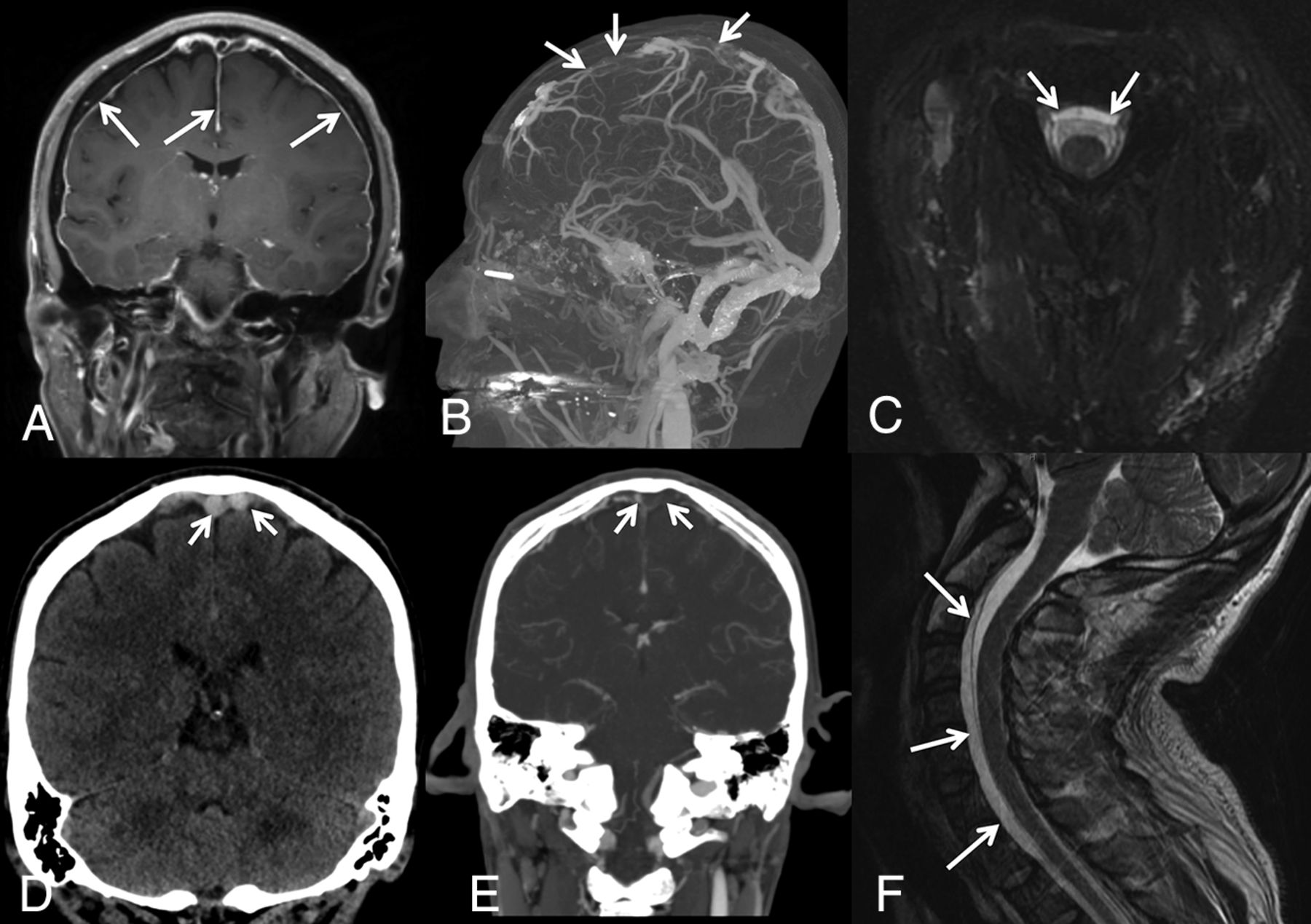
SIH presenting with venous sinus thrombosis. A 43-year-old man presented with sudden onset of headaches, dizziness, and nausea clinically suspected to represent migraines. A, Contrast-enhanced T1-weighted image demonstrates diffuse, smooth, nonspecific thin pachymeningeal enhancement (arrows). The patient presented to the emergency department 3 days later with a marked increase in his headache and sudden onset of right lower-extremity weakness. D, Unenhanced coronal head CT demonstrates high attenuation and expansile content within the superior sagittal sinus and adjacent cortical veins, compatible with acute dural venous sinus and cortical vein thrombosis (selected clot highlighted by arrows). Sagittal MIP (B) and coronal (E) images from a contrast-enhanced CT venogram confirm a long-segment acute, expansile thrombus as a filling defect within the superior sagittal sinus, as well as multiple bilateral left-greater-than-right superior cortical veins (arrows). While he was being treated for sinus thrombosis, he reported that his headaches were orthostatic in nature. His condition continued to deteriorate with progression of the thrombosis despite treatment and parenchymal hemorrhage. Axial (C) and sagittal (F) T2WI demonstrates a ventral extradural fluid collection. A hyperdynamic CT myelogram was performed, and a leak was found at a spiculated osteophyte at T1-2.

Frontotemporal Dementia.

Behavioral-variant frontotemporal dementia is characterized by disproportionate atrophy of the frontal and temporal lobes and classically has an early age of onset (younger than 60 years of age).73,74 Patients present with insidious personality changes, poor judgment, disinhibition, and apathy. Patients with SIH can also have behavioral changes, and there have been several reports of SIH presenting with frontotemporal dementia-like symptoms.75⇓⇓-78 The etiology of this relationship is not known but may be related to brain sag, obstruction of the venous outflow, and associated swelling of the diencephalon.78 In most cases, cognitive symptoms promptly improve almost immediately after treating the underlying SIH.75⇓⇓-78

Pituitary Tumor or Apoplexy.

As the volume of intracranial CSF declines in SIH, the compensatory enlargement of the cerebral venous system includes the hypophyseal veins. Thus, many patients with SIH have pituitary engorgement and upward bowing of the diaphragm sellae.79⇓-81 These findings may lead to misdiagnosis as a nonfunctioning pituitary adenoma (Fig 7).82,83 Furthermore, due to the uniquely dense hypophyseal portal system, venous congestion can predispose patients to apoplexy, a rare-but-emergent hemorrhage, or infarction of the pituitary gland. SIH-induced pituitary engorgement may also suppress dopamine release and thereby raise prolactin levels. Galactorrhea only occasionally results.

FIG 7.
  • Download figure
  • Open in new tab
  • Download powerpoint
FIG 7.

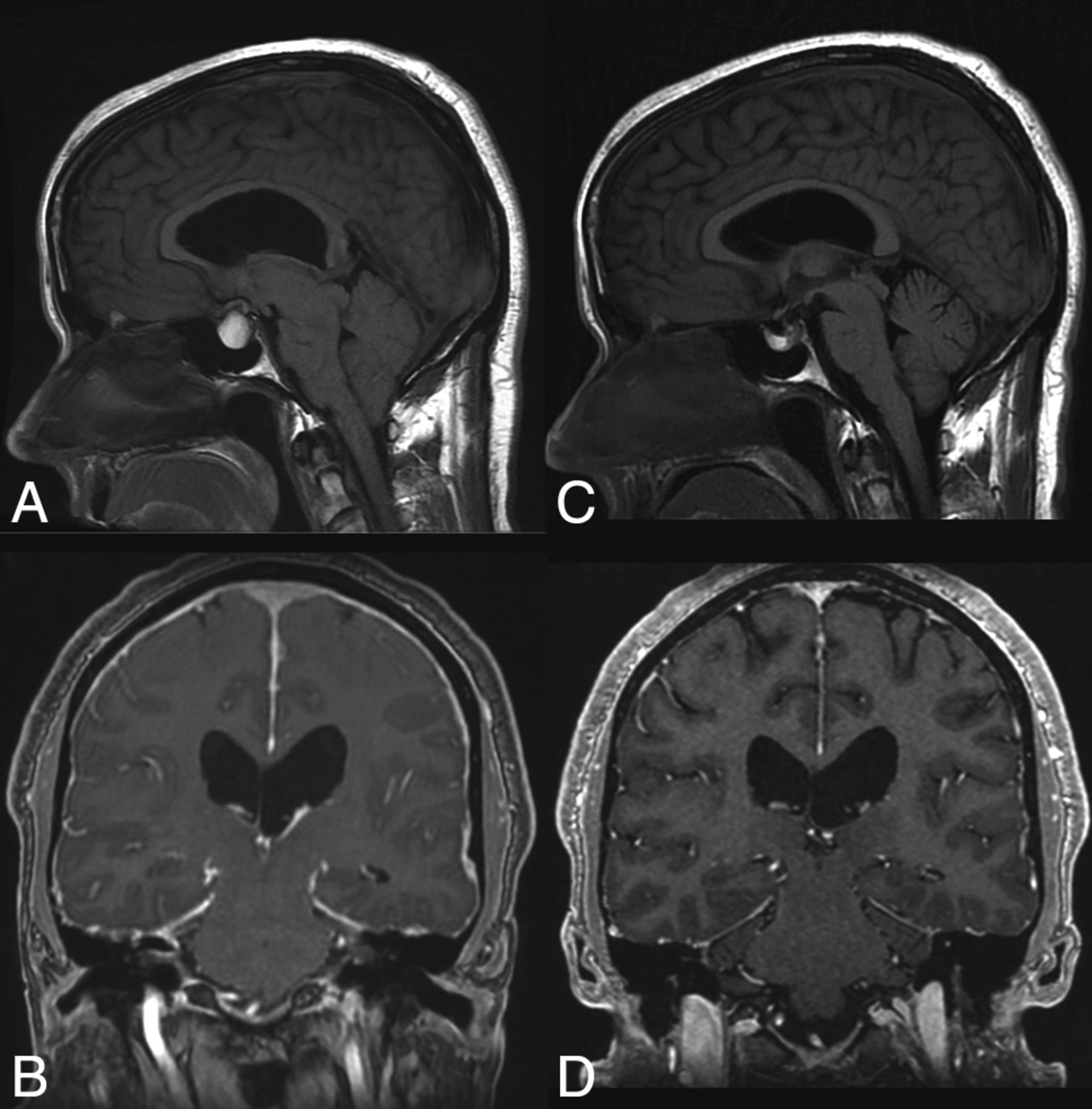
SIH presenting as pituitary apoplexy. Sagittal T1WI (A) and coronal postcontrast T1WI (B) images demonstrate a hyperintense lesion filling the sella and extending into the suprasellar cistern. The patient was diagnosed with pituitary apoplexy of a macroadenoma or a Rathke cleft cyst and a Chiari I malformation. However, the MR imaging actually demonstrates the expected intracranial stigmata of an SIH, including severe brain sag and dural enhancement. Despite extensive evaluation, the source of the leak was not found. The patient was treated with a nontargeted blood patch. His headaches and imaging findings (C and D) resolved.

Superficial Siderosis.

Superficial siderosis (SS) is a radiologic or pathologic diagnosis of hemosiderin deposition in the leptomeninges and subpial layer of the central nervous system. About half of the cases are attributed to idiopathic chronic subarachnoid hemorrhage, and the other half have a known cause such as trauma, tumor, or arteriovenous malformation.84 Classically, SS occurs infratentorially, and patients present with ataxia, pyramidal signs, or sensorineural hearing loss.13,84,85 Supratentorial bleeding can occur, and this is referred to as cortical superficial siderosis.86

There have been several reports of SIH presenting with or coinciding with the presence of SS (Fig 8).87⇓-89 One review of patients with SS noted a spinal extradural fluid collection in approximately 50% of patients.13 While SS is not reversible, repair of the inciting dural defect, often found via a dynamic CT myelogram, may prevent further bleeding and lead to stability or improvement of symptoms.90,91

FIG 8.
  • Download figure
  • Open in new tab
  • Download powerpoint
FIG 8.

SIH presenting as superficial siderosis. A 37-year-old man who presented with episodes of dizziness and loss of consciousness was found to have diffuse posterior fossa–predominant superficial siderosis on axial SWI (A and B). Subsequent imaging of the spine demonstrated a ventral extradural fluid collection (curved arrow, C; straight arrows, D). A subsequent hyperdynamic CT myelogram identified the source of the CSF leak, which was surgically repaired.

The etiologic mechanism that ties SS to SIH is uncertain. Some authors have suggested that the pathogenesis of SS is related to traction of the cerebellum and/or vermian veins on the skull base, leading to microhemorrhages and hemosiderin deposition.88 Others, noting the association between extradural spinal fluid collections and SS, have suggested that bleeding may occur at the site of the dural defect.92 The latter hypothesis has been supported by recent case reports of patients with hemorrhage within either the extradural fluid collection or thecal sac.93

CONCLUSIONS

Spontaneous intracranial hypotension is caused by various types of defects in the spinal dura and subsequent CSF extravasation. Patients classically present with orthostatic headache, low or normal CSF opening pressure on lumbar puncture, and brain sag with diffuse pachymeningeal enhancement on MR imaging. Many radiologic and clinical findings may mimic these classic findings, and conversely, secondary changes from SIH can give rise to symptoms that imitate other conditions. Because SIH is a curable condition, it is important for physicians to recognize its nonclassic presentations and be familiar with the differential diagnoses of its radiologic and clinical findings.

ACKNOWLEDGMENTS

The authors acknowledge the assistance of Sonia Watson, PhD, in editing the manuscript.

Footnotes

  • Disclosures: Jeremy K. Cutsforth-Gregory—UNRELATED: Royalties: Oxford University Press, Comments: Mayo Clinic Medical Neurosciences (textbook).

Indicates open access to non-subscribers at www.ajnr.org

References

  1. 1.↵
    1. Kranz PG,
    2. Gray L,
    3. Amrhein TJ
    . Spontaneous intracranial hypotension: 10 myths and misperceptions. Headache 2018;58:948–59 doi:10.1111/head.13328 pmid:29797515
    CrossRefPubMed
  2. 2.↵
    1. Schievink WI
    . Spontaneous spinal cerebrospinal fluid leaks and intracranial hypotension. JAMA 2006;295:2286–96 doi:10.1001/jama.295.19.2286 pmid:16705110
    CrossRefPubMed
  3. 3.↵
    1. Schievink WI,
    2. Meyer FB,
    3. Atkinson JL, et al
    . Spontaneous spinal cerebrospinal fluid leaks and intracranial hypotension. J Neurosurg 1996;84:598–60 doi:10.3171/jns.1996.84.4.0598 pmid:8613851
    CrossRefPubMed
  4. 4.↵
    1. Kranz PG,
    2. Amrhein TJ,
    3. Gray L
    . CSF venous fistulas in spontaneous intracranial hypotension: imaging characteristics on dynamic and CT myelography. AJR Am J Roentgenol 2017;209:1360–66 doi:10.2214/AJR.17.18351 pmid:29023155
    CrossRefPubMed
  5. 5.↵
    1. Schievink WI,
    2. Moser FG,
    3. Maya MM
    . CSF-venous fistula in spontaneous intracranial hypotension. Neurology 2014;83:472–73 doi:10.1212/WNL.0000000000000639 pmid:24951475
    CrossRefPubMed
  6. 6.↵
    1. Kranz PG,
    2. Tanpitukpongse TP,
    3. Choudhury KR, et al
    . Imaging signs in spontaneous intracranial hypotension: prevalence and relationship to CSF pressure. AJNR Am J Neuroradiol 2016;37:1374–78 doi:10.3174/ajnr.A4689 pmid:26869465
    Abstract/FREE Full Text
  7. 7.↵
    1. Sencakova D,
    2. Mokri B,
    3. McClelland RL
    . The efficacy of epidural blood patch in spontaneous CSF leaks. Neurology 2001;57:1921–23 doi:10.1212/wnl.57.10.1921 pmid:11723293
    Abstract/FREE Full Text
  8. 8.↵
    1. Schievink WI
    . A novel technique for treatment of intractable spontaneous intracranial hypotension: lumbar dural reduction surgery. Headache 2009;49:1047–51 doi:10.1111/j.1526-4610.2009.01450.x pmid:19473279
    CrossRefPubMed
  9. 9.↵
    1. Schievink WI
    . Misdiagnosis of spontaneous intracranial hypotension. Arch Neurol 2003;60:1713–18 doi:10.1001/archneur.60.12.1713 pmid:14676045
    CrossRefPubMed
  10. 10.↵
    1. Kranz PG,
    2. Amrhein TJ,
    3. Schievink WI, et al
    . The “hyperdense paraspinal vein” sign: a marker of CSF-venous fistula. AJNR Am J Neuroradiol 2016;37:1379–81 doi:10.3174/ajnr.A4682 pmid:26869470
    Abstract/FREE Full Text
  11. 11.↵
    1. Mokri B,
    2. Piepgras DG,
    3. Miller GM
    . Syndrome of orthostatic headaches and diffuse pachymeningeal gadolinium enhancement. Mayo Clin Proc 1997;72:400–13 doi:10.4065/72.5.400 pmid:9146681
    CrossRefPubMed
  12. 12.↵
    1. Dobrocky T,
    2. Grunder L,
    3. Breiding PS, et al
    . Assessing spinal cerebrospinal fluid leaks in spontaneous intracranial hypotension with a scoring system based on brain magnetic resonance imaging findings. JAMA Neurol 2019;76:580–87 doi:10.1001/jamaneurol.2018.4921 pmid:30776059
    CrossRefPubMed
  13. 13.↵
    1. Kumar N,
    2. Cohen-Gadol AA,
    3. Wright RA, et al
    . Superficial siderosis. Neurology 2006;66:1144–52 doi:10.1212/01.wnl.0000208510.76323.5b pmid:16636229
    Abstract/FREE Full Text
  14. 14.↵
    1. Spelle L,
    2. Boulin A,
    3. Pierot L, et al
    . Spontaneous intracranial hypotension: MRI and radionuclide cisternography findings. J Neurol Neurosurg Psychiatry 1997;62:291–92 doi:10.1136/jnnp.62.3.291 pmid:9069493
    FREE Full Text
  15. 15.↵
    1. Sehgal AK,
    2. Sethi RS,
    3. Raghavan S, et al
    . Radionuclide cisternography: a prudent investigation in diagnosing spontaneous intracranial hypotension. Indian J Nucl Med 2013;28:42–44 doi:10.4103/0972-3919.116815 pmid:24019678
    CrossRefPubMed
  16. 16.↵
    1. Yoo HM,
    2. Kim SJ,
    3. Choi CG, et al
    . Detection of CSF leak in spinal CSF leak syndrome using MR myelography: correlation with radioisotope cisternography. AJNR Am J Neuroradiol 2008;29:649–54 doi:10.3174/ajnr.A0920 pmid:18202233
    Abstract/FREE Full Text
  17. 17.↵
    1. Watanabe A,
    2. Horikoshi T,
    3. Uchida M, et al
    . Diagnostic value of spinal MR imaging in spontaneous intracranial hypotension syndrome. AJNR Am J Neuroradiol 2009;30:147–51 doi:10.3174/ajnr.A1277 pmid:18768717
    Abstract/FREE Full Text
  18. 18.↵
    1. Kranz PG,
    2. Stinnett SS,
    3. Huang KT, et al
    . Spinal meningeal diverticula in spontaneous intracranial hypotension: analysis of prevalence and myelographic appearance. AJNR Am J Neuroradiol 2013;34:1284–89 doi:10.3174/ajnr.A3359 pmid:23221945
    Abstract/FREE Full Text
  19. 19.↵
    1. Meadows J,
    2. Kraut M,
    3. Guarnieri M, et al
    . Asymptomatic Chiari type I malformations identified on magnetic resonance imaging. J Neurosurg 2000;92:920–26 doi:10.3171/jns.2000.92.6.0920 pmid:10839250
    CrossRefPubMed
  20. 20.↵
    1. Morris Z,
    2. Whiteley WN,
    3. Longstreth WT, Jr., et al
    . Incidental findings on brain magnetic resonance imaging: systematic review and meta-analysis. BMJ 2009;339:b3016–16 doi:10.1136/bmj.b3016 pmid:19687093
    Abstract/FREE Full Text
  21. 21.↵
    1. Langridge B,
    2. Phillips E,
    3. Choi D
    . Chiari malformation type 1: a systematic review of natural history and conservative management. World Neurosurg 2017;104:213–19 doi:10.1016/j.wneu.2017.04.082 pmid:28435116
    CrossRefPubMed
  22. 22.↵
    1. Atkinson JL,
    2. Weinshenker BG,
    3. Miller GM, et al
    . Acquired Chiari I malformation secondary to spontaneous spinal cerebrospinal fluid leakage and chronic intracranial hypotension syndrome in seven cases. J Neurosurg 1998;88:237–42 doi:10.3171/jns.1998.88.2.0237 pmid:9452230
    CrossRefPubMed
  23. 23.↵
    1. Holbrook J,
    2. Saindane AM
    . Imaging of intracranial pressure disorders. Neurosurgery 2017;80:341–54 doi:10.1227/NEU.0000000000001362 pmid:27471977
    CrossRefPubMed
  24. 24.↵
    1. Sainani NI,
    2. Lawande MA,
    3. Pungavkar SA, et al
    . Spontaneous intracranial hypotension: a study of six cases with MR findings and literature review. Australas Radiol 2006;50:419–23 doi:10.1111/j.1440-1673.2006.01615.x pmid:16981936
    CrossRefPubMed
  25. 25.↵
    1. Wald JT,
    2. Diehn FE
    . Spontaneous intracranial hypotension. Applied Radiol 2018;47:18–22
  26. 26.↵
    1. Smith RM,
    2. Garza I,
    3. Robertson CE
    . Chronic CSF leak causing syringomyelia and pseudo-Arnold-Chiari malformation. Neurology 2015;85:1994 doi:10.1212/WNL.0000000000002178 pmid:26628487
    Abstract/FREE Full Text
  27. 27.↵
    1. Luxon LM,
    2. Harrison MJ
    . Chronic subdural haematoma. Q J Med 1979;48:43–53 pmid:482590
    PubMed
  28. 28.↵
    1. Schievink WI,
    2. Maya MM,
    3. Pikul BK, et al
    . Spontaneous spinal cerebrospinal fluid leaks as the cause of subdural hematomas in elderly patients on anticoagulation: report of 3 cases. J Neurosurg 2010;112:295–99 doi:10.3171/2008.10.JNS08428 pmid:19199465
    CrossRefPubMed
  29. 29.↵
    1. Huang YH,
    2. Yang KY,
    3. Lee TC, et al
    . Bilateral chronic subdural hematoma: what is the clinical significance? Int J Surg 2013;11:544–48 doi:10.1016/j.ijsu.2013.05.007 pmid:23707986
    CrossRefPubMed
  30. 30.↵
    1. Schievink WI,
    2. Maya MM,
    3. Moser FG, et al
    . Spectrum of subdural fluid collections in spontaneous intracranial hypotension. J Neurosurg 2005;103:608–13 doi:10.3171/jns.2005.103.4.0608 pmid:16266041
    CrossRefPubMed
  31. 31.↵
    1. de Noronha RJ,
    2. Sharrack B,
    3. Hadjivassiliou M, et al
    . Subdural haematoma: a potentially serious consequence of spontaneous intracranial hypotension. J Neurol Neurosurg Psychiatry 2003;74:752–55 doi:10.1136/jnnp.74.6.752 pmid:12754345
    Abstract/FREE Full Text
  32. 32.↵
    1. Chen YC,
    2. Wang YF,
    3. Li JY, et al
    . Treatment and prognosis of subdural hematoma in patients with spontaneous intracranial hypotension. Cephalalgia 2016;36:225–31 doi:10.1177/0333102415585095 pmid:25944817
    CrossRefPubMed
  33. 33.↵
    1. Rettenmaier LA,
    2. Park BJ,
    3. Holland MT, et al
    . Value of targeted epidural blood patch and management of subdural hematoma in spontaneous intracranial hypotension: case report and review of the literature. World Neurosurg 2017;97:27–38 doi:10.1016/j.wneu.2016.09.076 pmid:27693247
    CrossRefPubMed
  34. 34.↵
    1. Stone JH,
    2. Zen Y,
    3. Deshpande V
    . IgG4-related disease. N Engl J Med 2012;366:539–51 doi:10.1056/NEJMra1104650 pmid:22316447
    CrossRefPubMed
  35. 35.↵
    1. Lu LX,
    2. Della-Torre E,
    3. Stone JH, et al
    . IgG4-related hypertrophic pachymeningitis: clinical features, diagnostic criteria, and treatment. JAMA Neurol 2014;71:785–93 doi:10.1001/jamaneurol.2014.243 pmid:24733677
    CrossRefPubMed
  36. 36.↵
    1. Smith JK,
    2. Matheus MG,
    3. Castillo M
    . Imaging manifestations of neurosarcoidosis. AJR Am J Roentgenol 2004;182:289–95 doi:10.2214/ajr.182.2.1820289 pmid:14736648
    CrossRefPubMed
  37. 37.↵
    1. Pickuth D,
    2. Spielmann RP,
    3. Heywang-Köbrunner SH
    . Role of radiology in the diagnosis of neurosarcoidosis. Eur Radiol 2000;10:941–44 doi:10.1007/s003300051042 pmid:10879708
    CrossRefPubMed
  38. 38.↵
    1. Zajicek JP,
    2. Scolding NJ,
    3. Foster O, et al
    . Central nervous system sarcoidosis: diagnosis and management. QJM 1999;92:103–17 doi:10.1093/qjmed/92.2.103 pmid:10209662
    CrossRefPubMed
  39. 39.↵
    1. Otsuka M,
    2. Fujiwara T,
    3. Kuwata Y, et al
    . A case of rheumatoid pachymeningitis [in Japanese]. Rinsho Shinkeigaku 1997;37:834–40 pmid:9431000
    PubMed
  40. 40.↵
    1. Micheli F,
    2. Scorticati MC,
    3. Pikielny R, et al
    . Pachymeningeal thickening in rheumatoid arthritis. Eur Neurol 1993;33:397–98 doi:10.1159/000116980 pmid:8243519
    CrossRefPubMed
  41. 41.↵
    1. Marano E,
    2. D'Armiento FP,
    3. Scarano V, et al
    . Focal hypertrophic cranial pachymeningitis associated with temporal arteritis: a new case report. J Neurol 2003;250:98–100 doi:10.1007/s00415-003-0914-6 pmid:12528001
    CrossRefPubMed
  42. 42.↵
    1. Wen JH,
    2. Wang C,
    3. Jin YY, et al
    . Radiological and clinical findings of isolated meningeal Rosai-Dorfman disease of the central nervous system. Medicine (Baltimore) 2019;98:e15365 doi:10.1097/MD.0000000000015365 pmid:31083164
    CrossRefPubMed
  43. 43.↵
    1. Z'Graggen WJ,
    2. Sturzenegger M,
    3. Mariani L, et al
    . Isolated Rosai-Dorfman disease of intracranial meninges. Pathol Res Pract 2006;202:165–70 doi:10.1016/j.prp.2005.11.004 pmid:16377100
    CrossRefPubMed
  44. 44.↵
    1. Kumar P,
    2. Singh A,
    3. Gamanagatti S, et al
    . Imaging findings in Erdheim-Chester disease: what every radiologist needs to know. Pol J Radiol 2018;83:E54–62 doi:10.5114/pjr.2018.73290 pmid:30038679
    CrossRefPubMed
  45. 45.↵
    1. Parks NE,
    2. Goyal G,
    3. Go RS, et al
    . Neuroradiologic manifestations of Erdheim-Chester disease. Neurol Clin Pract 2018;8:15–20 doi:10.1212/CPJ.0000000000000422 pmid:29517068
    Abstract/FREE Full Text
  46. 46.↵
    1. Chin JH
    . Tuberculous meningitis: diagnostic and therapeutic challenges. Neurol Clin Pract 2014;4:199–205 doi:10.1212/CPJ.0000000000000023 pmid:25110618
    Abstract/FREE Full Text
  47. 47.↵
    1. Thurtell MJ,
    2. Keed AB,
    3. Yan M, et al
    . Tuberculous cranial pachymeningitis. Neurology 2007;68:298–300 doi:10.1212/01.wnl.0000252367.99393.34 pmid:17242338
    Abstract/FREE Full Text
  48. 48.↵
    1. Fonseka CL,
    2. Kanakkahewa TE,
    3. Singhapura S, et al
    . Tuberculous pachymeningitis presenting as a diffused dural thickening in a patient with chronic headache and recurrent neurological abnormalities for more than a decade: a case report and a review of the literature. Case Rep Infect Dis 2018;2018:3012034 doi:10.1155/2018/3012034 pmid:30364129
    CrossRefPubMed
  49. 49.↵
    1. Goyal M,
    2. Sharma A,
    3. Mishra NK, et al
    . Imaging appearance of pachymeningeal tuberculosis. AJR Am J Roentgenol 1997;169:1421–24 doi:10.2214/ajr.169.5.9353472 pmid:9353472
    CrossRefPubMed
  50. 50.↵
    1. Blazekovic A,
    2. Ozretic D,
    3. Habek M, et al
    . Neurosyphilis: the shape of a rising threat. Int J Infect Dis 2018;76:1–3 doi:10.1016/j.ijid.2018.07.022 pmid:30081207
    CrossRefPubMed
  51. 51.↵
    1. Rosa M Jr.,
    2. de Almeida Cacador T,
    3. Biasutti C, et al
    . Teaching NeuroImages: skull and dural lesions in neurosyphilis. Neurology 2016;87:e129–30 doi:10.1212/WNL.0000000000003133 pmid:27647589
    Abstract/FREE Full Text
  52. 52.↵
    1. Mekinian A,
    2. Maisonobe L,
    3. Boukari L, et al
    . Characteristics, outcome and treatments with cranial pachymeningitis: a multicenter French retrospective study of 60 patients. Medicine (Baltimore) 2018;97:e11413 doi:10.1097/MD.0000000000011413 pmid:30045263
    CrossRefPubMed
  53. 53.↵
    1. Cutsforth-Gregory JK,
    2. Sandroni P
    . Clinical neurophysiology of postural tachycardia syndrome. Handb Clin Neurol 2019;161:429–45 doi:10.1016/B978-0-444-64142-7.00066-7 pmid:31307619
    CrossRefPubMed
  54. 54.↵
    1. Graf N,
    2. Fernandes Santos AM,
    3. Ulrich CT, et al
    . Clinical symptoms and results of autonomic function testing overlap in spontaneous intracranial hypotension and postural tachycardia syndrome. Cephalalgia Reports 2018;1:251581631877377 doi:10.1177/2515816318773774
    CrossRef
  55. 55.↵
    1. Mokri B,
    2. Low PA
    . Orthostatic headaches without CSF leak in postural tachycardia syndrome. Neurology 2003;61:980–82 doi:10.1212/01.wnl.0000085868.37963.7d pmid:14557573
    Abstract/FREE Full Text
  56. 56.↵
    1. Raj SR
    . The postural tachycardia syndrome (POTS): pathophysiology, diagnosis and management. Indian Pacing Electrophysiol J 2006;6:84–99 pmid:16943900
    PubMed
  57. 57.↵
    1. Bleasdale-Barr KM,
    2. Mathias CJ
    . Neck and other muscle pains in autonomic failure: their association with orthostatic hypotension. J R Soc Med 1998;91:355–59 doi:10.1177/014107689809100704 pmid:9771493
    CrossRefPubMed
  58. 58.↵
    1. Naschitz JE,
    2. Rosner I
    . Orthostatic hypotension: framework of the syndrome. Postgrad Med J 2007;83:568–74 doi:10.1136/pgmj.2007.058198 pmid:17823222
    Abstract/FREE Full Text
  59. 59.↵
    Headache Classification Committee of the International Headache Society (IHS) The International Classification of Headache Disorders, 3rd edition. Cephalalgia 2018; 38:1–211 doi:10.1177/0333102417738202 pmid:29368949
    CrossRefPubMed
  60. 60.↵
    1. Furman JM,
    2. Marcus DA,
    3. Balaban CD
    . Vestibular migraine: clinical aspects and pathophysiology. Lancet Neurol 2013;12:706–15 doi:10.1016/S1474-4422(13)70107-8 pmid:23769597
    CrossRefPubMed
  61. 61.↵
    1. Montenegro MM,
    2. Cutsforth-Gregory JK
    . Orthostatic headache after suboccipital craniectomy without CSF leak: two case reports. Headache 2018;58:1238–43 doi:10.1111/head.13346 pmid:29920678
    CrossRefPubMed
  62. 62.↵
    1. Schievink WI,
    2. Maya MM
    . Cerebral venous thrombosis in spontaneous intracranial hypotension. Headache 2008;48:1511–19 doi:10.1111/j.1526-4610.2008.01251.x pmid:19076649
    CrossRefPubMed
  63. 63.↵
    1. Stam J
    . Thrombosis of the cerebral veins and sinuses. N Engl J Med 2005;352:1791–98 doi:10.1056/NEJMra042354 pmid:15858188
    CrossRefPubMed
  64. 64.↵
    1. Zhang D,
    2. Wang J,
    3. Zhang Q, et al
    . Cerebral venous thrombosis in spontaneous intracranial hypotension: a report on 4 cases and a review of the literature. Headache 2018;58:1244–55 doi:10.1111/head.13413 pmid:30238694
    CrossRefPubMed
  65. 65.↵
    1. Kataoka H,
    2. Tanizawa E,
    3. Ueno S
    . Spontaneous intracranial hypotension is associated with a risk of venous sinus thrombosis and subdural hematoma. Cerebrovasc Dis 2007;23:315–17 doi:10.1159/000098446 pmid:17213717
    CrossRefPubMed
  66. 66.↵
    1. Mokri B
    . The Monro-Kellie hypothesis: applications in CSF volume depletion. Neurology 2001;56:1746–48 doi:10.1212/wnl.56.12.1746 pmid:11425944
    Abstract/FREE Full Text
  67. 67.↵
    1. Rice CM,
    2. Renowden SA,
    3. Sandeman DR, et al
    . Spontaneous intracranial hypotension and venous sinus thrombosis. Pract Neurol 2013;13:120–24 doi:10.1136/practneurol-2012-000257 pmid:23468563
    FREE Full Text
  68. 68.↵
    1. Yoon KW,
    2. Cho MK,
    3. Kim YJ, et al
    . Sinus thrombosis in a patient with intracranial hypotension: a suggested hypothesis of venous stasis. A case report. Interv Neuroradiol 2011;17:248–51 doi:10.1177/159101991101700218 pmid:21696667
    CrossRefPubMed
  69. 69.↵
    1. Costa P,
    2. Del Zotto E,
    3. Giossi A, et al
    . Headache due to spontaneous intracranial hypotension and subsequent cerebral vein thrombosis. Headache 2012;52:1592–96 doi:10.1111/j.1526-4610.2012.02261.x pmid:23046074
    CrossRefPubMed
  70. 70.↵
    1. Savoiardo M,
    2. Armenise S,
    3. Spagnolo P, et al
    . Dural sinus thrombosis in spontaneous intracranial hypotension: hypotheses on possible mechanisms. J Neurol 2006;253:1197–1202 doi:10.1007/s00415-006-0194-z pmid:16680559
    CrossRefPubMed
  71. 71.↵
    1. Richard S,
    2. Kremer S,
    3. Lacour JC, et al
    . Cerebral venous thrombosis caused by spontaneous intracranial hypotension: two cases. Eur J Neurol 2007;14:1296–98 doi:10.1111/j.1468-1331.2007.01947.x pmid:17868278
    CrossRefPubMed
  72. 72.↵
    1. Berroir S,
    2. Grabli D,
    3. Heran F, et al
    . Cerebral sinus venous thrombosis in two patients with spontaneous intracranial hypotension. Cerebrovasc Dis 2004;17:9–12 doi:10.1159/000073892 pmid:14530632
    CrossRefPubMed
  73. 73.↵
    1. Neary D,
    2. Snowden JS,
    3. Gustafson L, et al
    . Frontotemporal lobar degeneration: a consensus on clinical diagnostic criteria. Neurology 1998;51:1546–54 doi:10.1212/wnl.51.6.1546 pmid:9855500
    Abstract/FREE Full Text
  74. 74.↵
    1. Neary D,
    2. Snowden JS,
    3. Northen B, et al
    . Dementia of frontal lobe type. J Neurol Neurosurg Psychiatry 1988;51:353–61 doi:10.1136/jnnp.51.3.353 pmid:3258902
    Abstract/FREE Full Text
  75. 75.↵
    1. Hong M,
    2. Shah GV,
    3. Adams KM, et al
    . Spontaneous intracranial hypotension causing reversible frontotemporal dementia. Neurology 2002;58:1285–87 doi:10.1212/wnl.58.8.1285 pmid:11971102
    Abstract/FREE Full Text
  76. 76.↵
    1. Mostofi E,
    2. Schievink WI,
    3. Sim VL
    . Dural reduction surgery: a treatment option for frontotemporal brain sagging syndrome. Can J Neurol Sci 2016;43:593–95 doi:10.1017/cjn.2016.3 pmid:26972054
    CrossRefPubMed
  77. 77.↵
    1. Wicklund MR,
    2. Mokri B,
    3. Drubach DA, et al
    . Frontotemporal brain sagging syndrome: an SIH-like presentation mimicking FTD. Neurology 2011;76:1377–82 doi:10.1212/WNL.0b013e3182166e42 pmid:21502595
    Abstract/FREE Full Text
  78. 78.↵
    1. Schievink WI,
    2. Maya MM,
    3. Barnard ZR, et al
    . Behavioral variant frontotemporal dementia as a serious complication of spontaneous intracranial hypotension. Oper Neurosurg (Hagerstown) 2018;15:505–15 doi:10.1093/ons/opy029 pmid:29534203
    CrossRefPubMed
  79. 79.↵
    1. Firat AK,
    2. Karakas HM,
    3. Firat Y, et al
    . Spontaneous intracranial hypotension with pituitary adenoma. J Headache Pain 2006;7:47–50 doi:10.1007/s10194-006-0269-3 pmid:16485075
    CrossRefPubMed
  80. 80.↵
    1. Tapia DQ,
    2. Pardo J,
    3. Serrano JM, et al
    . Spontaneous intracranial hypotension: use of unenhanced MRI. Neuroradiology 2000;42:529–31 doi:10.1007/s002340000327 pmid:10952188
    CrossRefPubMed
  81. 81.↵
    1. Tsui EY,
    2. Ng SH,
    3. Cheung YK, et al
    . Spontaneous intracranial hypotension with diffuse dural enhancement of the spinal canal and transient enlargement of the pituitary gland. Eur J Radiol 2001;38:59–63 doi:10.1016/s0720-048x(01)00288-1 pmid:11287167
    CrossRefPubMed
  82. 82.↵
    1. Leung GK,
    2. Ho J,
    3. Pu JK
    . Pituitary enlargement in spontaneous intracranial hypotension: a diagnostic pitfall. Acta Neurochir (Wien) 2011;153:2445–46 doi:10.1007/s00701-011-1099-x pmid:21800104
    CrossRefPubMed
  83. 83.↵
    1. Luna J,
    2. Khanna I,
    3. Cook FJ, et al
    . Sagging brain masquerading as a pituitary adenoma. J Clin Endocrinol Metab 2014;99:3043–43 doi:10.1210/jc.2014-1397 pmid:24947035
    CrossRefPubMed
  84. 84.↵
    1. Fearnley JM,
    2. Stevens JM,
    3. Rudge P
    . Superficial siderosis of the central nervous system. Brain 1995;118(Pt 4):1051–66 doi:10.1093/brain/118.4.1051 pmid:7655881
    CrossRefPubMed
  85. 85.↵
    1. Levy M,
    2. Turtzo C,
    3. Llinas RH
    . Superficial siderosis: a case report and review of the literature. Nat Clin Pract Neurol 2007;3:54–58 doi:10.1038/ncpneuro0356 pmid:17205075
    CrossRefPubMed
  86. 86.↵
    1. Linn J,
    2. Herms J,
    3. Dichgans M, et al
    . Subarachnoid hemosiderosis and superficial cortical hemosiderosis in cerebral amyloid angiopathy. AJNR Am J Neuroradiol 2008;29:184–86 doi:10.3174/ajnr.A0783 pmid:17947366
    Abstract/FREE Full Text
  87. 87.↵
    1. Beck J,
    2. Häni L,
    3. Ulrich CT, et al
    . Diagnostic challenges and therapeutic possibilities in spontaneous intracranial hypotension. Clinical and Translational Neuroscience 2018;2:2514183X1878737 doi:10.1177/2514183X18787371
    CrossRef
  88. 88.↵
    1. Webb AJ,
    2. Flossmann E,
    3. Armstrong RJ
    . Superficial siderosis following spontaneous intracranial hypotension. Pract Neurol 2015;15:382–84 doi:10.1136/practneurol-2015-001169 pmid:26139598
    FREE Full Text
  89. 89.↵
    1. Schievink WI,
    2. Maya MM
    . Spinal meningeal diverticula, spontaneous intracranial hypotension, and superficial siderosis. Neurology 2017;88:916–17 doi:10.1212/WNL.0000000000003665 pmid:28130464
    Abstract/FREE Full Text
  90. 90.↵
    1. Kumar N
    . Neuroimaging in superficial siderosis: an in-depth look. AJNR Am J Neuroradiol 2010;31:5–14 doi:10.3174/ajnr.A1628 pmid:19729538
    Abstract/FREE Full Text
  91. 91.↵
    1. Kumar N,
    2. Lane JI,
    3. Piepgras DG
    . Superficial siderosis: sealing the defect. Neurology 2009;72:671–73 doi:10.1212/01.wnl.0000342457.22536.af pmid:19221303
    FREE Full Text
  92. 92.↵
    1. Kumar N,
    2. McKeon A,
    3. Rabinstein AA, et al
    . Superficial siderosis and CSF hypovolemia: the defect (dural) in the link. Neurology 2007;69:925–26 doi:10.1212/01.wnl.0000267847.69896.be pmid:17724297
    FREE Full Text
  93. 93.↵
    1. Schievink WI,
    2. Wasserstein P,
    3. Maya MM
    . Intraspinal hemorrhage in spontaneous intracranial hypotension: link to superficial siderosis? Report of 2 cases. J Neurosurg Spine 2016;24:454–56 doi:10.3171/2015.6.SPINE15428 pmid:26588500
    CrossRefPubMed
  • Received March 2, 2020.
  • Accepted after revision April 30, 2020.
  • © 2020 by American Journal of Neuroradiology
View Abstract
PreviousNext
Back to top
Advertisement
Print
Download PDF
Email Article

Thank you for your interest in spreading the word on American Journal of Neuroradiology.

NOTE: We only request your email address so that the person you are recommending the page to knows that you wanted them to see it, and that it is not junk mail. We do not capture any email address.

Enter multiple addresses on separate lines or separate them with commas.
Spontaneous Intracranial Hypotension: Atypical Radiologic Appearances, Imaging Mimickers, and Clinical Look-Alikes
(Your Name) has sent you a message from American Journal of Neuroradiology
(Your Name) thought you would like to see the American Journal of Neuroradiology web site.
CAPTCHA
This question is for testing whether or not you are a human visitor and to prevent automated spam submissions.
Cite this article
K.M. Bond, J.C. Benson, J.K. Cutsforth-Gregory, D.K. Kim, F.E. Diehn, C.M. Carr
Spontaneous Intracranial Hypotension: Atypical Radiologic Appearances, Imaging Mimickers, and Clinical Look-Alikes
American Journal of Neuroradiology Jul 2020, DOI: 10.3174/ajnr.A6637

Citation Manager Formats

  • BibTeX
  • Bookends
  • EasyBib
  • EndNote (tagged)
  • EndNote 8 (xml)
  • Medlars
  • Mendeley
  • Papers
  • RefWorks Tagged
  • Ref Manager
  • RIS
  • Zotero
0 Responses
Respond to this article
Share
Bookmark this article
Spontaneous Intracranial Hypotension: Atypical Radiologic Appearances, Imaging Mimickers, and Clinical Look-Alikes
K.M. Bond, J.C. Benson, J.K. Cutsforth-Gregory, D.K. Kim, F.E. Diehn, C.M. Carr
American Journal of Neuroradiology Jul 2020, DOI: 10.3174/ajnr.A6637
del.icio.us logo Twitter logo Facebook logo Mendeley logo
  • Tweet Widget
  • Facebook Like
  • Google Plus One
Purchase

Jump to section

  • Article
    • Abstract
    • ABBREVIATIONS:
    • CONCLUSIONS
    • ACKNOWLEDGMENTS
    • Footnotes
    • References
  • Figures & Data
  • Info & Metrics
  • Responses
  • References
  • PDF

Related Articles

  • No related articles found.
  • PubMed
  • Google Scholar

Cited By...

  • Spinal CSF Leaks: The Neuroradiologist Transforming Care
  • Skull Base CSF Leaks: Potential Underlying Pathophysiology and Evaluation of Brain MR Imaging Findings Associated with Spontaneous Intracranial Hypotension
  • Natural history of spontaneous intracranial hypotension: a clinical and imaging study
  • Likelihood of Discovering a CSF Leak Based on Intracranial MRI Findings in Patients without a Spinal Longitudinal Extradural Collection: A New Probabilistic Scoring System
  • The Monro-Kellie Doctrine: A Review and Call for Revision
  • Case of spontaneous intracranial hypotension: clinical, neuroimaging and treatment approach
  • Crossref (29)
  • Google Scholar

This article has been cited by the following articles in journals that are participating in Crossref Cited-by Linking.

  • Spontaneous intracranial hypotension: searching for the CSF leak
    Tomas Dobrocky, Patrick Nicholson, Levin Häni, Pasquale Mordasini, Timo Krings, Waleed Brinjikji, Jeremy K Cutsforth-Gregory, Ralph Schär, Christoph Schankin, Jan Gralla, Vitor M Pereira, Andreas Raabe, Richard Farb, Jürgen Beck, Eike I Piechowiak
    The Lancet Neurology 2022 21 4
  • International Consensus Statement: Spontaneous Cerebrospinal Fluid Rhinorrhea
    Christos Georgalas, Amanda Oostra, Shahzada Ahmed, Paolo Castelnuovo, Iacopo Dallan, Wouter van Furth, Richard J. Harvey, Philippe Herman, Dimitrios Kombogiorgas, Davide Locatelli, Cem Meco, Jim N. Palmer, Otavio Piltcher, Anshul M. Sama, Hesham Saleh, Raj Sindwani, Thibaut Van Zele, Bradford A. Woodworth
    International Forum of Allergy & Rhinology 2021 11 4
  • The Monro-Kellie Doctrine: A Review and Call for Revision
    J.C. Benson, A.A. Madhavan, J.K. Cutsforth-Gregory, D.R. Johnson, C.M. Carr
    American Journal of Neuroradiology 2023 44 1
  • Skull Base CSF Leaks: Potential Underlying Pathophysiology and Evaluation of Brain MR Imaging Findings Associated with Spontaneous Intracranial Hypotension
    Ian T. Mark, Jeremy Cutsforth-Gregory, Patrick Luetmer, Ajay A. Madhavan, Michael Oien, Paul Farnsworth, Girish Bathla, Steve Messina, Michael Link, Jamie Van Gompel
    American Journal of Neuroradiology 2024 45 10
  • ACR Appropriateness Criteria® Headache: 2022 Update
    Pallavi S. Utukuri, Robert Y. Shih, Amna A. Ajam, Kathryn E. Callahan, Doris Chen, Jeffrey W. Dunkle, Christopher H. Hunt, Jana Ivanidze, Luke N. Ledbetter, Ryan K. Lee, Omojo Malu, Jeffrey S. Pannell, Jeffrey M. Pollock, Sonja X. Potrebic, Michael Setzen, Richard D. Shih, Bruno P. Soares, Michael D. Staudt, Lily L. Wang, Judah Burns
    Journal of the American College of Radiology 2023 20 5
  • Cerebrospinal Fluid Leaks, Spontaneous Intracranial Hypotension, and Chiari I Malformation
    Rahul Kumar, Jeremy K. Cutsforth-Gregory, Waleed Brinjikji
    Neurosurgery Clinics of North America 2023 34 1
  • Likelihood of Discovering a CSF Leak Based on Intracranial MRI Findings in Patients without a Spinal Longitudinal Extradural Collection: A New Probabilistic Scoring System
    John C. Benson, Ajay A. Madhavan, Ian T. Mark, Jeremy K. Cutsforth-Gregory, Waleed Brinjikji, Jared T. Verdoorn
    American Journal of Neuroradiology 2023 44 11
  • Natural history of spontaneous intracranial hypotension: a clinical and imaging study
    James Williams, Waleed Brinjikji, Jeremy K Cutsforth-Gregory
    Journal of NeuroInterventional Surgery 2023 15 11
  • Outcome of epidural blood patch for imaging-negative spontaneous intracranial hypotension
    So Youn Choi, Minjung Seong, Eung Yeop Kim, Michelle Sojung Youn, Soohyun Cho, Hyemin Jang, Mi Ji Lee
    Cephalalgia 2023 43 2
  • Spontaneous Intracranial Hypotension - A Dilemma
    Dhaval Shukla, Nishanth Sadashiva, Jitender Saini, Sriganesh Kamath
    Neurology India 2021 69 Suppl 2

More in this TOC Section

REVIEW ARTICLE

  • An Atlas of Neonatal Neurovascular Imaging Anatomy as Depicted with Microvascular Imaging: The Intracranial Arteries
  • An Atlas of Neonatal Neurovascular Imaging Anatomy as Depicted with Microvascular Imaging: The Intracranial Veins
  • Clinical Translation of Hyperpolarized 13C Metabolic Probes for Glioma Imaging
Show more REVIEW ARTICLE

ADULT BRAIN

  • Diagnostic Neuroradiology of Monoclonal Antibodies
  • Clinical Outcomes After Chiari I Decompression
  • Segmentation of Brain Metastases with BLAST
Show more ADULT BRAIN

Similar Articles

Advertisement

Indexed Content

  • Current Issue
  • Accepted Manuscripts
  • Article Preview
  • Past Issues
  • Editorials
  • Editor's Choice
  • Fellows' Journal Club
  • Letters to the Editor
  • Video Articles

Cases

  • Case Collection
  • Archive - Case of the Week
  • Archive - Case of the Month
  • Archive - Classic Case

Special Collections

  • AJNR Awards
  • ASNR Foundation Special Collection
  • Most Impactful AJNR Articles
  • Photon-Counting CT
  • Spinal CSF Leak Articles (Jan 2020-June 2024)

More from AJNR

  • Trainee Corner
  • Imaging Protocols
  • MRI Safety Corner

Multimedia

  • AJNR Podcasts
  • AJNR Scantastics

Resources

  • Turnaround Time
  • Submit a Manuscript
  • Submit a Video Article
  • Submit an eLetter to the Editor/Response
  • Manuscript Submission Guidelines
  • Statistical Tips
  • Fast Publishing of Accepted Manuscripts
  • Graphical Abstract Preparation
  • Imaging Protocol Submission
  • Evidence-Based Medicine Level Guide
  • Publishing Checklists
  • Author Policies
  • Become a Reviewer/Academy of Reviewers
  • News and Updates

About Us

  • About AJNR
  • Editorial Board
  • Editorial Board Alumni
  • Alerts
  • Permissions
  • Not an AJNR Subscriber? Join Now
  • Advertise with Us
  • Librarian Resources
  • Feedback
  • Terms and Conditions
  • AJNR Editorial Board Alumni

American Society of Neuroradiology

  • Not an ASNR Member? Join Now

© 2025 by the American Society of Neuroradiology All rights, including for text and data mining, AI training, and similar technologies, are reserved.
Print ISSN: 0195-6108 Online ISSN: 1936-959X

Powered by HighWire