
Article Figures & Data
Figures
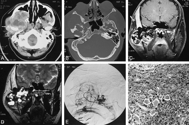
- fig 1.
37-year-old man with rapidly growing right-sided periauricular mass.
A, Axial CT scan of the skull base shows a large heterogeneous mass in the region of the right TMJ. The tumor has extended into the right infratemporal fossa and bowed the posterior wall of the maxillary sinus anteriorly. Multiple areas of low attenuation are seen, which could represent cysts or necrosis. There is diffuse increased attenuation surrounding the right TMJ with speckled areas of high attenuation, suggestive of hemosiderin deposition and calcification (arrows).
B, Axial CT scan of the skull base (bone windows) shows destruction of the right sphenoid and temporal bones.
C, Contrast-enhanced coronal T1-weighted MR image (700/20/2 [TR/TE/excitations]) shows areas of low signal intensity associated with mostly peripheral enhancement within the large tumor. The tumor has invaded the temporal bone and is slightly compressing the inferior right temporal lobe. There is thickening and enhancement in the right temporalis muscle (arrow).
D, Coronal T2-weighted MR image (2600/100/1) shows blooming of the abnormal low signal intensity within the tumor, consistent with hemosiderin deposition. Multiple areas of focal high signal intensity are seen also, suggestive of cysts or multifocal necrosis. The right temporalis muscle is again noted to be markedly thickened, with diffuse increase in signal.
E, Superselective digital subtraction angiogram of the right internal maxillary artery shows extensive hypervascularity of the tumor with a prominent neovascular blush and focal areas of unusual puddling of contrast material (arrow).
F, Photomicrograph of a surgical pathologic specimen shows extensive iron deposition (black stain) within histiocytes (arrows). Note also the presence of multinucleated giant cells (not staining) and a fibrous background (Prussian blue stain, original magnification ×20).

In this issue


Jump to section
Related Articles
Cited By...
- No citing articles found.