Advertisement

Main menu

  • Home
  • Content
    • Current Issue
    • Accepted Manuscripts
    • Article Preview
    • Past Issue Archive
    • Video Articles
    • AJNR Case Collection
    • Case of the Week Archive
    • Case of the Month Archive
    • Classic Case Archive
  • Special Collections
    • AJNR Awards
    • ASNR Foundation Special Collection
    • Most Impactful AJNR Articles
    • Photon-Counting CT
    • Spinal CSF Leak Articles (Jan 2020-June 2024)
  • Multimedia
    • AJNR Podcasts
    • AJNR SCANtastic
    • Trainee Corner
    • MRI Safety Corner
    • Imaging Protocols
  • For Authors
    • Submit a Manuscript
    • Submit a Video Article
    • Submit an eLetter to the Editor/Response
    • Manuscript Submission Guidelines
    • Statistical Tips
    • Fast Publishing of Accepted Manuscripts
    • Graphical Abstract Preparation
    • Imaging Protocol Submission
    • Author Policies
  • About Us
    • About AJNR
    • Editorial Board
    • Editorial Board Alumni
  • More
    • Become a Reviewer/Academy of Reviewers
    • Subscribers
    • Permissions
    • Alerts
    • Feedback
    • Advertisers
    • ASNR Home

User menu

  • Alerts
  • Log in

Search

  • Advanced search
American Journal of Neuroradiology

American Journal of Neuroradiology

ASHNR American Society of Functional Neuroradiology ASHNR American Society of Pediatric Neuroradiology ASSR
  • Alerts
  • Log in

Advanced Search

  • Home
  • Content
    • Current Issue
    • Accepted Manuscripts
    • Article Preview
    • Past Issue Archive
    • Video Articles
    • AJNR Case Collection
    • Case of the Week Archive
    • Case of the Month Archive
    • Classic Case Archive
  • Special Collections
    • AJNR Awards
    • ASNR Foundation Special Collection
    • Most Impactful AJNR Articles
    • Photon-Counting CT
    • Spinal CSF Leak Articles (Jan 2020-June 2024)
  • Multimedia
    • AJNR Podcasts
    • AJNR SCANtastic
    • Trainee Corner
    • MRI Safety Corner
    • Imaging Protocols
  • For Authors
    • Submit a Manuscript
    • Submit a Video Article
    • Submit an eLetter to the Editor/Response
    • Manuscript Submission Guidelines
    • Statistical Tips
    • Fast Publishing of Accepted Manuscripts
    • Graphical Abstract Preparation
    • Imaging Protocol Submission
    • Author Policies
  • About Us
    • About AJNR
    • Editorial Board
    • Editorial Board Alumni
  • More
    • Become a Reviewer/Academy of Reviewers
    • Subscribers
    • Permissions
    • Alerts
    • Feedback
    • Advertisers
    • ASNR Home
  • Follow AJNR on Twitter
  • Visit AJNR on Facebook
  • Follow AJNR on Instagram
  • Join AJNR on LinkedIn
  • RSS Feeds

AJNR is seeking candidates for the AJNR Podcast Editor. Read the position description.

Research ArticleBRAIN

Brain Arteriovenous Malformations: Assessment with Dynamic MR Digital Subtraction Angiography

Paul D. Griffiths, Nigel Hoggard, Daniel J. Warren, Iain D. Wilkinson, Bob Anderson and Charles A. Romanowski
American Journal of Neuroradiology November 2000, 21 (10) 1892-1899;
Paul D. Griffiths
  • Find this author on Google Scholar
  • Find this author on PubMed
  • Search for this author on this site
Nigel Hoggard
  • Find this author on Google Scholar
  • Find this author on PubMed
  • Search for this author on this site
Daniel J. Warren
  • Find this author on Google Scholar
  • Find this author on PubMed
  • Search for this author on this site
Iain D. Wilkinson
  • Find this author on Google Scholar
  • Find this author on PubMed
  • Search for this author on this site
Bob Anderson
  • Find this author on Google Scholar
  • Find this author on PubMed
  • Search for this author on this site
Charles A. Romanowski
  • Find this author on Google Scholar
  • Find this author on PubMed
  • Search for this author on this site
  • Article
  • Figures & Data
  • Info & Metrics
  • Responses
  • References
  • PDF
Loading

Article Figures & Data

Figures

  • Tables
  • fig 1.
    • Download figure
    • Open in new tab
    • Download powerpoint
    fig 1.

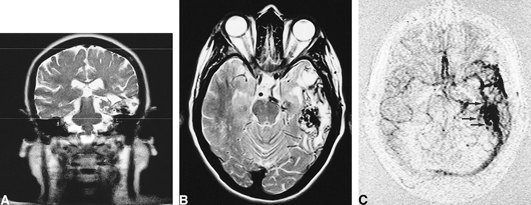
    54-year-old patient with Wyburn-Mason syndrome and bilateral mirror-image parietal AVMs.

    A, Axial T2-weighted (2900/87.5/2) image shows flow voids in both AVMs and the set-up acquisition block for the anteroposterior projection MR-DSA. Note that the anterior cerebral arteries are not included.

    B, Right carotid selective conventional catheter angiogram shows the right-sided AVM.

    C–E, Several stages of MR-DSA during the passage of a contrast bolus in a Towne projection. Early arterial phase (C), late arterial phase (D), and early venous phase (E).

  • fig 2.
    • Download figure
    • Open in new tab
    • Download powerpoint
    fig 2.

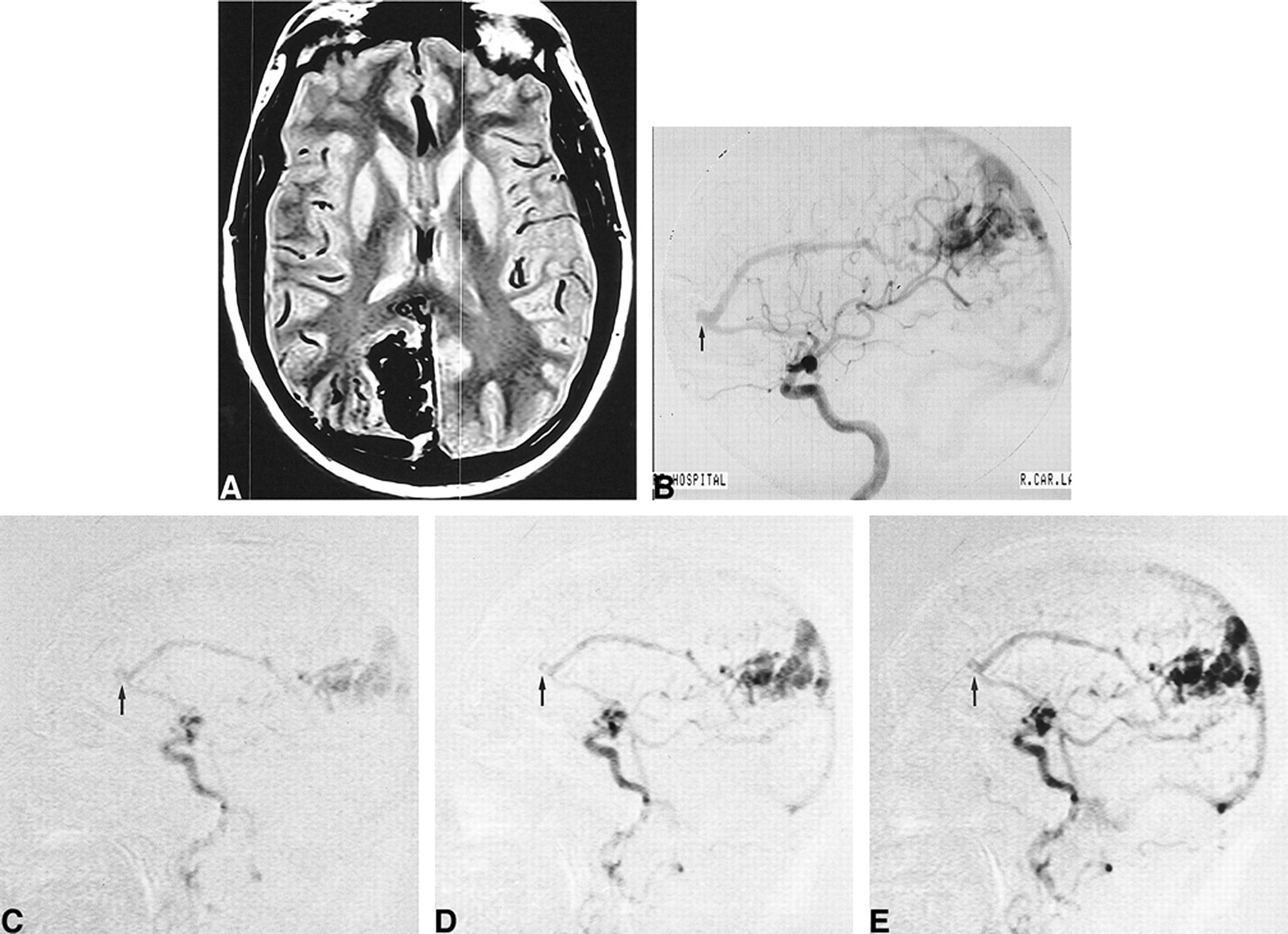
    36-year-old man with a corpus callosum AVM that extends into the left lateral ventricle.

    A, Axial T2-weighted (2900/87.5/2) image though the level of the nidus with set-up acquisition block for lateral projection MR-DSA.

    B, T2-weighted image shows the nidus extending into the left lateral ventricle.

    C, Left carotid selective conventional catheter angiogram shows the AVM.

    D–F, Several stages of MR-DSA during passage of a contrast bolus in a lateral projection. Early arterial phase (D), late arterial phase (E), and early venous phase (F).
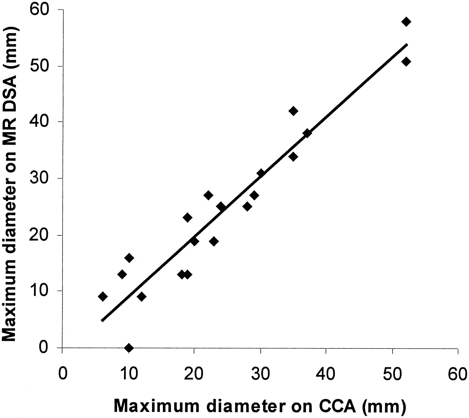
  • fig 3.
    • Download figure
    • Open in new tab
    • Download powerpoint
    fig 3.

    Scatter plot of maximum measured diameter of AVM nidus on MR-DSA against that measured on CCA with the line of best fit shown; calculated Pearson correlation coefficient, r = .953, P = .01

  • fig 4.
    • Download figure
    • Open in new tab
    • Download powerpoint
    fig 4.

    31-year-old woman with a left temporal lobe AVM.

    A, Coronal T2-weighted (2900/75/1) single-shot echo-planar image shows the set-up acquisition block for the axial projection MR-DSA.

    B, T2-weighted 2900/87.5/2 axial section through the level of the nidus.

    C, Early arterial phase axial projection MR-DSA shows the contour of medial border (arrows). This contour cannot be depicted with CCA.

  • fig 5.
    • Download figure
    • Open in new tab
    • Download powerpoint
    fig 5.

    62-year-old man with an occipitoparietal AVM.

    A, Axial T2-weighted (2900/87.5/2) image shows the set-up acquisition block for the lateral projection MR-DSA at the level of the nidus.

    B, Lateral projection from a selective right internal carotid conventional catheter angiogram shows a flow-related aneurysm on the pericallosal artery (arrow).

    C–E, This projection not only shows the nidus in the early arterial phase but also the flow-related aneurysm (arrow, C). In addition, there is an anterior communicating artery aneurysm, not well shown on this projection, which was noted by consensus at review. Late arterial phase (D), early venous phase (E).

Tables

  • Figures
  • TABLE 1:
    • View popup
    • Download powerpoint
    TABLE 1:

    Anatomic locations of 20 arteriovenous malformations

  • TABLE 2:
    • View popup
    • Download powerpoint
    TABLE 2:

    Summary of findings in 20 patients with arteriovenous malformations

PreviousNext
Back to top

In this issue

American Journal of Neuroradiology
Vol. 21, Issue 10
1 Nov 2000
  • Table of Contents
  • Index by author
Advertisement
Print
Download PDF
Email Article

Thank you for your interest in spreading the word on American Journal of Neuroradiology.

NOTE: We only request your email address so that the person you are recommending the page to knows that you wanted them to see it, and that it is not junk mail. We do not capture any email address.

Enter multiple addresses on separate lines or separate them with commas.
Brain Arteriovenous Malformations: Assessment with Dynamic MR Digital Subtraction Angiography
(Your Name) has sent you a message from American Journal of Neuroradiology
(Your Name) thought you would like to see the American Journal of Neuroradiology web site.
CAPTCHA
This question is for testing whether or not you are a human visitor and to prevent automated spam submissions.
Cite this article
Paul D. Griffiths, Nigel Hoggard, Daniel J. Warren, Iain D. Wilkinson, Bob Anderson, Charles A. Romanowski
Brain Arteriovenous Malformations: Assessment with Dynamic MR Digital Subtraction Angiography
American Journal of Neuroradiology Nov 2000, 21 (10) 1892-1899;

Citation Manager Formats

  • BibTeX
  • Bookends
  • EasyBib
  • EndNote (tagged)
  • EndNote 8 (xml)
  • Medlars
  • Mendeley
  • Papers
  • RefWorks Tagged
  • Ref Manager
  • RIS
  • Zotero
0 Responses
Respond to this article
Share
Bookmark this article
Brain Arteriovenous Malformations: Assessment with Dynamic MR Digital Subtraction Angiography
Paul D. Griffiths, Nigel Hoggard, Daniel J. Warren, Iain D. Wilkinson, Bob Anderson, Charles A. Romanowski
American Journal of Neuroradiology Nov 2000, 21 (10) 1892-1899;
del.icio.us logo Twitter logo Facebook logo Mendeley logo
  • Tweet Widget
  • Facebook Like
  • Google Plus One

Jump to section

  • Article
    • Abstract
    • Methods
    • Results
    • Discussion
    • Conclusion
    • Acknowledgments
    • References
  • Figures & Data
  • Info & Metrics
  • Responses
  • References
  • PDF

Related Articles

  • No related articles found.
  • PubMed
  • Google Scholar

Cited By...

  • Time-Resolved MR Angiography: Optimal Parallel Imaging Method
  • In Re: Griffiths PD, Hoggard N, Warren DJ, Wilkinson ID, Anderson B, Romanowski CA. Brain Arteriovenous Malformations: Assessment with Dynamic MR Digital Subtraction Angiography. AJNR Am J Neuroradiol 21:1892-1899
  • Crossref
  • Google Scholar

This article has not yet been cited by articles in journals that are participating in Crossref Cited-by Linking.

More in this TOC Section

  • Usefulness of Quantitative Susceptibility Mapping for the Diagnosis of Parkinson Disease
  • White Matter Alterations in the Brains of Patients with Active, Remitted, and Cured Cushing Syndrome: A DTI Study
  • Qualitative and Quantitative Analysis of MR Imaging Findings in Patients with Middle Cerebral Artery Stroke Implanted with Mesenchymal Stem Cells
Show more BRAIN

Similar Articles

Advertisement

Indexed Content

  • Current Issue
  • Accepted Manuscripts
  • Article Preview
  • Past Issues
  • Editorials
  • Editor's Choice
  • Fellows' Journal Club
  • Letters to the Editor
  • Video Articles

Cases

  • Case Collection
  • Archive - Case of the Week
  • Archive - Case of the Month
  • Archive - Classic Case

Special Collections

  • AJNR Awards
  • ASNR Foundation Special Collection
  • Most Impactful AJNR Articles
  • Photon-Counting CT
  • Spinal CSF Leak Articles (Jan 2020-June 2024)

More from AJNR

  • Trainee Corner
  • Imaging Protocols
  • MRI Safety Corner

Multimedia

  • AJNR Podcasts
  • AJNR Scantastics

Resources

  • Turnaround Time
  • Submit a Manuscript
  • Submit a Video Article
  • Submit an eLetter to the Editor/Response
  • Manuscript Submission Guidelines
  • Statistical Tips
  • Fast Publishing of Accepted Manuscripts
  • Graphical Abstract Preparation
  • Imaging Protocol Submission
  • Evidence-Based Medicine Level Guide
  • Publishing Checklists
  • Author Policies
  • Become a Reviewer/Academy of Reviewers
  • News and Updates

About Us

  • About AJNR
  • Editorial Board
  • Editorial Board Alumni
  • Alerts
  • Permissions
  • Not an AJNR Subscriber? Join Now
  • Advertise with Us
  • Librarian Resources
  • Feedback
  • Terms and Conditions
  • AJNR Editorial Board Alumni

American Society of Neuroradiology

  • Not an ASNR Member? Join Now

© 2025 by the American Society of Neuroradiology All rights, including for text and data mining, AI training, and similar technologies, are reserved.
Print ISSN: 0195-6108 Online ISSN: 1936-959X

Powered by HighWire