
Article Figures & Data
Figures
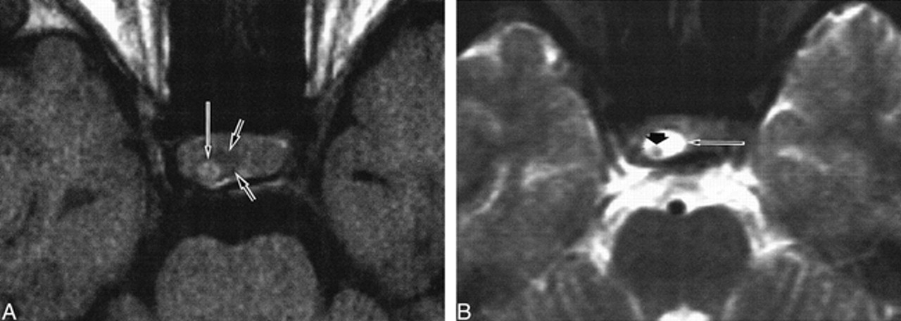
- fig 1.
Patient 9. Rathke's cleft cyst with intracystic nodule.
A, Nodule (long arrow) in the cyst shows high signal intensity. Cystic fluid (small arrow) shows low signal intensity on axial T1-weighted image (600/25/2).
B, High signal intensity of surrounding fluid (long arrow) and low signal intensity of intracystic nodule (small arrow) are well delineated on axial T2-weighted image (2300/120/2).

Tables

Clinical, MR imaging, and intraoperative findings in Rathke's cleft cysts

In this issue


Jump to section
Related Articles
Cited By...
- Uncommon discovery: infrasellar intrasphenoidal Rathkes cleft cyst
- MR Imaging Appearance of Ruptured Rathke Cleft Cyst and Associated Bone Marrow Enhancement
- Follicular carcinoma of the thyroid presenting as metastasis in the wall of an arachnoid cyst
- Usefulness of Contrast-Enhanced 3D-FLAIR MR Imaging for Differentiating Rathke Cleft Cyst from Cystic Craniopharyngioma
- MANAGEMENT OF ENDOCRINE DISEASE: Pituitary 'incidentaloma: neuroradiological assessment and differential diagnosis
- Differentiation between Cystic Pituitary Adenomas and Rathke Cleft Cysts: A Diagnostic Model Using MRI
- Quantitative investigation and classification by MRI of residual cleft cysts in the pituitary glands of cows