
Article Figures & Data
Figures
FIG 1. Neonatal MR score versus initial sonographic (US) score (n = 50). Solid circles represent infants with the same initial and end sonographic score; open circles, infants with a difference between initial and end sonographic scores
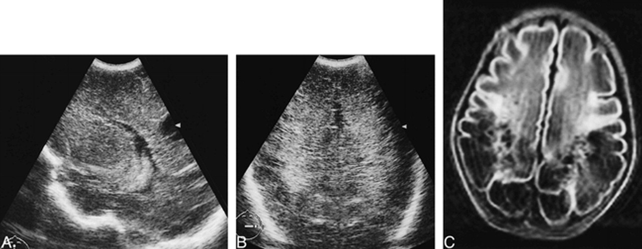
FIG 2. Infant born at 28 4/7 weeks.
A–D, Transient homogeneous periventricular densities are seen in the frontoparietal region on coronal and parasagittal sonograms at a PCA of 29 2/7 weeks (A). Coronal and parasagittal sonograms at a PCA of 34 4/7 weeks (B) show normalized echogenicity of the white matter with a homogeneous aspect. Axial T1-weighted (C) and T2-weighted (D) MR images on the same day show a frontal and parietal periventricular zone of mildly changed signal intensity.
FIG 3. Infant born at 34 2/7 weeks.
A–C, Parasagittal sonogram at a PCA of 35 5/7 weeks (A) shows periventricular densities with an inhomogeneous aspect in the frontoparietal region. T1-weighted MR images on the same day in the parasagittal (B) and axial (C) planes show multiple, small, punctate hemorrhages, occurring in the periventricular white matter on both sides.
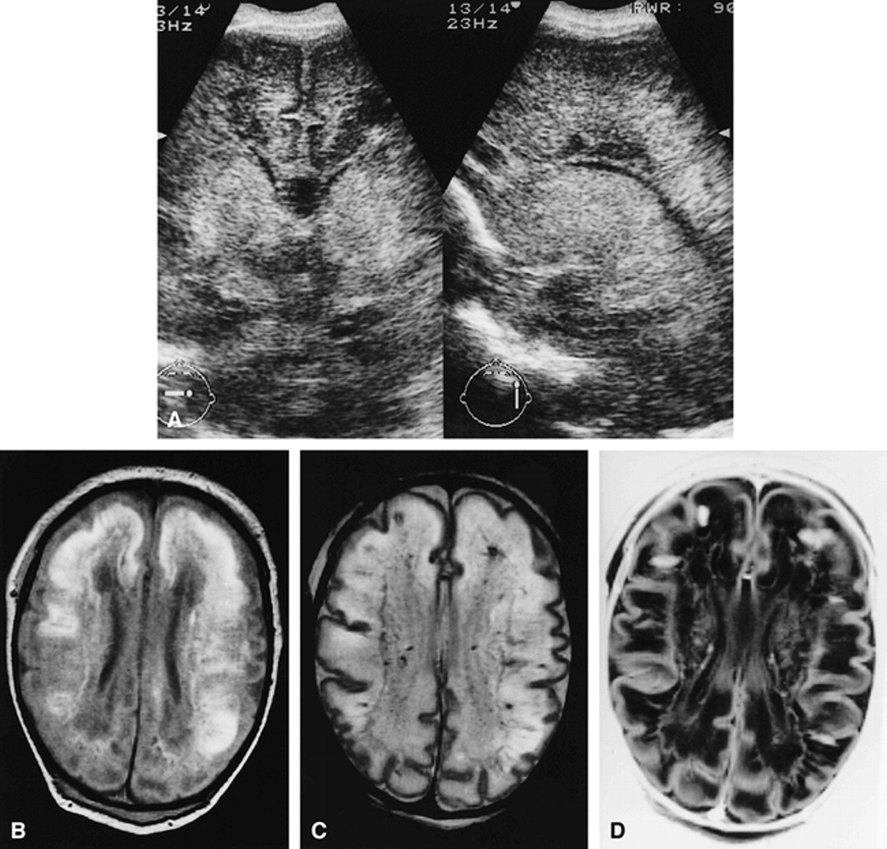
FIG 4. Infant born at 37 3/7 weeks.
A–D, Coronal and parasagittal sonograms (A) at a PCA of 38 2/7 weeks show a diffuse increased echogenicity throughout the white matter and basal ganglia with an inhomogeneous, partly precystic aspect in the frontal region. Proton density–weighted (B), T2-weighted (C), and inversion recovery (D) MR images in the axial plane show large areas of high signal on the proton density–weighted image (B) and combined high and low signal changes on the T2-weighted and inversion recovery images throughout the white matter (C, D) and basal ganglia (D). In comparison with sonograms, MR images show more extensive, small, partly cystic and partly precystic periventricular lesions in the frontal and parietal region, with signal intensities identical or close to CSF (B–D). The hemorrhagic ischemic areas correspond to the large cystic lesions detected at autopsy performed 4 days later.
FIG 5. Infant born at 37 6/7 weeks.
A and B, Coronal sonogram (A) at a PCA of 39 weeks shows a strikingly asymmetrical increased echogenicity of the periventricular white matter, predominantly in the left parietal region, with an inhomogeneous aspect. In addition, intraventricular and germinal-layer hemorrhages were present. T1-weighted MR image on the same day in the axial plane (B) shows extensive signal intensity changes and hemorrhagic lesions throughout the left hemisphere and a small periventricular hemorrhage on the other side.
FIG 6. Infant born at 32 weeks.
A and B, Parasagittal sonogram (A) at a PCA of 33 4/7 weeks shows a (stable) posthemorrhagic ventricular dilatation and a moderately increased echogenicity of the white matter in the parietooccipital region with an inhomogeneous aspect and few small cystic lesions. Axial T1-weighted MR image on the same day (B) shows extensive white matter signal intensity changes with multiple hemorrhagic lesions and more numerous cystic lesions.
FIG 7. Infant born at 34 5/7 weeks.
A and B, Parasagittal (A) and coronal (B) sonograms at a PCA of 36 5/7 weeks show a moderately increased echogenicity of the periventricular white matter with a homogeneous aspect and one large cyst in the parietal region. Axial T1-weighted MR image on the same day (C) shows more numerous, bilateral, large periventricular cysts on MR images, with focal extension into the subcortical white matter in the entire parietooccipital region.
FIG 8. Neonatal MR score versus sonographic (US) end score (n = 48). Solid circles represent infants with the same initial and end sonographic score; open circles, infants with a difference between initial and end sonographic scores; cross, infants who did not survive the neonatal period


Tables

Modified sonographic grading system*

In this issue


Jump to section
Related Articles
Cited By...
- Cerebral ultrasound abnormalities in infants born to mothers with autoimmune disease
- Magnetic resonance imaging of preterm brain injury
- Limitations of ultrasonography for diagnosing white matter damage in preterm infants
- Practice parameter: Neuroimaging of the neonate: Report of the Quality Standards Subcommittee of the American Academy of Neurology and the Practice Committee of the Child Neurology Society
- Ultrasound diagnosis and neurodevelopmental outcome of localised and extensive cystic periventricular leucomalacia