Advertisement

Main menu

  • Home
  • Content
    • Current Issue
    • Accepted Manuscripts
    • Article Preview
    • Past Issue Archive
    • Video Articles
    • AJNR Case Collection
    • Case of the Week Archive
    • Case of the Month Archive
    • Classic Case Archive
  • Special Collections
    • AJNR Awards
    • ASNR Foundation Special Collection
    • Most Impactful AJNR Articles
    • Photon-Counting CT
    • Spinal CSF Leak Articles (Jan 2020-June 2024)
  • Multimedia
    • AJNR Podcasts
    • AJNR SCANtastic
    • Trainee Corner
    • MRI Safety Corner
    • Imaging Protocols
  • For Authors
    • Submit a Manuscript
    • Submit a Video Article
    • Submit an eLetter to the Editor/Response
    • Manuscript Submission Guidelines
    • Statistical Tips
    • Fast Publishing of Accepted Manuscripts
    • Graphical Abstract Preparation
    • Imaging Protocol Submission
    • Author Policies
  • About Us
    • About AJNR
    • Editorial Board
    • Editorial Board Alumni
  • More
    • Become a Reviewer/Academy of Reviewers
    • Subscribers
    • Permissions
    • Alerts
    • Feedback
    • Advertisers
    • ASNR Home

User menu

  • Alerts
  • Log in

Search

  • Advanced search
American Journal of Neuroradiology

American Journal of Neuroradiology

ASHNR American Society of Functional Neuroradiology ASHNR American Society of Pediatric Neuroradiology ASSR
  • Alerts
  • Log in

Advanced Search

  • Home
  • Content
    • Current Issue
    • Accepted Manuscripts
    • Article Preview
    • Past Issue Archive
    • Video Articles
    • AJNR Case Collection
    • Case of the Week Archive
    • Case of the Month Archive
    • Classic Case Archive
  • Special Collections
    • AJNR Awards
    • ASNR Foundation Special Collection
    • Most Impactful AJNR Articles
    • Photon-Counting CT
    • Spinal CSF Leak Articles (Jan 2020-June 2024)
  • Multimedia
    • AJNR Podcasts
    • AJNR SCANtastic
    • Trainee Corner
    • MRI Safety Corner
    • Imaging Protocols
  • For Authors
    • Submit a Manuscript
    • Submit a Video Article
    • Submit an eLetter to the Editor/Response
    • Manuscript Submission Guidelines
    • Statistical Tips
    • Fast Publishing of Accepted Manuscripts
    • Graphical Abstract Preparation
    • Imaging Protocol Submission
    • Author Policies
  • About Us
    • About AJNR
    • Editorial Board
    • Editorial Board Alumni
  • More
    • Become a Reviewer/Academy of Reviewers
    • Subscribers
    • Permissions
    • Alerts
    • Feedback
    • Advertisers
    • ASNR Home
  • Follow AJNR on Twitter
  • Visit AJNR on Facebook
  • Follow AJNR on Instagram
  • Join AJNR on LinkedIn
  • RSS Feeds

AJNR is seeking candidates for the AJNR Podcast Editor. Read the position description.

Research ArticleBRAIN

Gross-total Surgery of Supratentorial Low-grade Gliomas under Intraoperative MR Guidance

Jens P. Schneider, Thomas Schulz, Frank Schmidt, Jürgen Dietrich, Siegbert Lieberenz, Christos Trantakis, Volker Seifert, Steffen Kellermann, Ralf Schober, Lutz Schaffranietz, Mario Laufer and Thomas Kahn
American Journal of Neuroradiology January 2001, 22 (1) 89-98;
Jens P. Schneider
  • Find this author on Google Scholar
  • Find this author on PubMed
  • Search for this author on this site
Thomas Schulz
  • Find this author on Google Scholar
  • Find this author on PubMed
  • Search for this author on this site
Frank Schmidt
  • Find this author on Google Scholar
  • Find this author on PubMed
  • Search for this author on this site
Jürgen Dietrich
  • Find this author on Google Scholar
  • Find this author on PubMed
  • Search for this author on this site
Siegbert Lieberenz
  • Find this author on Google Scholar
  • Find this author on PubMed
  • Search for this author on this site
Christos Trantakis
  • Find this author on Google Scholar
  • Find this author on PubMed
  • Search for this author on this site
Volker Seifert
  • Find this author on Google Scholar
  • Find this author on PubMed
  • Search for this author on this site
Steffen Kellermann
  • Find this author on Google Scholar
  • Find this author on PubMed
  • Search for this author on this site
Ralf Schober
  • Find this author on Google Scholar
  • Find this author on PubMed
  • Search for this author on this site
Lutz Schaffranietz
  • Find this author on Google Scholar
  • Find this author on PubMed
  • Search for this author on this site
Mario Laufer
  • Find this author on Google Scholar
  • Find this author on PubMed
  • Search for this author on this site
Thomas Kahn
  • Find this author on Google Scholar
  • Find this author on PubMed
  • Search for this author on this site
  • Article
  • Figures & Data
  • Info & Metrics
  • Responses
  • References
  • PDF
Loading

Article Figures & Data

Figures

  • Tables
  • fig 1.
    • Download figure
    • Open in new tab
    • Download powerpoint
    fig 1.

    Intraoperative MR imaging environment.

    A, The patient is positioned in the magnet by using the side entry of the table. The neurosurgeon can stand behind the head of the patient by using an MR-compatible microscope (white arrow). The assisting nurse and the surgical instrumentation table are located behind the neurosurgeon. Black arrow indicates in-bore monitor.

    B, The Flashpoint Position Encoder (open arrow) with the three light-emitting diodes (asterisks) in situ. The MR Track Pointer (long thin arrow) is mounted on the Flashpoint Position Encoder.
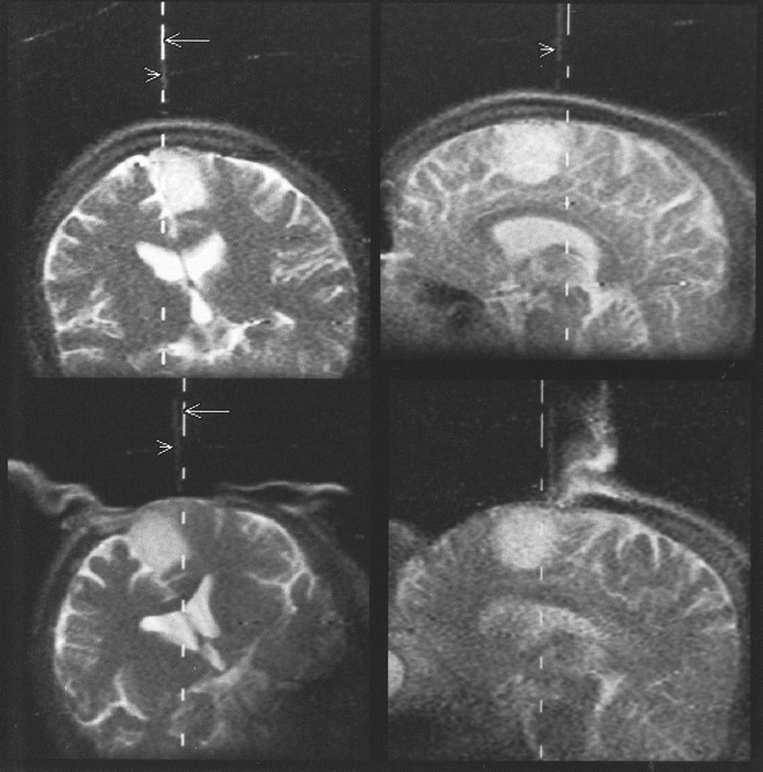
  • fig 2.
    • Download figure
    • Open in new tab
    • Download powerpoint
    fig 2.

    Patient 2 (astrocytoma, WHO grade II).

    Top left and top right, The Flashpoint Position Encoder is used to localize the borders of the tumor in projection onto the skull before craniotomy. The dotted lines (arrows) mark the calculated actual position of the hand-held device. Additionally, an MR Track Pointer (arrowheads) mounted on the hand-held device is used. On T2-weighted single-shot fast spin-echo sequences, the instrument, filled with a sodium chloride solution, is displayed with high signal intensity. The sagittal view (top right) shows a slight deviation between the calculated and imaged positions caused by a short delay between image update (every 3 seconds) and optical tracking update (four per second).

    Bottom left and bottom right, After craniotomy, this approach is used again to define the borders of the tumor in projection to the dura.
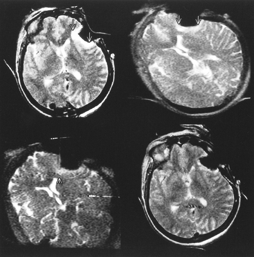
  • fig 3.
    • Download figure
    • Open in new tab
    • Download powerpoint
    fig 3.

    Patient 9 (astrocytoma, WHO grade II).

    Top left and top right, T2-weighted axial fast spin-echo images were used to determine the tumor area before resection.

    Middle left and middle right, Comparable slices obtained at the point at which the neurosurgeon considered the resection to be complete (first control) were used to calculate the residual tumor volume.

    Bottom left and bottom right, T2-weighted images at the same position were used to measure very small areas of residual tumor tissue at the end of the operation before closing the craniotomy.

  • fig 4.
    • Download figure
    • Open in new tab
    • Download powerpoint
    fig 4.

    Patient 10 (astrocytoma, WHO grade II).

    A, First control at the point at which the resection was considered complete by the neurosurgeon. T2-weighted fast spin-echo image shows two hyperintense areas suspicious for tumor (arrows) at the border of resection.

    B, Using the interactive scan mode (T2-weighted single-shot fast spin-echo, 3 seconds per image) and the MR Track Pointer (arrowhead) filled with sodium chloride solution, one lesion is marked (asterisks). Fluid (F) is at the bottom of the resection cavity while the patient is lying on the right side.

    C, The last control (T2-weighted fast spin-echo image) shows complete removal of all tumor-suspicious tissue. There is no surgically induced edema evident in the bed of resection.

  • fig 5.
    • Download figure
    • Open in new tab
    • Download powerpoint
    fig 5.

    Patient 4 (oligodendroglioma, WHO grade II).

    Top left, The first control image (T2-weighted fast spin-echo) shows hyperintense tumor-suspicious tissue (arrows) in the bed of resection.

    Top right and bottom left, Abnormalities are also seen on the interactively guided single-shot fast spin-echo images (arrows). The MR Track Pointer is used to localize the suspicious area (arrowhead).

    Bottom right, Before closure of the craniotomy, T2-weighted fast spin-echo image shows the final result of the resection. The tumor-suspicious tissue was reduced; nevertheless, some nodular areas are still suggestive of residual tumor. Because of the proximity of this region to the language cortex, the resection was stopped at this point. The patient had no postoperative neurologic deficit.

  • fig 6.
    • Download figure
    • Open in new tab
    • Download powerpoint
    fig 6.

    Patient 1 (astrocytoma, WHO grade II).

    Top left, T2-weighted fast spin-echo image before craniotomy shows high signal intensity of the temporal astrocytoma (arrow).

    Top right, T2-weighted fast spin-echo image after corticotomy for surgical access to the tumor.

    Bottom left, T2-weighted fast spin-echo image at the point at which resection was considered complete by the neurosurgeon and by the radiologist reveals no residual tumor at this slice.

    Bottom right, After closure of the craniotomy, the bed of resection is displayed with high signal intensity, probably caused by a mixture of blood, CSF, saline, and hemostatic material.
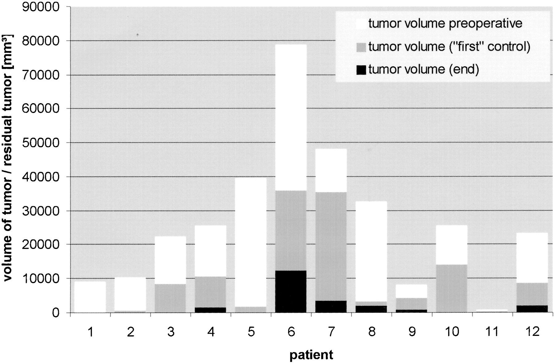
  • fig 7.
    • Download figure
    • Open in new tab
    • Download powerpoint
    fig 7.

    Volume of tumor/residual tumor detected on MR images in all 12 patients at three different points of resection: 1) preoperatively (before craniotomy/corticotomy), 2) at the point of first control condition (when resection was considered complete by inspection of the operative field), and 3) at the conclusion of surgery (before closing craniotomy)

Tables

  • Figures
  • Table 1:
    • View popup
    • Download powerpoint
    Table 1:

    Imaging protocol

  • Table 2:
    • View popup
    • Download powerpoint
    Table 2:

    Tumor volume at different points of the operation

PreviousNext
Back to top

In this issue

American Journal of Neuroradiology
Vol. 22, Issue 1
1 Jan 2001
  • Table of Contents
  • Index by author
Advertisement
Print
Download PDF
Email Article

Thank you for your interest in spreading the word on American Journal of Neuroradiology.

NOTE: We only request your email address so that the person you are recommending the page to knows that you wanted them to see it, and that it is not junk mail. We do not capture any email address.

Enter multiple addresses on separate lines or separate them with commas.
Gross-total Surgery of Supratentorial Low-grade Gliomas under Intraoperative MR Guidance
(Your Name) has sent you a message from American Journal of Neuroradiology
(Your Name) thought you would like to see the American Journal of Neuroradiology web site.
CAPTCHA
This question is for testing whether or not you are a human visitor and to prevent automated spam submissions.
Cite this article
Jens P. Schneider, Thomas Schulz, Frank Schmidt, Jürgen Dietrich, Siegbert Lieberenz, Christos Trantakis, Volker Seifert, Steffen Kellermann, Ralf Schober, Lutz Schaffranietz, Mario Laufer, Thomas Kahn
Gross-total Surgery of Supratentorial Low-grade Gliomas under Intraoperative MR Guidance
American Journal of Neuroradiology Jan 2001, 22 (1) 89-98;

Citation Manager Formats

  • BibTeX
  • Bookends
  • EasyBib
  • EndNote (tagged)
  • EndNote 8 (xml)
  • Medlars
  • Mendeley
  • Papers
  • RefWorks Tagged
  • Ref Manager
  • RIS
  • Zotero
0 Responses
Respond to this article
Share
Bookmark this article
Gross-total Surgery of Supratentorial Low-grade Gliomas under Intraoperative MR Guidance
Jens P. Schneider, Thomas Schulz, Frank Schmidt, Jürgen Dietrich, Siegbert Lieberenz, Christos Trantakis, Volker Seifert, Steffen Kellermann, Ralf Schober, Lutz Schaffranietz, Mario Laufer, Thomas Kahn
American Journal of Neuroradiology Jan 2001, 22 (1) 89-98;
del.icio.us logo Twitter logo Facebook logo Mendeley logo
  • Tweet Widget
  • Facebook Like
  • Google Plus One

Jump to section

  • Article
    • Abstract
    • Methods
    • Results
    • Discussion
    • Conclusion
    • Acknowledgments
    • Footnotes
    • References
  • Figures & Data
  • Info & Metrics
  • Responses
  • References
  • PDF

Related Articles

  • No related articles found.
  • PubMed
  • Google Scholar

Cited By...

  • No citing articles found.
  • Crossref
  • Google Scholar

This article has not yet been cited by articles in journals that are participating in Crossref Cited-by Linking.

More in this TOC Section

  • Usefulness of Quantitative Susceptibility Mapping for the Diagnosis of Parkinson Disease
  • White Matter Alterations in the Brains of Patients with Active, Remitted, and Cured Cushing Syndrome: A DTI Study
  • Qualitative and Quantitative Analysis of MR Imaging Findings in Patients with Middle Cerebral Artery Stroke Implanted with Mesenchymal Stem Cells
Show more BRAIN

Similar Articles

Advertisement

Indexed Content

  • Current Issue
  • Accepted Manuscripts
  • Article Preview
  • Past Issues
  • Editorials
  • Editor's Choice
  • Fellows' Journal Club
  • Letters to the Editor
  • Video Articles

Cases

  • Case Collection
  • Archive - Case of the Week
  • Archive - Case of the Month
  • Archive - Classic Case

Special Collections

  • AJNR Awards
  • ASNR Foundation Special Collection
  • Most Impactful AJNR Articles
  • Photon-Counting CT
  • Spinal CSF Leak Articles (Jan 2020-June 2024)

More from AJNR

  • Trainee Corner
  • Imaging Protocols
  • MRI Safety Corner

Multimedia

  • AJNR Podcasts
  • AJNR Scantastics

Resources

  • Turnaround Time
  • Submit a Manuscript
  • Submit a Video Article
  • Submit an eLetter to the Editor/Response
  • Manuscript Submission Guidelines
  • Statistical Tips
  • Fast Publishing of Accepted Manuscripts
  • Graphical Abstract Preparation
  • Imaging Protocol Submission
  • Evidence-Based Medicine Level Guide
  • Publishing Checklists
  • Author Policies
  • Become a Reviewer/Academy of Reviewers
  • News and Updates

About Us

  • About AJNR
  • Editorial Board
  • Editorial Board Alumni
  • Alerts
  • Permissions
  • Not an AJNR Subscriber? Join Now
  • Advertise with Us
  • Librarian Resources
  • Feedback
  • Terms and Conditions
  • AJNR Editorial Board Alumni

American Society of Neuroradiology

  • Not an ASNR Member? Join Now

© 2025 by the American Society of Neuroradiology All rights, including for text and data mining, AI training, and similar technologies, are reserved.
Print ISSN: 0195-6108 Online ISSN: 1936-959X

Powered by HighWire