
Article Figures & Data
Figures
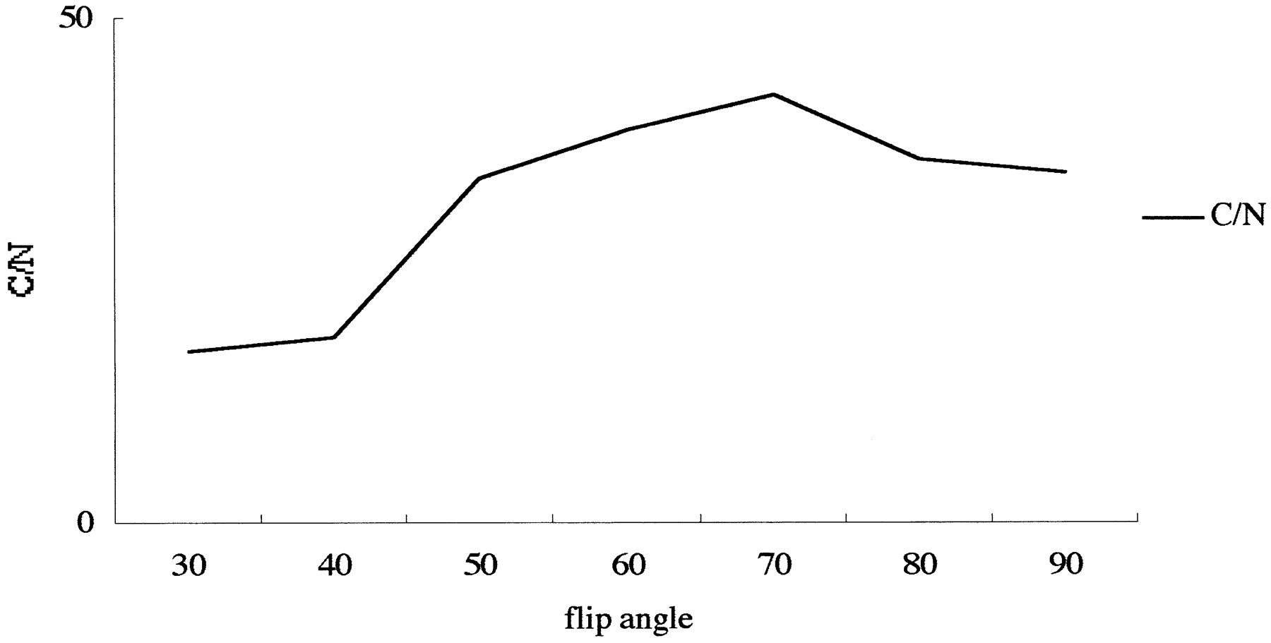
- fig 1.
Contrast-to-noise ratio (C/N) between CSF and the cerebellum on 3D-CISS images obtained at various flip angles in a volunteer. The maximum C/N was obtained at a flip angle of 70°. In this particular subject, the C/N on 3D-FASE images was 50.3
- fig 2.
Phantom experiment to evaluate the effect of TE on 3D-CISS sequences. Water-filled polyethylene tube is coiled and placed on cubic water phantom.
A–C, 3D-CISS (12/6/1) (A), 3D-CISS (16/8/1) (B), and 3D-FASE (5000/241.6/1) (C) sequences. Bandlike susceptibility artifact is seen on A and B (arrows); however, it is slightly more prominent on B. There is no artifact on C (arrow). Note that the cubic water phantom signal is uniform in C. This experiment shows that shorter TE has some ability to reduce susceptibility artifacts on 3D-CISS studies, but 3D-FASE has far fewer susceptibility artifacts.
- fig 3.
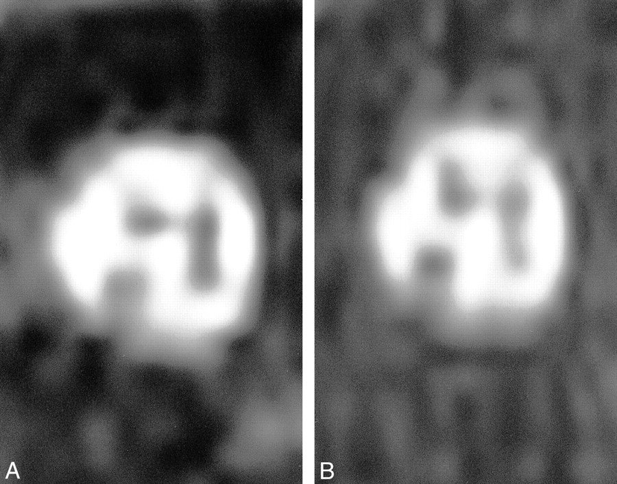
Multiplanar reformatted images of the internal auditory canal of a volunteer.
A and B, Four nerve branches are clearly depicted by both 3D-CISS (16/8/1) (A) and 3D-FASE (5000/241.6/1) (B) sequences.
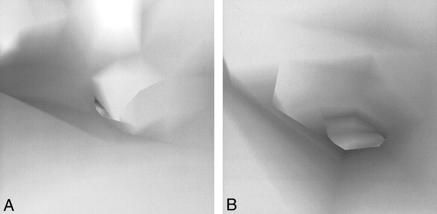
- fig 4.
A and B, Flow ghosts of CSF are pronounced on 3D-CISS (16/8/1) sequence (white arrows, A); however, no ghosts are seen on 3D-FASE (5000/241.6/1) image (B) in this volunteer. Note that the shape of the vestibule near the oval window is deformed on the 3D-CISS image (black arrow, A) but not on the 3D-FASE image (black arrow, B)
- fig 5.
A and B, Low-signal bands due to susceptibility artifacts are prominent from the vestibular area just above the oval window to the crus of the lateral semicircular canal on 3D-CISS (16/8/1) image (arrow, A), whereas no low-signal bands are seen on 3D-FASE (5000/241.6/1) image (B) in this volunteer. Intracochlear anatomy, such as modiolus (M) and osseous spiral lamina (O), is depicted slightly better on 3D-CISS image, probably because of the susceptibility effect from these bony structures. Endolymphatic duct (E) is visible only on 3D-CISS image in this particular subject
- fig 6.
Virtual endoscopic images of the superior semicircular canal from the vestibule in a volunteer.
A and B, Pseudoocclusion is seen on 3D-CISS image (16/8/1) (A). On 3D-FASE (5000/241.6/1) image (B), it is possible to navigate through the three semicircular canals.
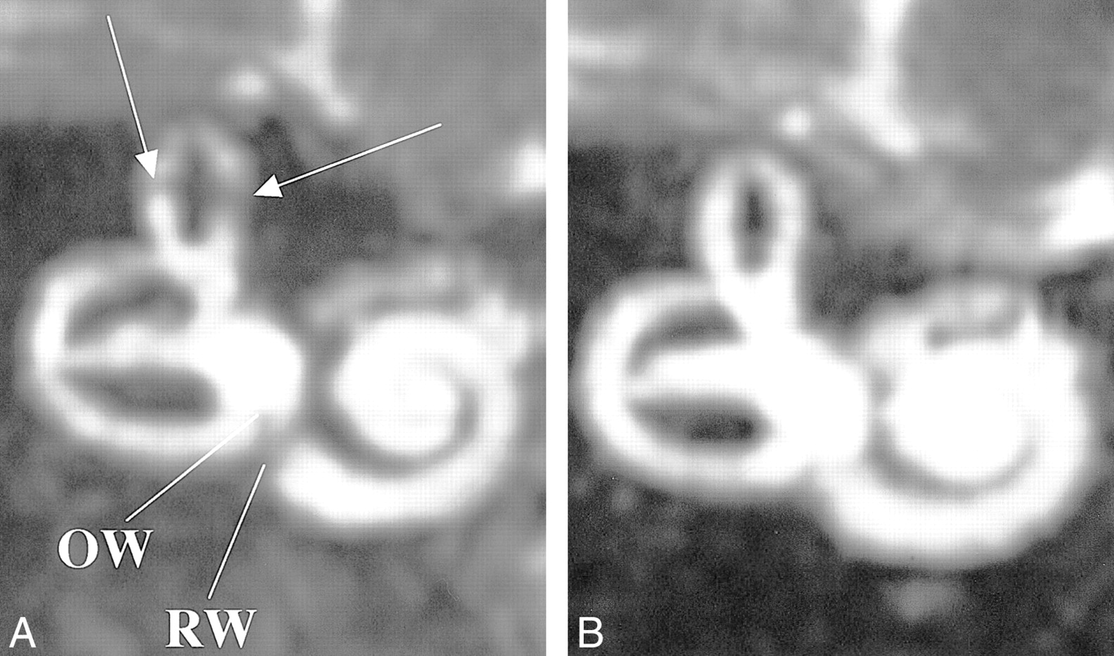
- fig 7.
MIP image of the labyrinth in a volunteer.
A and B, Low-signal artifacts are seen in superior semicircular canal (arrows, A) as well as in the area of the oval window (OW) and round window (RW) on 3D-CISS image (16/8/1) (A), but not on the 3D-FASE image (5000/241.6/1) (B).
- fig 8.
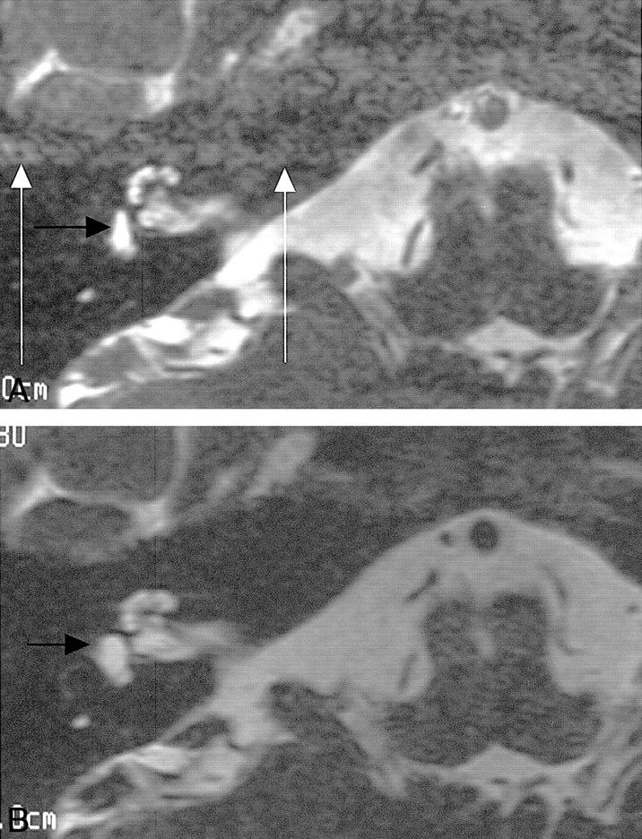
63-year-old man with right intracanalicular acoustic schwannoma.
A–E, A mass (arrow) is seen on unenhanced (A) and contrast-enhanced (B) 3D-FASE (5000/241.6/1) images, on unenhanced (C) and contrast-enhanced (D) 3D-CISS (16/8/1) images, and on contrast-enhanced 3D-SPGR (23/10/1) image (E). Note that the signal of the mass is not influenced by contrast material on the 3D-FASE image, but enhancement is present on the 3D-CISS sequence.
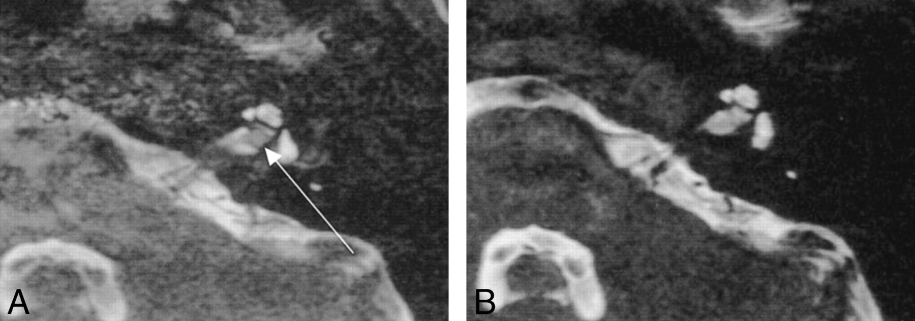
- fig 9.
False-positive finding on 3D-CISS image in an 11-year-old girl.
A and B, A small masslike nodule is seen at the fundus of the left internal auditory canal on 3D-CISS (16/8/1) image (arrow, A), although no mass is seen on 3D-FASE (5000/241.6/1) image (B). This artifact was assumed to be due to the combination of susceptibility and flow artifacts.


Tables


In this issue


Jump to section
Related Articles
Cited By...
- Does CISS MRI Reliably Depict the Endolymphatic Duct in Children with and without Vestibular Aqueduct Enlargement?
- Correlation between Histopathology and Signal Loss on Spin-Echo T2-Weighted MR Images of the Inner Ear: Distinguishing Artifacts from Anatomy
- Measuring 3D Cochlear Duct Length on MRI: Is It Accurate and Reliable?
- Visualization of the Peripheral Branches of the Mandibular Division of the Trigeminal Nerve on 3D Double-Echo Steady-State with Water Excitation Sequence
- 3D Double-Echo Steady-State with Water Excitation MR Imaging of the Intraparotid Facial Nerve at 1.5T: A Pilot Study