Advertisement

Main menu

  • Home
  • Content
    • Current Issue
    • Accepted Manuscripts
    • Article Preview
    • Past Issue Archive
    • Video Articles
    • AJNR Case Collection
    • Case of the Week Archive
    • Case of the Month Archive
    • Classic Case Archive
  • Special Collections
    • AJNR Awards
    • ASNR Foundation Special Collection
    • Most Impactful AJNR Articles
    • Photon-Counting CT
    • Spinal CSF Leak Articles (Jan 2020-June 2024)
  • Multimedia
    • AJNR Podcasts
    • AJNR SCANtastic
    • Trainee Corner
    • MRI Safety Corner
    • Imaging Protocols
  • For Authors
    • Submit a Manuscript
    • Submit a Video Article
    • Submit an eLetter to the Editor/Response
    • Manuscript Submission Guidelines
    • Statistical Tips
    • Fast Publishing of Accepted Manuscripts
    • Graphical Abstract Preparation
    • Imaging Protocol Submission
    • Author Policies
  • About Us
    • About AJNR
    • Editorial Board
    • Editorial Board Alumni
  • More
    • Become a Reviewer/Academy of Reviewers
    • Subscribers
    • Permissions
    • Alerts
    • Feedback
    • Advertisers
    • ASNR Home

User menu

  • Alerts
  • Log in

Search

  • Advanced search
American Journal of Neuroradiology

American Journal of Neuroradiology

ASHNR American Society of Functional Neuroradiology ASHNR American Society of Pediatric Neuroradiology ASSR
  • Alerts
  • Log in

Advanced Search

  • Home
  • Content
    • Current Issue
    • Accepted Manuscripts
    • Article Preview
    • Past Issue Archive
    • Video Articles
    • AJNR Case Collection
    • Case of the Week Archive
    • Case of the Month Archive
    • Classic Case Archive
  • Special Collections
    • AJNR Awards
    • ASNR Foundation Special Collection
    • Most Impactful AJNR Articles
    • Photon-Counting CT
    • Spinal CSF Leak Articles (Jan 2020-June 2024)
  • Multimedia
    • AJNR Podcasts
    • AJNR SCANtastic
    • Trainee Corner
    • MRI Safety Corner
    • Imaging Protocols
  • For Authors
    • Submit a Manuscript
    • Submit a Video Article
    • Submit an eLetter to the Editor/Response
    • Manuscript Submission Guidelines
    • Statistical Tips
    • Fast Publishing of Accepted Manuscripts
    • Graphical Abstract Preparation
    • Imaging Protocol Submission
    • Author Policies
  • About Us
    • About AJNR
    • Editorial Board
    • Editorial Board Alumni
  • More
    • Become a Reviewer/Academy of Reviewers
    • Subscribers
    • Permissions
    • Alerts
    • Feedback
    • Advertisers
    • ASNR Home
  • Follow AJNR on Twitter
  • Visit AJNR on Facebook
  • Follow AJNR on Instagram
  • Join AJNR on LinkedIn
  • RSS Feeds

AJNR is seeking candidates for the AJNR Podcast Editor. Read the position description.

Research ArticleINTERVENTIONAL

Dural Arteriovenous Fistula in Children: Endovascular Treatment and Outcomes in Seven Cases

Patricia K. Kincaid, Gary R. Duckwiler, Y. Pierre Gobin and Fernando Viñuela
American Journal of Neuroradiology June 2001, 22 (6) 1217-1225;
Patricia K. Kincaid
  • Find this author on Google Scholar
  • Find this author on PubMed
  • Search for this author on this site
Gary R. Duckwiler
  • Find this author on Google Scholar
  • Find this author on PubMed
  • Search for this author on this site
Y. Pierre Gobin
  • Find this author on Google Scholar
  • Find this author on PubMed
  • Search for this author on this site
Fernando Viñuela
  • Find this author on Google Scholar
  • Find this author on PubMed
  • Search for this author on this site
  • Article
  • Figures & Data
  • Info & Metrics
  • Responses
  • References
  • PDF
Loading

Article Figures & Data

Figures

  • Tables
  • fig 1.
    • Download figure
    • Open in new tab
    • Download powerpoint
    fig 1.

    Patient 7 was a 10-year-old girl with a complex, multifocal dural AVF (infantile type dural arteriovenous shunt), who presented late with advanced neurologic symptoms.

    A, Axial view contrast-enhanced CT scan obtained at the time of referral shows dystrophic calcification in the brain parenchyma, hydrocephalus, thinning of white matter that is abnormally hypodense, and enlarged right sphenoparietal and superior sagittal sinuses. This patient had already developed irreversible brain injury related to her dural AVF.

    B, Sagittal view T1-weighted MR image shows a significantly enlarged flow void in the superior sagittal sinus and torcular herophili, some of the sites of arteriovenous shunting in this patient. Also note the prolapse of the cerebellar tonsils at the craniocervical junction, the so-called acquired Chiari I malformation that can also be seen with the vein of Galen malformation.

  • fig 2.
    • Download figure
    • Open in new tab
    • Download powerpoint
    fig 2.

    Patient 1 was a newborn with severe congestive heart failure.

    A, Anteroposterior view angiogram of the right external carotid artery shows a large left transverse and sigmoid sinus dural AVF (dural sinus malformation) supplied by enlarged right occipital artery branches, subsequently embolized using coils and N-butylcyanoacrylate. Note the coil mass already present in the left transverse sinus that was placed via direct puncture at an outside institution at 1 day of age.

    B, Anteroposterior view shows left common carotid artery injection, venous phase, with prominent venous collateral channels draining to the right transverse sinus.

    C, Lateral view shows left external carotid artery injection before embolization. The large left transverse and sigmoid sinus dural AVF is supplied by a markedly enlarged left middle meningeal artery that arises from the left ophthalmic artery. There is abnormal enlargement of the artery of the foramen rotundum (arrowhead) that supplies an enlarged middle meningeal artery (large arrows). The middle meningeal artery has a variant origin from the ophthalmic artery (small arrows).

    D, Lateral view shows left common carotid artery injection after embolization. After complete embolization using microcoils and N-butylcyanoacrylate, there is stagnation of flow in the stump of the middle meningeal artery (open arrow), no evidence of arteriovenous shunting, and good filling of the normal intracranial circulation.

  • fig 3.
    • Download figure
    • Open in new tab
    • Download powerpoint
    fig 3.

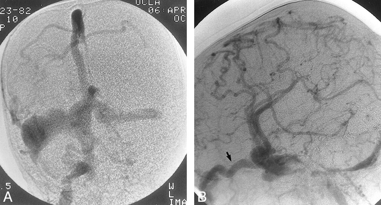
    Patient 5, with a dural sinus malformation, was an 18-month-old who presented with seizures and prominent facial veins.

    A, Anteroposterior view angiogram of the right external carotid artery, venous phase, shows bilateral sigmoid sinus occlusions and an enlarged occipital vein. The arterial phase (not shown) showed enlarged right occipital artery feeders to a large right transverse sinus dural fistula.

    B, Lateral view angiogram of the right common carotid artery, obtained after complete embolization of the dural AVF, shows development of collateral venous drainage, ultimately draining through the superior ophthalmic veins (arrow), which is seen to be markedly dilated.

Tables

  • Figures
  • TABLE 1:
    • View popup
    • Download powerpoint
    TABLE 1:

    Clinical and radiologic findings

  • TABLE 2:
    • View popup
    • Download powerpoint
    TABLE 2:

    Treatment and outcome

PreviousNext
Back to top

In this issue

American Journal of Neuroradiology: 22 (6)
American Journal of Neuroradiology
Vol. 22, Issue 6
1 Jun 2001
  • Table of Contents
  • Index by author
Advertisement
Print
Download PDF
Email Article

Thank you for your interest in spreading the word on American Journal of Neuroradiology.

NOTE: We only request your email address so that the person you are recommending the page to knows that you wanted them to see it, and that it is not junk mail. We do not capture any email address.

Enter multiple addresses on separate lines or separate them with commas.
Dural Arteriovenous Fistula in Children: Endovascular Treatment and Outcomes in Seven Cases
(Your Name) has sent you a message from American Journal of Neuroradiology
(Your Name) thought you would like to see the American Journal of Neuroradiology web site.
CAPTCHA
This question is for testing whether or not you are a human visitor and to prevent automated spam submissions.
Cite this article
Patricia K. Kincaid, Gary R. Duckwiler, Y. Pierre Gobin, Fernando Viñuela
Dural Arteriovenous Fistula in Children: Endovascular Treatment and Outcomes in Seven Cases
American Journal of Neuroradiology Jun 2001, 22 (6) 1217-1225;

Citation Manager Formats

  • BibTeX
  • Bookends
  • EasyBib
  • EndNote (tagged)
  • EndNote 8 (xml)
  • Medlars
  • Mendeley
  • Papers
  • RefWorks Tagged
  • Ref Manager
  • RIS
  • Zotero
0 Responses
Respond to this article
Share
Bookmark this article
Dural Arteriovenous Fistula in Children: Endovascular Treatment and Outcomes in Seven Cases
Patricia K. Kincaid, Gary R. Duckwiler, Y. Pierre Gobin, Fernando Viñuela
American Journal of Neuroradiology Jun 2001, 22 (6) 1217-1225;
del.icio.us logo Twitter logo Facebook logo Mendeley logo
  • Tweet Widget
  • Facebook Like
  • Google Plus One

Jump to section

  • Article
    • Abstract
    • Methods
    • Results
    • Discussion
    • Conclusion
    • Footnotes
    • References
  • Figures & Data
  • Info & Metrics
  • Responses
  • References
  • PDF

Related Articles

  • No related articles found.
  • PubMed
  • Google Scholar

Cited By...

  • Paediatric intracranial dural arteriovenous fistulas: clinical characteristics, treatment outcomes and prognosis
  • Endovascular management of torcular dural sinus malformations in children: the role of straight sinus occlusion
  • Progressive versus Nonprogressive Intracranial Dural Arteriovenous Fistulas: Characteristics and Outcomes
  • Occlusion of a clival dural arteriovenous fistula using a novel approach through the foramen ovale
  • Dural arteriovenous fistulae in pediatric patients: associated conditions and treatment outcomes
  • Occlusion of a clival dural arteriovenous fistula using a novel approach through the foramen ovale
  • Complications Related to Percutaneous Transarterial Embolization of Intracranial Dural Arteriovenous Fistulas in 40 Patients
  • Crossref
  • Google Scholar

This article has not yet been cited by articles in journals that are participating in Crossref Cited-by Linking.

More in this TOC Section

  • SAVE vs. Solumbra Techniques for Thrombectomy
  • Contrast-Induced Encephalopathy after NeuroIR
  • CT Perfusion&Reperfusion in Acute Ischemic Stroke
Show more Interventional

Similar Articles

Advertisement

Indexed Content

  • Current Issue
  • Accepted Manuscripts
  • Article Preview
  • Past Issues
  • Editorials
  • Editor's Choice
  • Fellows' Journal Club
  • Letters to the Editor
  • Video Articles

Cases

  • Case Collection
  • Archive - Case of the Week
  • Archive - Case of the Month
  • Archive - Classic Case

Special Collections

  • AJNR Awards
  • ASNR Foundation Special Collection
  • Most Impactful AJNR Articles
  • Photon-Counting CT
  • Spinal CSF Leak Articles (Jan 2020-June 2024)

More from AJNR

  • Trainee Corner
  • Imaging Protocols
  • MRI Safety Corner

Multimedia

  • AJNR Podcasts
  • AJNR Scantastics

Resources

  • Turnaround Time
  • Submit a Manuscript
  • Submit a Video Article
  • Submit an eLetter to the Editor/Response
  • Manuscript Submission Guidelines
  • Statistical Tips
  • Fast Publishing of Accepted Manuscripts
  • Graphical Abstract Preparation
  • Imaging Protocol Submission
  • Evidence-Based Medicine Level Guide
  • Publishing Checklists
  • Author Policies
  • Become a Reviewer/Academy of Reviewers
  • News and Updates

About Us

  • About AJNR
  • Editorial Board
  • Editorial Board Alumni
  • Alerts
  • Permissions
  • Not an AJNR Subscriber? Join Now
  • Advertise with Us
  • Librarian Resources
  • Feedback
  • Terms and Conditions
  • AJNR Editorial Board Alumni

American Society of Neuroradiology

  • Not an ASNR Member? Join Now

© 2025 by the American Society of Neuroradiology All rights, including for text and data mining, AI training, and similar technologies, are reserved.
Print ISSN: 0195-6108 Online ISSN: 1936-959X

Powered by HighWire