Advertisement

Main menu

  • Home
  • Content
    • Current Issue
    • Accepted Manuscripts
    • Article Preview
    • Past Issue Archive
    • Video Articles
    • AJNR Case Collection
    • Case of the Week Archive
    • Case of the Month Archive
    • Classic Case Archive
  • Special Collections
    • AJNR Awards
    • ASNR Foundation Special Collection
    • Most Impactful AJNR Articles
    • Photon-Counting CT
    • Spinal CSF Leak Articles (Jan 2020-June 2024)
  • Multimedia
    • AJNR Podcasts
    • AJNR SCANtastic
    • Trainee Corner
    • MRI Safety Corner
    • Imaging Protocols
  • For Authors
    • Submit a Manuscript
    • Submit a Video Article
    • Submit an eLetter to the Editor/Response
    • Manuscript Submission Guidelines
    • Statistical Tips
    • Fast Publishing of Accepted Manuscripts
    • Graphical Abstract Preparation
    • Imaging Protocol Submission
    • Author Policies
  • About Us
    • About AJNR
    • Editorial Board
    • Editorial Board Alumni
  • More
    • Become a Reviewer/Academy of Reviewers
    • Subscribers
    • Permissions
    • Alerts
    • Feedback
    • Advertisers
    • ASNR Home

User menu

  • Alerts
  • Log in

Search

  • Advanced search
American Journal of Neuroradiology

American Journal of Neuroradiology

ASHNR American Society of Functional Neuroradiology ASHNR American Society of Pediatric Neuroradiology ASSR
  • Alerts
  • Log in

Advanced Search

  • Home
  • Content
    • Current Issue
    • Accepted Manuscripts
    • Article Preview
    • Past Issue Archive
    • Video Articles
    • AJNR Case Collection
    • Case of the Week Archive
    • Case of the Month Archive
    • Classic Case Archive
  • Special Collections
    • AJNR Awards
    • ASNR Foundation Special Collection
    • Most Impactful AJNR Articles
    • Photon-Counting CT
    • Spinal CSF Leak Articles (Jan 2020-June 2024)
  • Multimedia
    • AJNR Podcasts
    • AJNR SCANtastic
    • Trainee Corner
    • MRI Safety Corner
    • Imaging Protocols
  • For Authors
    • Submit a Manuscript
    • Submit a Video Article
    • Submit an eLetter to the Editor/Response
    • Manuscript Submission Guidelines
    • Statistical Tips
    • Fast Publishing of Accepted Manuscripts
    • Graphical Abstract Preparation
    • Imaging Protocol Submission
    • Author Policies
  • About Us
    • About AJNR
    • Editorial Board
    • Editorial Board Alumni
  • More
    • Become a Reviewer/Academy of Reviewers
    • Subscribers
    • Permissions
    • Alerts
    • Feedback
    • Advertisers
    • ASNR Home
  • Follow AJNR on Twitter
  • Visit AJNR on Facebook
  • Follow AJNR on Instagram
  • Join AJNR on LinkedIn
  • RSS Feeds

AJNR is seeking candidates for the AJNR Podcast Editor. Read the position description.

Research ArticleBRAIN

Acute Postischemic Renormalization of the Apparent Diffusion Coefficient of Water is not Associated with Reversal of Astrocytic Swelling and Neuronal Shrinkage in Rats

Fuhai Li, Kai-Feng Liu, Matthew D. Silva, Xiangjun Meng, Tibo Gerriets, Karl G. Helmer, Joseph D. Fenstermacher, Christopher H. Sotak and Marc Fisher
American Journal of Neuroradiology February 2002, 23 (2) 180-188;
Fuhai Li
  • Find this author on Google Scholar
  • Find this author on PubMed
  • Search for this author on this site
Kai-Feng Liu
  • Find this author on Google Scholar
  • Find this author on PubMed
  • Search for this author on this site
Matthew D. Silva
  • Find this author on Google Scholar
  • Find this author on PubMed
  • Search for this author on this site
Xiangjun Meng
  • Find this author on Google Scholar
  • Find this author on PubMed
  • Search for this author on this site
Tibo Gerriets
  • Find this author on Google Scholar
  • Find this author on PubMed
  • Search for this author on this site
Karl G. Helmer
  • Find this author on Google Scholar
  • Find this author on PubMed
  • Search for this author on this site
Joseph D. Fenstermacher
  • Find this author on Google Scholar
  • Find this author on PubMed
  • Search for this author on this site
Christopher H. Sotak
  • Find this author on Google Scholar
  • Find this author on PubMed
  • Search for this author on this site
Marc Fisher
  • Find this author on Google Scholar
  • Find this author on PubMed
  • Search for this author on this site
  • Article
  • Figures & Data
  • Info & Metrics
  • Responses
  • References
  • PDF
Loading

Article Figures & Data

Figures

  • Fig 1.
    • Download figure
    • Open in new tab
    • Download powerpoint
    Fig 1.

    Representative ADC maps from each group. ROIs (□) in the center of the lesion of the ipsilateral ischemic hemisphere and in a homologous region of the contralateral nonischemic hemisphere were chosen for measurement of ADC values and labeled for histologic analysis.

  • Fig 2.
    • Download figure
    • Open in new tab
    • Download powerpoint
    Fig 2.

    Bar graph shows changes in ADC values over time in the center of the lesion of the ipsilateral caudoputamen (white bars) and the nonischemic homologous region of the contralateral caudoputamen (striped bars). In all groups, ADC values in the ischemic lesion center decreased significantly (* indicates P < .001) during 30 minutes of ischemia, compared with the contralateral hemisphere. In group B, the reduced ADC values during ischemia recovered to normal 90 minutes (1.5 h) after reperfusion. In group C, the previously renormalized ADC values declined secondarily at 12 hours (12 h) after reperfusion. Occ indicates occlusion.

  • Fig 3.
    • Download figure
    • Open in new tab
    • Download powerpoint
    Fig 3.

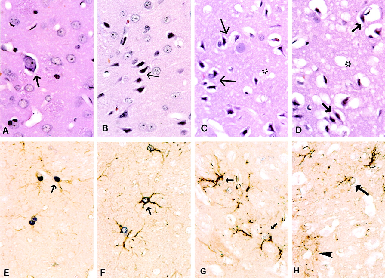
    Photomicrographs of neurons and astrocytes (original magnification ×160). A–D, Neuronal morphology indicated by hematoxylin-eosin staining, and E–H, astrocytic changes indicated by double staining with GFAP plus S-100 protein.

    A, Many normal neurons and one dark neuron with perineural vacuoles (arrow) are present in the contralateral nonischemic caudoputamen.

    B, At the end of 30 minutes of ischemia when ADC had initially decreased, some neurons show slightly shrunken perikarya and nuclei (arrow).

    C, After 90 minutes of reperfusion and recovery of ADC to normal, most neurons are moderately shrunken and surrounded by swollen cellular processes and are scalloped in appearance (arrows). Neuropil shows marked vacuolation (∗), indicative of swollen dendrites and astrocytic processes.

    D, After 12 hours of reperfusion and a subsequent or second decrease in ADC, most neurons are severely shrunken (arrows), and the neuropil shows extensive vacuolation, and possibly some vasogenic edema (∗).

    E, Dark purple reaction product demarcates S-100 protein in astrocytic nuclei; brown reaction product indicates GFAP in the cytoplasm and processes of astrocytes (arrow) in the contralateral nonischemic caudoputamen.

    F, At the end of 30 minutes of ischemia, astrocytes are moderately swollen (arrows).

    G, After 90 minutes of reperfusion, astrocytes are severely swollen (arrow) with less intranuclear S-100 reactivity and watery appearance.

    H, After 12 hours of reperfusion, astrocytes have begun to disintegrate and lose nearly all S-100 immunoreactivity within their nuclei (arrow). At this time, some GFAP has moved into the extracellular space (arrowhead).

  • Fig 4.
    • Download figure
    • Open in new tab
    • Download powerpoint
    Fig 4.

    Electron micrographs of neurons (original magnification ×3000) and astrocytes (original magnification ×4500).

    A and B, At the end of 30 minutes of ischemia. A, Shrunken neurons (N) with condensed nuclei and cytoplasm are surrounded by a ring of swollen astrocytic processes (a). B, Swollen astrocyte with clumps of heterochromatin around the edge of its nucleus and an abnormally wide rim of watery perikaryal cytoplasm, which contained a few swollen mitochondria (m).

    C and D, At the end of 90 minutes of reperfusion. C, Two markedly shrunken neurons with highly condensed nuclei and cytoplasm; these neurons are surrounded by severely swollen astrocytic processes (a). D, Greatly swollen astrocyte with extensive and watery perikaryal cytoplasm and a few contracted mitochondria (m).

    E and F, At the end of 12 hours of reperfusion. E, Two necrotic neurons show cytoplasmic and nuclear disintegration with marked chromatin clumping and discontinuous cellular membranes. F, An irreversibly injured astrocyte with breaks in the nuclear membrane (arrows), watery cytoplasm, and severely swollen mitochondria (M).

  • Fig 5.
    • Download figure
    • Open in new tab
    • Download powerpoint
    Fig 5.

    Bar graph indicates percentage of injured neurons in the ipsilateral ROI in the three groups (mean ± SD). Group A, at end of 30 minutes of MCAO; group B, at 90 minutes of reperfusion after 30 minutes of transient MCAO; and group C, at 12 hours of reperfusion after 30 minutes of transient MCAO.

  • Fig 6.
    • Download figure
    • Open in new tab
    • Download powerpoint
    Fig 6.

    Bar graph indicates combined area of expression of two astrocytic proteins, GFAP and nuclear S-100, in both ipsilateral and contralateral hemispheres of the three groups (mean ± SD). Group A, at end of 30 minutes of MCAO; group B, at 90 minutes of reperfusion after 30 minutes of transient MCAO; and group C, at 12 hours of reperfusion after 30 minutes of transient MCAO.

PreviousNext
Back to top

In this issue

American Journal of Neuroradiology: 23 (2)
American Journal of Neuroradiology
Vol. 23, Issue 2
1 Feb 2002
  • Table of Contents
  • Index by author
Advertisement
Print
Download PDF
Email Article

Thank you for your interest in spreading the word on American Journal of Neuroradiology.

NOTE: We only request your email address so that the person you are recommending the page to knows that you wanted them to see it, and that it is not junk mail. We do not capture any email address.

Enter multiple addresses on separate lines or separate them with commas.
Acute Postischemic Renormalization of the Apparent Diffusion Coefficient of Water is not Associated with Reversal of Astrocytic Swelling and Neuronal Shrinkage in Rats
(Your Name) has sent you a message from American Journal of Neuroradiology
(Your Name) thought you would like to see the American Journal of Neuroradiology web site.
CAPTCHA
This question is for testing whether or not you are a human visitor and to prevent automated spam submissions.
Cite this article
Fuhai Li, Kai-Feng Liu, Matthew D. Silva, Xiangjun Meng, Tibo Gerriets, Karl G. Helmer, Joseph D. Fenstermacher, Christopher H. Sotak, Marc Fisher
Acute Postischemic Renormalization of the Apparent Diffusion Coefficient of Water is not Associated with Reversal of Astrocytic Swelling and Neuronal Shrinkage in Rats
American Journal of Neuroradiology Feb 2002, 23 (2) 180-188;

Citation Manager Formats

  • BibTeX
  • Bookends
  • EasyBib
  • EndNote (tagged)
  • EndNote 8 (xml)
  • Medlars
  • Mendeley
  • Papers
  • RefWorks Tagged
  • Ref Manager
  • RIS
  • Zotero
0 Responses
Respond to this article
Share
Bookmark this article
Acute Postischemic Renormalization of the Apparent Diffusion Coefficient of Water is not Associated with Reversal of Astrocytic Swelling and Neuronal Shrinkage in Rats
Fuhai Li, Kai-Feng Liu, Matthew D. Silva, Xiangjun Meng, Tibo Gerriets, Karl G. Helmer, Joseph D. Fenstermacher, Christopher H. Sotak, Marc Fisher
American Journal of Neuroradiology Feb 2002, 23 (2) 180-188;
del.icio.us logo Twitter logo Facebook logo Mendeley logo
  • Tweet Widget
  • Facebook Like
  • Google Plus One

Jump to section

  • Article
    • Abstract
    • Methods
    • Results
    • Discussion
    • Conclusion
    • Acknowledgments
    • Footnotes
    • References
  • Figures & Data
  • Info & Metrics
  • Responses
  • References
  • PDF

Related Articles

  • No related articles found.
  • PubMed
  • Google Scholar

Cited By...

  • The Effect of Age and Cerebral Ischemia on Diffusion-Weighted Proton MR Spectroscopy of the Human Brain
  • Correlation between CT and Diffusion-Weighted Imaging of Acute Cerebral Ischemia in a Rat Model
  • Neurite beading is sufficient to decrease the apparent diffusion coefficient after ischemic stroke
  • Modest MRI Signal Intensity Changes Precede Delayed Cortical Necrosis After Transient Focal Ischemia in the Rat
  • Editorial Comment--ADC and Metabolites in Stroke: Even More Confusion About Diffusion?
  • Guidelines and Recommendations for Perfusion Imaging in Cerebral Ischemia: A Scientific Statement for Healthcare Professionals by the Writing Group on Perfusion Imaging, From the Council on Cardiovascular Radiology of the American Heart Association
  • Crossref
  • Google Scholar

This article has not yet been cited by articles in journals that are participating in Crossref Cited-by Linking.

More in this TOC Section

  • Usefulness of Quantitative Susceptibility Mapping for the Diagnosis of Parkinson Disease
  • White Matter Alterations in the Brains of Patients with Active, Remitted, and Cured Cushing Syndrome: A DTI Study
  • Qualitative and Quantitative Analysis of MR Imaging Findings in Patients with Middle Cerebral Artery Stroke Implanted with Mesenchymal Stem Cells
Show more BRAIN

Similar Articles

Advertisement

Indexed Content

  • Current Issue
  • Accepted Manuscripts
  • Article Preview
  • Past Issues
  • Editorials
  • Editor's Choice
  • Fellows' Journal Club
  • Letters to the Editor
  • Video Articles

Cases

  • Case Collection
  • Archive - Case of the Week
  • Archive - Case of the Month
  • Archive - Classic Case

Special Collections

  • AJNR Awards
  • ASNR Foundation Special Collection
  • Most Impactful AJNR Articles
  • Photon-Counting CT
  • Spinal CSF Leak Articles (Jan 2020-June 2024)

More from AJNR

  • Trainee Corner
  • Imaging Protocols
  • MRI Safety Corner

Multimedia

  • AJNR Podcasts
  • AJNR Scantastics

Resources

  • Turnaround Time
  • Submit a Manuscript
  • Submit a Video Article
  • Submit an eLetter to the Editor/Response
  • Manuscript Submission Guidelines
  • Statistical Tips
  • Fast Publishing of Accepted Manuscripts
  • Graphical Abstract Preparation
  • Imaging Protocol Submission
  • Evidence-Based Medicine Level Guide
  • Publishing Checklists
  • Author Policies
  • Become a Reviewer/Academy of Reviewers
  • News and Updates

About Us

  • About AJNR
  • Editorial Board
  • Editorial Board Alumni
  • Alerts
  • Permissions
  • Not an AJNR Subscriber? Join Now
  • Advertise with Us
  • Librarian Resources
  • Feedback
  • Terms and Conditions
  • AJNR Editorial Board Alumni

American Society of Neuroradiology

  • Not an ASNR Member? Join Now

© 2025 by the American Society of Neuroradiology All rights, including for text and data mining, AI training, and similar technologies, are reserved.
Print ISSN: 0195-6108 Online ISSN: 1936-959X

Powered by HighWire