Advertisement

Main menu

  • Home
  • Content
    • Current Issue
    • Accepted Manuscripts
    • Article Preview
    • Past Issue Archive
    • Video Articles
    • AJNR Case Collection
    • Case of the Week Archive
    • Case of the Month Archive
    • Classic Case Archive
  • Special Collections
    • AJNR Awards
    • ASNR Foundation Special Collection
    • Most Impactful AJNR Articles
    • Photon-Counting CT
    • Spinal CSF Leak Articles (Jan 2020-June 2024)
  • Multimedia
    • AJNR Podcasts
    • AJNR SCANtastic
    • Trainee Corner
    • MRI Safety Corner
    • Imaging Protocols
  • For Authors
    • Submit a Manuscript
    • Submit a Video Article
    • Submit an eLetter to the Editor/Response
    • Manuscript Submission Guidelines
    • Statistical Tips
    • Fast Publishing of Accepted Manuscripts
    • Graphical Abstract Preparation
    • Imaging Protocol Submission
    • Author Policies
  • About Us
    • About AJNR
    • Editorial Board
    • Editorial Board Alumni
  • More
    • Become a Reviewer/Academy of Reviewers
    • Subscribers
    • Permissions
    • Alerts
    • Feedback
    • Advertisers
    • ASNR Home

User menu

  • Alerts
  • Log in

Search

  • Advanced search
American Journal of Neuroradiology

American Journal of Neuroradiology

ASHNR American Society of Functional Neuroradiology ASHNR American Society of Pediatric Neuroradiology ASSR
  • Alerts
  • Log in

Advanced Search

  • Home
  • Content
    • Current Issue
    • Accepted Manuscripts
    • Article Preview
    • Past Issue Archive
    • Video Articles
    • AJNR Case Collection
    • Case of the Week Archive
    • Case of the Month Archive
    • Classic Case Archive
  • Special Collections
    • AJNR Awards
    • ASNR Foundation Special Collection
    • Most Impactful AJNR Articles
    • Photon-Counting CT
    • Spinal CSF Leak Articles (Jan 2020-June 2024)
  • Multimedia
    • AJNR Podcasts
    • AJNR SCANtastic
    • Trainee Corner
    • MRI Safety Corner
    • Imaging Protocols
  • For Authors
    • Submit a Manuscript
    • Submit a Video Article
    • Submit an eLetter to the Editor/Response
    • Manuscript Submission Guidelines
    • Statistical Tips
    • Fast Publishing of Accepted Manuscripts
    • Graphical Abstract Preparation
    • Imaging Protocol Submission
    • Author Policies
  • About Us
    • About AJNR
    • Editorial Board
    • Editorial Board Alumni
  • More
    • Become a Reviewer/Academy of Reviewers
    • Subscribers
    • Permissions
    • Alerts
    • Feedback
    • Advertisers
    • ASNR Home
  • Follow AJNR on Twitter
  • Visit AJNR on Facebook
  • Follow AJNR on Instagram
  • Join AJNR on LinkedIn
  • RSS Feeds

AJNR is seeking candidates for the AJNR Podcast Editor. Read the position description.

Research ArticleBRAIN

Prognostic Value of Subacute Crossed Cerebellar Diaschisis: Single-Photon Emission CT Study in Patients with Middle Cerebral Artery Territory Infarct

Masashi Takasawa, Manabu Watanabe, Shiro Yamamoto, Taku Hoshi, Tsutomu Sasaki, Kazuo Hashikawa, Masayasu Matsumoto and Naokazu Kinoshita
American Journal of Neuroradiology February 2002, 23 (2) 189-193;
Masashi Takasawa
  • Find this author on Google Scholar
  • Find this author on PubMed
  • Search for this author on this site
Manabu Watanabe
  • Find this author on Google Scholar
  • Find this author on PubMed
  • Search for this author on this site
Shiro Yamamoto
  • Find this author on Google Scholar
  • Find this author on PubMed
  • Search for this author on this site
Taku Hoshi
  • Find this author on Google Scholar
  • Find this author on PubMed
  • Search for this author on this site
Tsutomu Sasaki
  • Find this author on Google Scholar
  • Find this author on PubMed
  • Search for this author on this site
Kazuo Hashikawa
  • Find this author on Google Scholar
  • Find this author on PubMed
  • Search for this author on this site
Masayasu Matsumoto
  • Find this author on Google Scholar
  • Find this author on PubMed
  • Search for this author on this site
Naokazu Kinoshita
  • Find this author on Google Scholar
  • Find this author on PubMed
  • Search for this author on this site
  • Article
  • Figures & Data
  • Info & Metrics
  • Responses
  • References
  • PDF
Loading

Article Figures & Data

Figures

  • Tables
  • Fig 1.
    • Download figure
    • Open in new tab
    • Download powerpoint
    Fig 1.

    ROIs on a SPECT image of the cerebellum.

  • Fig 2.
    • Download figure
    • Open in new tab
    • Download powerpoint
    Fig 2.

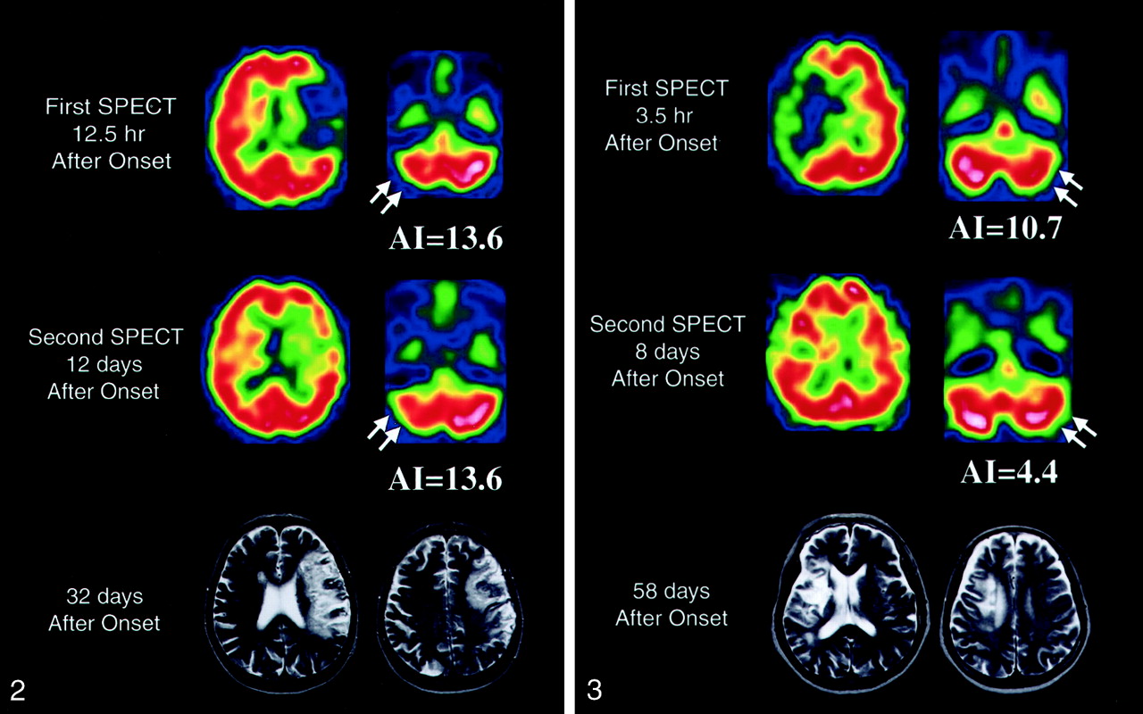
    SPECT images obtained in patient 1 show severe cerebellar hypoperfusion (arrows) in both the acute (top row) and subacute (middle row) stages. This patient had SSS scores of 13 at admission and 38 at 60 days after onset. MR images (bottom row) obtained at 32 days show a large infarct in the left hemisphere.

  • Fig 3.
    • Download figure
    • Open in new tab
    • Download powerpoint
    Fig 3.

    SPECT images obtained in patient 13 show cerebellar hypoperfusion (arrows) in both the acute (top row) and subacute (middle row) stages. This patient had SSS scores of 22 at admission and 55 at 60 days after onset. MR images (bottom row) obtained at 58 days show an infarct in the right basal ganglia and frontal lobe.

  • Fig 4.
    • Download figure
    • Open in new tab
    • Download powerpoint
    Fig 4.

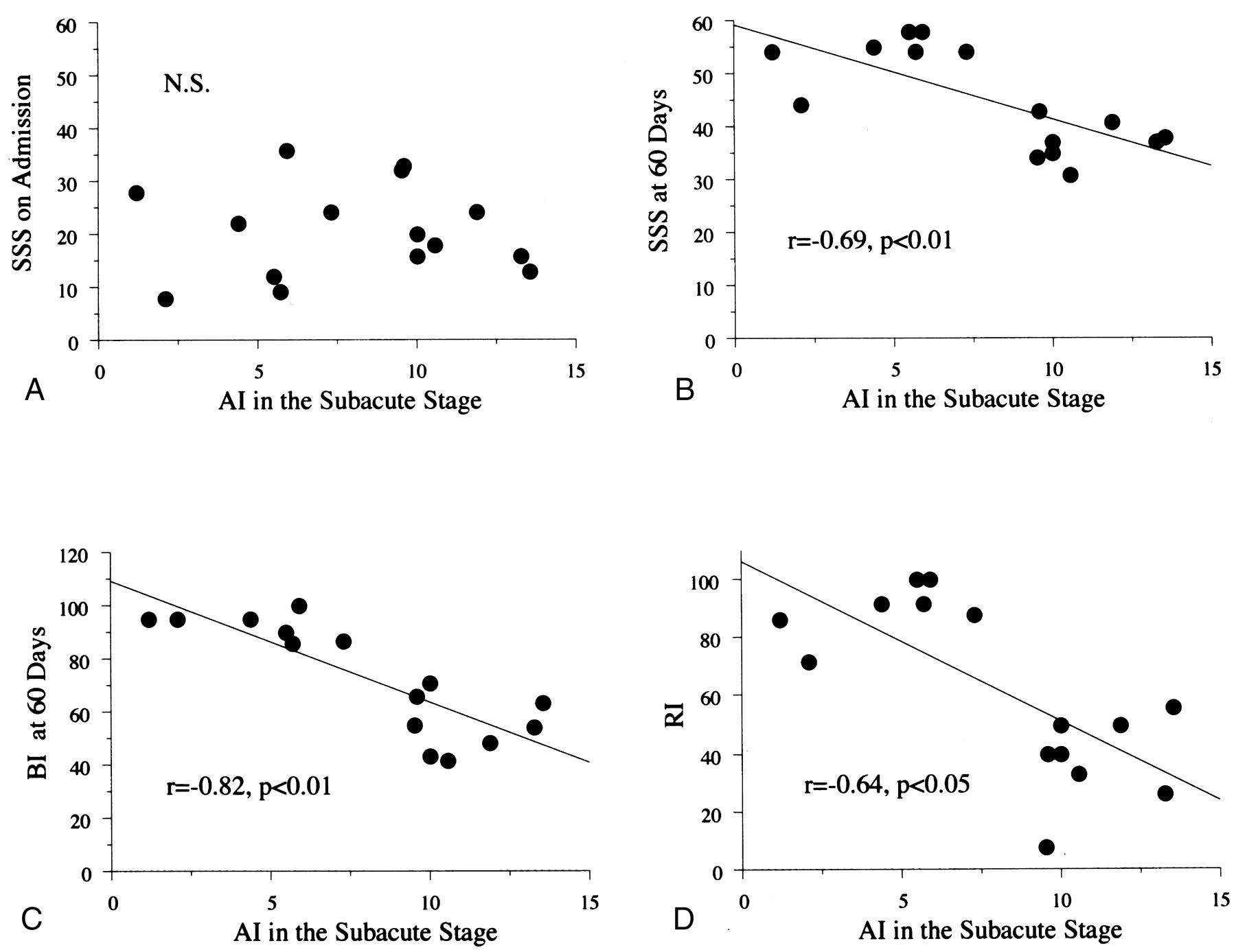
    Relationships between the AI in the subacute stage and neurologic state. No significant (N.S.) correlations between the AI in the subacute stage and SSS score at admission were found (top left). AI in the subacute stage was significantly associated with the final SSS score at 60 days (top right), the final BI score at 60 days (bottom left), and the RI (bottom right), as the results of the nonparametric Spearman rank test indicate.

Tables

  • Figures
    • View popup
    TABLE 1:

    Clinical characteristics and cerebellar AI

    Patient No./Age (y)/SexMCA SideInfarctAISSSRIBI at 60 Days
    Mechanism*Topography†AcuteAt 60 Days
    1/78/FLEF, T, P, d13.6133855.563
    2/72/MLEF, T, P13.3163726.254
    3/60/MREF, T, P11.9244150.048
    4/64/MLEF, T, P, d10.6186132.542
    5/81/MLAT, P10.0206539.543
    6/67/FLEF, T, P10.0166726.271
    7/88/MREF, T, P9.6334340.066
    8/75/MREP9.532347.755
    9/77/MLEF, d7.3245488.287
    10/59/FREd5.93658100100
    11/81/MLAP, d5.795491.886
    12/81/MLET, d5.5125810090
    13/67/FREF, d4.4225591.795
    14/72/MLAP, d2.184472.095
    15/82/MLEd1.2285486.795
    • * A indicates atherothrombotic; E, embolic.

    • † d indicates deep MCA territory; F, frontal; P, parietal; T, temporal.

PreviousNext
Back to top

In this issue

American Journal of Neuroradiology: 23 (2)
American Journal of Neuroradiology
Vol. 23, Issue 2
1 Feb 2002
  • Table of Contents
  • Index by author
Advertisement
Print
Download PDF
Email Article

Thank you for your interest in spreading the word on American Journal of Neuroradiology.

NOTE: We only request your email address so that the person you are recommending the page to knows that you wanted them to see it, and that it is not junk mail. We do not capture any email address.

Enter multiple addresses on separate lines or separate them with commas.
Prognostic Value of Subacute Crossed Cerebellar Diaschisis: Single-Photon Emission CT Study in Patients with Middle Cerebral Artery Territory Infarct
(Your Name) has sent you a message from American Journal of Neuroradiology
(Your Name) thought you would like to see the American Journal of Neuroradiology web site.
CAPTCHA
This question is for testing whether or not you are a human visitor and to prevent automated spam submissions.
Cite this article
Masashi Takasawa, Manabu Watanabe, Shiro Yamamoto, Taku Hoshi, Tsutomu Sasaki, Kazuo Hashikawa, Masayasu Matsumoto, Naokazu Kinoshita
Prognostic Value of Subacute Crossed Cerebellar Diaschisis: Single-Photon Emission CT Study in Patients with Middle Cerebral Artery Territory Infarct
American Journal of Neuroradiology Feb 2002, 23 (2) 189-193;

Citation Manager Formats

  • BibTeX
  • Bookends
  • EasyBib
  • EndNote (tagged)
  • EndNote 8 (xml)
  • Medlars
  • Mendeley
  • Papers
  • RefWorks Tagged
  • Ref Manager
  • RIS
  • Zotero
0 Responses
Respond to this article
Share
Bookmark this article
Prognostic Value of Subacute Crossed Cerebellar Diaschisis: Single-Photon Emission CT Study in Patients with Middle Cerebral Artery Territory Infarct
Masashi Takasawa, Manabu Watanabe, Shiro Yamamoto, Taku Hoshi, Tsutomu Sasaki, Kazuo Hashikawa, Masayasu Matsumoto, Naokazu Kinoshita
American Journal of Neuroradiology Feb 2002, 23 (2) 189-193;
del.icio.us logo Twitter logo Facebook logo Mendeley logo
  • Tweet Widget
  • Facebook Like
  • Google Plus One

Jump to section

  • Article
    • Abstract
    • Methods
    • Results
    • Discussion
    • Conclusion
    • Acknowledgments
    • References
  • Figures & Data
  • Info & Metrics
  • Responses
  • References
  • PDF

Related Articles

  • No related articles found.
  • PubMed
  • Google Scholar

Cited By...

  • Electrophysiological Correlates of Dentate Nucleus Deep Brain Stimulation for Poststroke Motor Recovery
  • Cortico-Cerebellar Connectivity Underlying Motor Control in Chronic Poststroke Individuals
  • Reader response: BOLD cerebrovascular reactivity as a novel marker for crossed cerebellar diaschisis
  • BOLD cerebrovascular reactivity as a novel marker for crossed cerebellar diaschisis
  • Cerebellar Hypoperfusion in Migraine Attack: Incidence and Significance
  • Correlation of Asymmetry Indices Measured by Arterial Spin-Labeling MR Imaging and SPECT in Patients with Crossed Cerebellar Diaschisis
  • Chronic Deep Cerebellar Stimulation Promotes Long-Term Potentiation, Microstructural Plasticity, and Reorganization of Perilesional Cortical Representation in a Rodent Model
  • Cerebellar Atrophy in Childhood Arterial Ischemic Stroke: Acute Diffusion MRI Biomarkers
  • Crossed Cerebellar Diaschisis in Acute Stroke Detected by Dynamic Susceptibility Contrast MR Perfusion Imaging
  • Crossref
  • Google Scholar

This article has not yet been cited by articles in journals that are participating in Crossref Cited-by Linking.

More in this TOC Section

  • Usefulness of Quantitative Susceptibility Mapping for the Diagnosis of Parkinson Disease
  • White Matter Alterations in the Brains of Patients with Active, Remitted, and Cured Cushing Syndrome: A DTI Study
  • Qualitative and Quantitative Analysis of MR Imaging Findings in Patients with Middle Cerebral Artery Stroke Implanted with Mesenchymal Stem Cells
Show more BRAIN

Similar Articles

Advertisement

Indexed Content

  • Current Issue
  • Accepted Manuscripts
  • Article Preview
  • Past Issues
  • Editorials
  • Editor's Choice
  • Fellows' Journal Club
  • Letters to the Editor
  • Video Articles

Cases

  • Case Collection
  • Archive - Case of the Week
  • Archive - Case of the Month
  • Archive - Classic Case

Special Collections

  • AJNR Awards
  • ASNR Foundation Special Collection
  • Most Impactful AJNR Articles
  • Photon-Counting CT
  • Spinal CSF Leak Articles (Jan 2020-June 2024)

More from AJNR

  • Trainee Corner
  • Imaging Protocols
  • MRI Safety Corner

Multimedia

  • AJNR Podcasts
  • AJNR Scantastics

Resources

  • Turnaround Time
  • Submit a Manuscript
  • Submit a Video Article
  • Submit an eLetter to the Editor/Response
  • Manuscript Submission Guidelines
  • Statistical Tips
  • Fast Publishing of Accepted Manuscripts
  • Graphical Abstract Preparation
  • Imaging Protocol Submission
  • Evidence-Based Medicine Level Guide
  • Publishing Checklists
  • Author Policies
  • Become a Reviewer/Academy of Reviewers
  • News and Updates

About Us

  • About AJNR
  • Editorial Board
  • Editorial Board Alumni
  • Alerts
  • Permissions
  • Not an AJNR Subscriber? Join Now
  • Advertise with Us
  • Librarian Resources
  • Feedback
  • Terms and Conditions
  • AJNR Editorial Board Alumni

American Society of Neuroradiology

  • Not an ASNR Member? Join Now

© 2025 by the American Society of Neuroradiology All rights, including for text and data mining, AI training, and similar technologies, are reserved.
Print ISSN: 0195-6108 Online ISSN: 1936-959X

Powered by HighWire