
Article Figures & Data
Figures
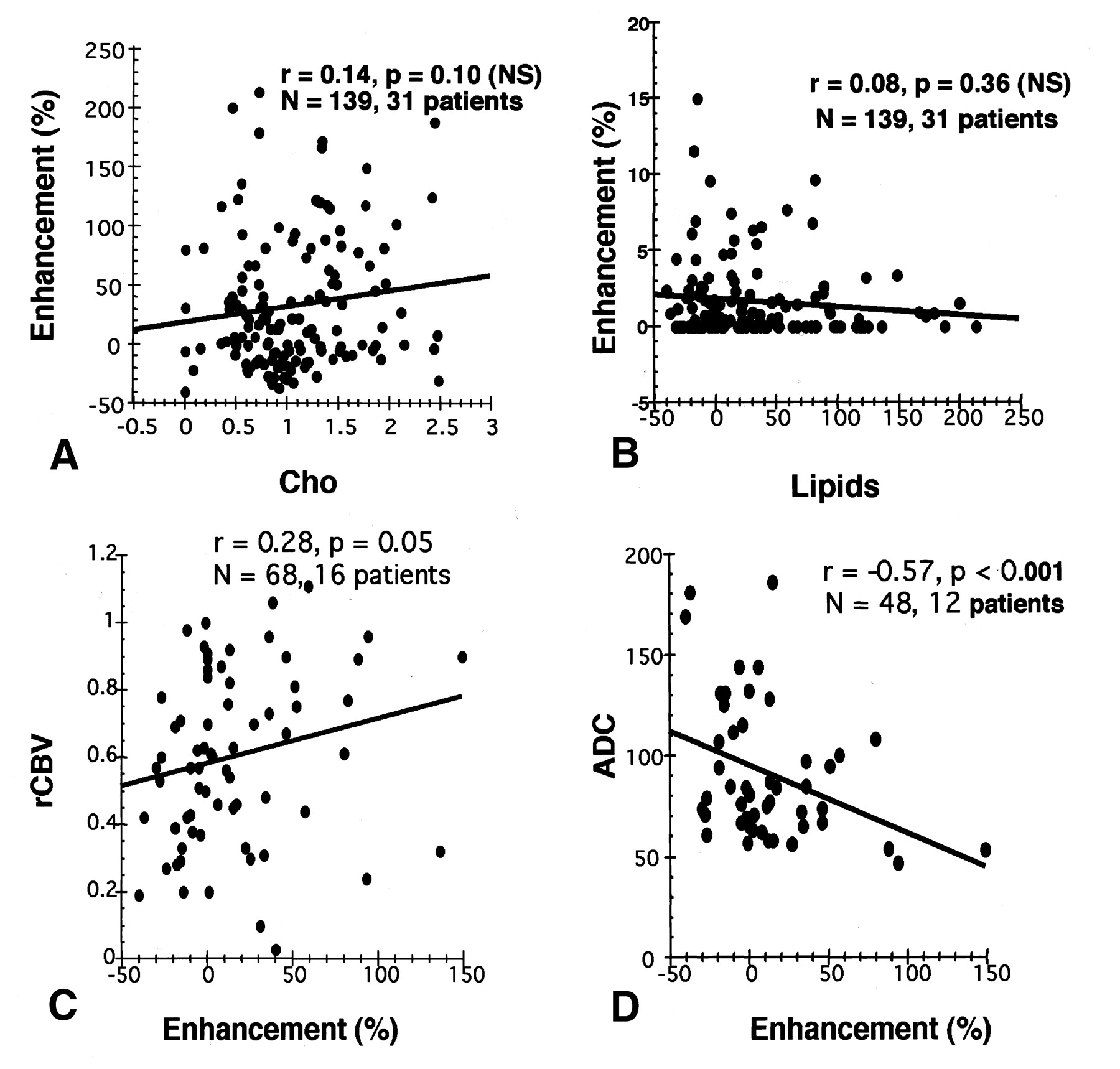
- Fig 1.
Plots show the relationships between percent enhancement and Cho (A), lipids (B), rCBV (C), and ADC (D). The number of measurements (N) and the number of patients are shown. The fitted regression line, Pearson correlation r, and P values are also shown.
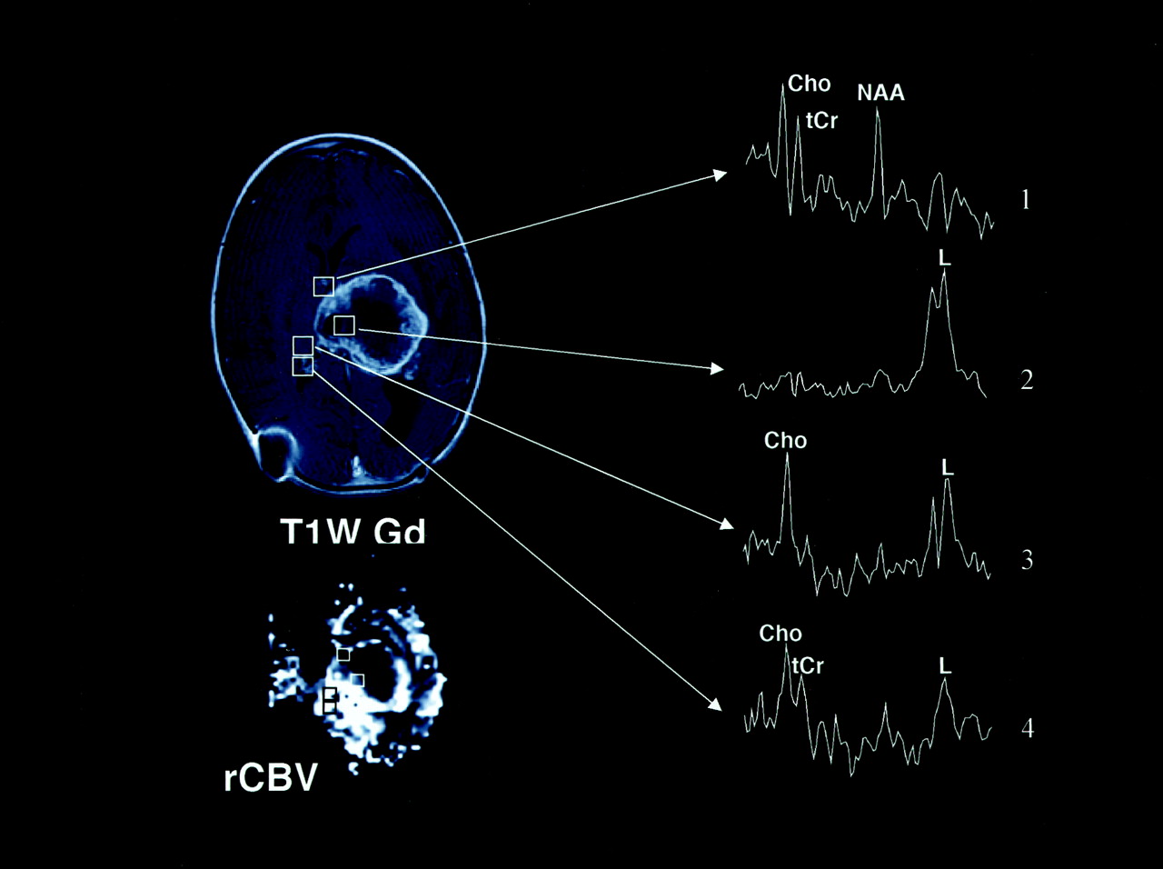
- Fig 2.
Axial T2-weighted (T2W), T1-weighted Gd-enhanced (T1W Gd), hemodynamic (rCBV), and diffusion (ADC) MR images and selected proton MR spectra (1–5) from a multivoxel spectroscopic data set in a 6-month-old male infant with a choroid plexus carcinoma. The lesion contains a large central area of low or normal signal intensity on the T2-weighted image, and it has inhomogeneous intense enhancement on the T1-weighted Gd-enhanced image, with nonenhancing areas that represent necrotic and/or cystic degeneration. The rCBV image shows increased perfusion (bright regions) in the areas of enhancement, whereas the corresponding areas on the ADC image appear hypointense. Selected proton MR spectra (1–4) show high Cho peaks, which are believed to correspond to areas of viable tumor. MR spectra 1 and 2 correspond to areas of low or normal signal intensity on the T2-weighted image that do not enhance on the T1-weighted; theses areas appear hypointense on both rCBV and ADC images. MR spectra 3 and 4 correspond to hypointense areas on the T2-weighted image that enhance; these appear hyperintense and hypointense on rCBV and ADC images. MR spectrum 5 shows only a high lipid (L) value, which is thought to indicate necrosis; this finding corresponds to a hypointense region on the T2-weighted image that does not enhance on the T1-weighted Gd-enhanced and rCBV images and appears hyperintense on the ADC image. High lipid values are also shown on MR spectra 3 and 4, in addition to high Cho values; this pattern indicates a mixture of viable tumor and necrosis. The Figure illustrates a positive relationship between Cho and rCBV values and an inverse relationship between Cho and ADC values. It shows that Cho (or active tumor) was detected in enhancing regions of the tumor and beyond.
- Fig 3.
Axial FLAIR, T2-weighted (T2W), and T1-weighted Gd-enhanced (T1WGd) MR images and selected proton MR spectra from a multivoxel MR spectroscopic data set in a 10-year-old girl with a cerebellar tumor. The lesion appears inhomogeneously hyperintense on the FLAIR and T2-weighted images and is not enhancing on the T1-weighted Gd-enhanced image. Prominent peaks corresponding to Cho are detected. Also, tCr and NAA peaks are occasionally detected. The Figure illustrates that no relationship existed between Cho detection and contrast enhancement on T1-weighted Gd-enhanced images.
- Fig 4.
Axial T2-weighted (T2W), T1-weighted Gd-enhanced (T1W Gd), hemodynamic (rCBV), diffusion (ADC) MR images and selected proton MR spectra (MRSI) from a multivoxel MR spectroscopic data set in a 16-year-old male adolescent with an enlarging inoperable brainstem lesion that was identified after the acute onset of left-sided nerve palsy. The lesion with high signal intensity on the T2-weighted image appears hyperintense on the T1-weighted Gd-enhanced and ADC images, and it is hypointense on the rCBV image. Multivoxel proton MR spectra (TE, 65 milliseconds) show a large lipid peak, in addition to Cho and tCr peaks, within the central portion of the mass; in the anterior voxel, the same peak was absent. These findings suggest that the lesion has a high neoplastic potential because increased lipid levels represent necrosis. Findings on both rCBV and ADC images are not consistent with the MR spectroscopic findings.
- Fig 5.
Images obtained at the first follow-up evaluation (after radiation therapy and chemotherapy) include an axial T1-weighted Gd-enhanced MR image and multivoxel MR spectra in a male patient with an anaplastic (malignant) astrocytoma in the left thalamus. Selected proton MR spectra (1–8) illustrate that tumor spectral patterns were detected beyond the contrast-enhancing region and that regions of tumor corresponded to necrotic spectral patterns. Spectrum 1 shows a pattern that is characteristic of normal tissue, because all three biologically important metabolites (NAA, Cho, tCr) are detected. The voxel corresponding to spectrum 2 includes enhancing tissue and shows increased Cho and prominent NAA levels; this spectral pattern is characteristic of mixed tumor and normal tissue. Spectrum 3 shows a spectral pattern with only Cho and lipids (L) values; this is consistent with tumor and necrosis and corresponds to both enhancing and nonenhancing tissue. Spectrum 4 shows extensive necrosis (prominent lipids), along with some normal and tumor tissue (Cho and NAA peaks). Spectrum 5 shows primarily necrosis (predominantly lipid) and, possibly, some tumor. Voxels corresponding to spectra 6 and 7 show minimal enhancement, whereas the voxel that corresponds to spectrum 8 includes areas of enhancement. Spectra 6–8 show tumor spectral patterns (prominent Cho and tCr) and correspond to regions of contrast enhancement (spectrum 8) and beyond (spectra 6 and 7).
- Fig 6.
Images obtained at the second follow-up evaluation (1 month later) in the patient in Figure 5 include conventional axial T1-weighted Gd-enhanced (T1W Gd) and hemodynamic (rCBV) MR images and multivoxel MR spectroscopic data. The increased size of the mass and mass effect implied tumor progression (Fig 5). Note that regions with high Cho levels that did not enhance in Figure 5 (spectra 6 and 7) are now enhancing. Neuropathologic analysis of stereotactic biopsy samples obtained from these regions confirmed the presence of tumor. Spectra 1–4 illustrate that hypointense regions of tumor corresponding mostly to normal tissue (spectrum 1) and central necrosis (spectrum 2). The high rCBV level corresponds to the ring of enhancement and regions of high Cho levels that are detected beyond the contrast-enhancing region (spectra 3 and 4). The complementary value of multivoxel MR spectroscopic imaging and hemodynamic MR imaging is illustrated.


In this issue


Jump to section
Related Articles
Cited By...
- Intravoxel Incoherent Motion MR Imaging of Pediatric Intracranial Tumors: Correlation with Histology and Diagnostic Utility
- Comparison of Perfusion, Diffusion, and MR Spectroscopy between Low-Grade Enhancing Pilocytic Astrocytomas and High-Grade Astrocytomas
- Clinical applications of imaging biomarkers. Part 1. The neuroradiologist's perspective
- Correlation of MR Relative Cerebral Blood Volume Measurements with Cellular Density and Proliferation in High-Grade Gliomas: An Image-Guided Biopsy Study
- Value and Limitations of Diffusion-Weighted Imaging in Grading and Diagnosis of Pediatric Posterior Fossa Tumors
- Magnetic Resonance As a Cancer Imaging Biomarker
- Noninvasive Magnetic Resonance Spectroscopic Imaging Biomarkers to Predict the Clinical Grade of Pediatric Brain Tumors
- Dynamic Magnetic Resonance Perfusion Imaging of Brain Tumors