Advertisement

Main menu

  • Home
  • Content
    • Current Issue
    • Accepted Manuscripts
    • Article Preview
    • Past Issue Archive
    • Video Articles
    • AJNR Case Collection
    • Case of the Week Archive
    • Case of the Month Archive
    • Classic Case Archive
  • Special Collections
    • AJNR Awards
    • ASNR Foundation Special Collection
    • Most Impactful AJNR Articles
    • Photon-Counting CT
    • Spinal CSF Leak Articles (Jan 2020-June 2024)
  • Multimedia
    • AJNR Podcasts
    • AJNR SCANtastic
    • Trainee Corner
    • MRI Safety Corner
    • Imaging Protocols
  • For Authors
    • Submit a Manuscript
    • Submit a Video Article
    • Submit an eLetter to the Editor/Response
    • Manuscript Submission Guidelines
    • Statistical Tips
    • Fast Publishing of Accepted Manuscripts
    • Graphical Abstract Preparation
    • Imaging Protocol Submission
    • Author Policies
  • About Us
    • About AJNR
    • Editorial Board
    • Editorial Board Alumni
  • More
    • Become a Reviewer/Academy of Reviewers
    • Subscribers
    • Permissions
    • Alerts
    • Feedback
    • Advertisers
    • ASNR Home

User menu

  • Alerts
  • Log in

Search

  • Advanced search
American Journal of Neuroradiology

American Journal of Neuroradiology

ASHNR American Society of Functional Neuroradiology ASHNR American Society of Pediatric Neuroradiology ASSR
  • Alerts
  • Log in

Advanced Search

  • Home
  • Content
    • Current Issue
    • Accepted Manuscripts
    • Article Preview
    • Past Issue Archive
    • Video Articles
    • AJNR Case Collection
    • Case of the Week Archive
    • Case of the Month Archive
    • Classic Case Archive
  • Special Collections
    • AJNR Awards
    • ASNR Foundation Special Collection
    • Most Impactful AJNR Articles
    • Photon-Counting CT
    • Spinal CSF Leak Articles (Jan 2020-June 2024)
  • Multimedia
    • AJNR Podcasts
    • AJNR SCANtastic
    • Trainee Corner
    • MRI Safety Corner
    • Imaging Protocols
  • For Authors
    • Submit a Manuscript
    • Submit a Video Article
    • Submit an eLetter to the Editor/Response
    • Manuscript Submission Guidelines
    • Statistical Tips
    • Fast Publishing of Accepted Manuscripts
    • Graphical Abstract Preparation
    • Imaging Protocol Submission
    • Author Policies
  • About Us
    • About AJNR
    • Editorial Board
    • Editorial Board Alumni
  • More
    • Become a Reviewer/Academy of Reviewers
    • Subscribers
    • Permissions
    • Alerts
    • Feedback
    • Advertisers
    • ASNR Home
  • Follow AJNR on Twitter
  • Visit AJNR on Facebook
  • Follow AJNR on Instagram
  • Join AJNR on LinkedIn
  • RSS Feeds

AJNR is seeking candidates for the AJNR Podcast Editor. Read the position description.

Research ArticleHEAD AND NECK

Comparative Study of MR Sialography and Digital Subtraction Sialography for Benign Salivary Gland Disorders

Marc Kalinowski, Johannes T. Heverhagen, Elisabeth Rehberg, Klaus Jochen Klose and Hans-Joachim Wagner
American Journal of Neuroradiology October 2002, 23 (9) 1485-1492;
Marc Kalinowski
  • Find this author on Google Scholar
  • Find this author on PubMed
  • Search for this author on this site
Johannes T. Heverhagen
  • Find this author on Google Scholar
  • Find this author on PubMed
  • Search for this author on this site
Elisabeth Rehberg
  • Find this author on Google Scholar
  • Find this author on PubMed
  • Search for this author on this site
Klaus Jochen Klose
  • Find this author on Google Scholar
  • Find this author on PubMed
  • Search for this author on this site
Hans-Joachim Wagner
  • Find this author on Google Scholar
  • Find this author on PubMed
  • Search for this author on this site
  • Article
  • Figures & Data
  • Info & Metrics
  • Responses
  • References
  • PDF
Loading

Article Figures & Data

Figures

  • Tables
  • Fig 1.
    • Download figure
    • Open in new tab
    • Download powerpoint
    Fig 1.

    A and B, Normal heavily T2-weighted turbo spin-echo (2800/1100 ms, flip angle 150°, acquisition time 7 seconds) MR sialograms obtained before (A) and after (B) salivation stimulation in a patient with status post acute parotiditis. Stensen duct (arrowheads in B) is better delineated after stimulation of salivation with a lemon mouth swab than before salivation stimulation.

  • Fig 2.
    • Download figure
    • Open in new tab
    • Download powerpoint
    Fig 2.

    59-year-old man with a history of chronic sialadenitis of the left parotid gland.

    A, Lateral digital subtraction sialogram shows multiple strictures, sialectasis, and prestenotic dilatation of the Stensen duct (arrows) and also of secondary and tertiary branching intraglandular ducts (arrowheads).

    B, Lateral MR sialogram shows the same abnormal findings but at lesser spatial resolution. Subtle strictures are more difficult to visualize and sialectasis is not as prominent (arrow). The enlargement of the ductal system is demonstrated up to secondary branching ducts.

  • Fig 3.
    • Download figure
    • Open in new tab
    • Download powerpoint
    Fig 3.

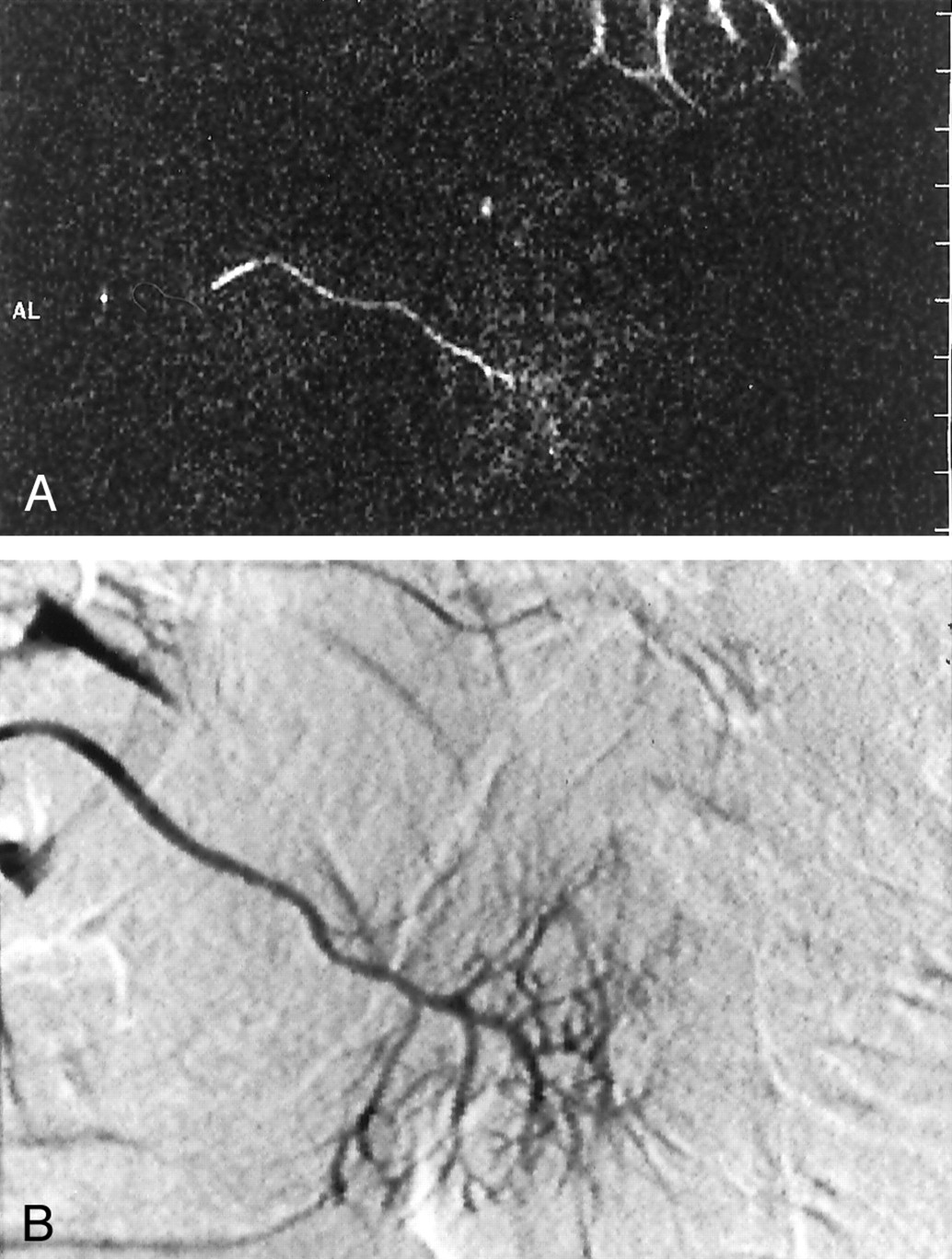
    61-year-old woman with status post acute sialadenitis.

    A and B, Oblique sagittal-coronal MR sialogram (A) and lateral digital subtraction sialogram (B) reveal normal ductal anatomy without abnormal findings. However, due to higher spatial resolution, the digital subtraction sialogram shows better delineation of ductal morphology and enables visualization of peripheral intraglandular ductal branches. Because of its normal small size, the Stensen duct shows some focal signal voids in A (compare with the enlarged duct in Fig 2B).

  • Fig 4.
    • Download figure
    • Open in new tab
    • Download powerpoint
    Fig 4.

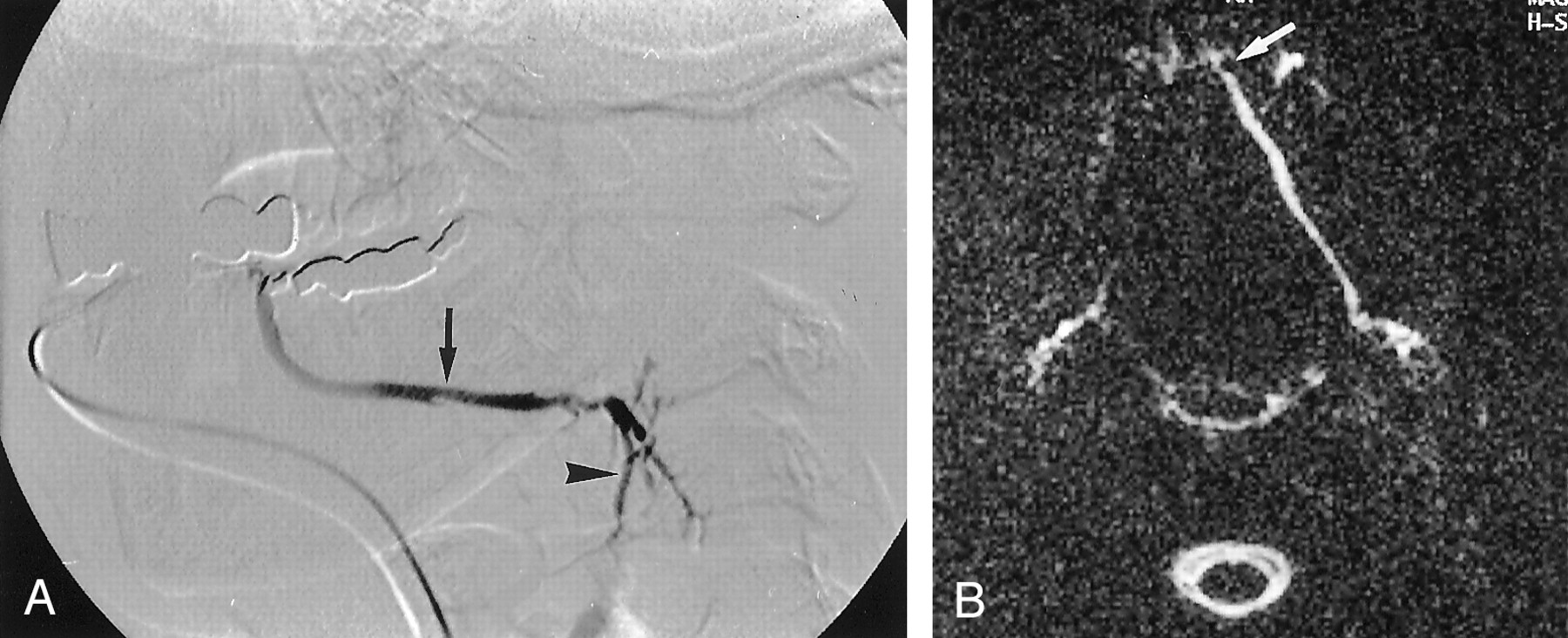
    64-year-old man with a large sialith in the main parotid duct.

    A, Lateral digital subtraction sialogram depicts a solid calculus in the proximal portion of the Stensen duct (arrow) and a prestenotic dilatation of intraglandular ductal structures (arrowheads). However, because of the near total obstruction of the main duct, visualization of prestenotic intraglandular structures is limited.

    B, MR sialogram (oblique sagittal to coronal view) compares favorably with the digital subtraction sialogram and shows a filling defect of the proximal main parotid duct (arrow) together with prestenotic dilatation of intraglandular ducts. However, because of surrounding fluid, the single calculus on the MR sialogram could be misinterpreted as two separate stones. This patient was successfully treated with extracorporal lithotripsy.

  • Fig 5.
    • Download figure
    • Open in new tab
    • Download powerpoint
    Fig 5.

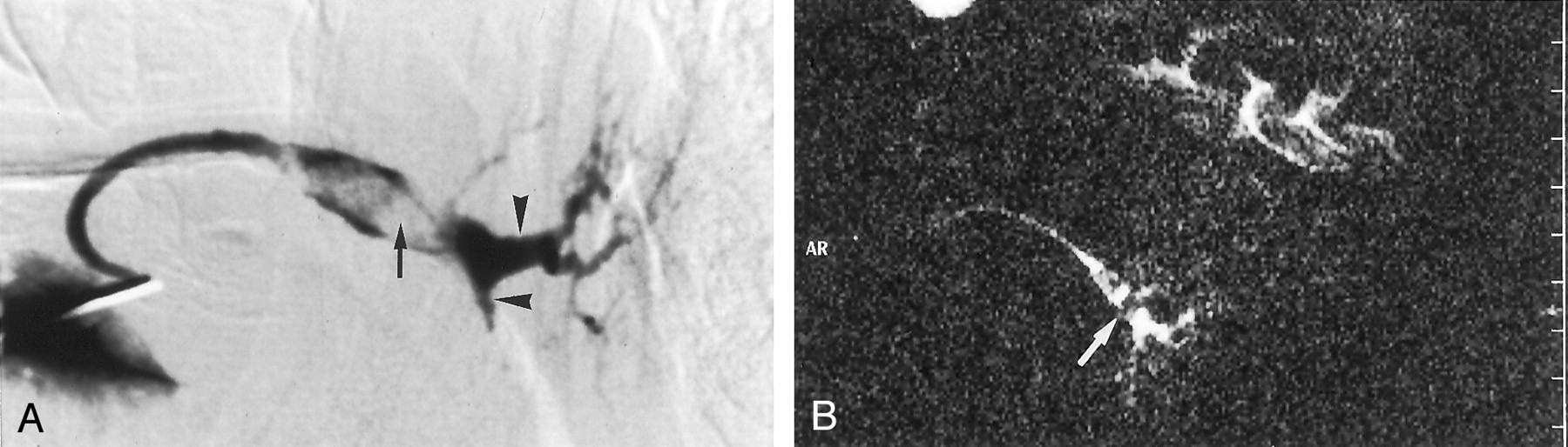
    36-year-old patient suspected to have sialolithiasis.

    A, Lateral digital subtraction sialogram shows a slightly dilated Wharton duct (arrow) and sialectasis in secondary duct branches (arrowhead). No filling defect in the main duct is seen.

    B, Transverse MR sialogram shows a filling defect near the orifice of the left Wharton duct (arrow). Note also the markedly dilated left Wharton duct compared with the right side. Patient underwent surgery, and a sialith was confirmed. The false-negative digital subtraction sialographic result may be due to dental hardware or to the fact that the sialographic catheter already passed the distal stone.

  • Fig 6.
    • Download figure
    • Open in new tab
    • Download powerpoint
    Fig 6.

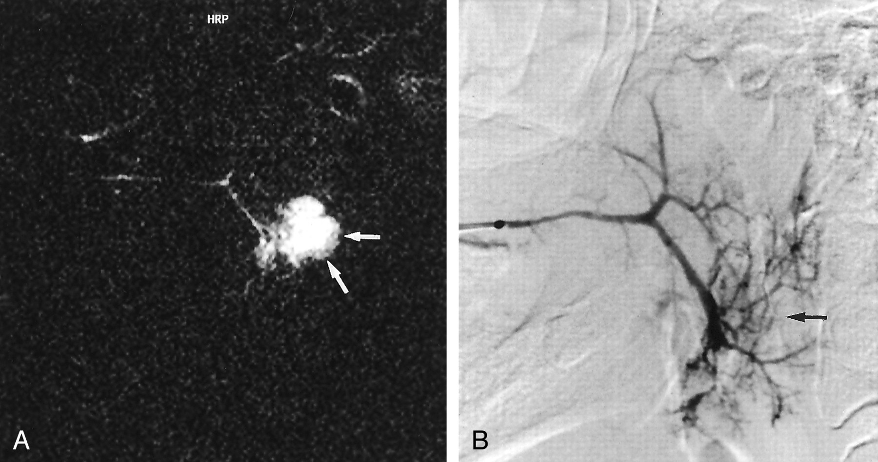
    55-year-old patient suspected to have an intraparenchymal cyst.

    A, Oblique sagittal-coronal MR sialogram shows a fluid-filled cystic mass lesion (arrows) in the lower parotid parenchyma.

    B, Lateral digital subtraction sialogram shows no connection between the ductal structures and the cyst. However, displacement of the lower pole ducts is depicted (arrow).

Tables

  • Figures
    • View popup
    TABLE 1:

    Diagnostic Discrimination for MR Sialography and Digital Subtraction Sialography for the Three Most Frequent Diagnoses

    Performance MeasureChronic SialadenitisAcute SialadenitisSialolithiasis
    MRSDSSMRSDSSMRSDSS
    Sensitivity (%)70961001008090
    Specificity (%)981001001009898
    PPV (%)951001001008990
    NPV (%)86981001009798
    • Note.—MRS indicates MR sialography; DSS, digital subtraction sialography; PPV, positive predictive value; NPV, negative predictive value.

    • View popup
    TABLE 2:

    Diagnostic Discrimination for MR Sialography and Digital Subtraction Sialography for the Three Most Frequent Diagnoses Stratified for the Investigated Gland

    Performance MeasureChronic SialadenitisAcute SialadenitisSialolithiasis
    MRSDSSMRSDSSMRSDSS
    Parotid gland
     Sensitivity (%)79100100100100100
     Specificity (%)9610010010098100
     PPV (%)7910010010075100
     NPV (%)88100100100100100
    Submandibular gland
     Sensitivity (%)50881001007186
     Specificity (%)100100100100100100
     PPV (%)100100100100100100
     NPV (%)86961001009396
    • Note.—MRS indicates MR sialography; DSS, digital subtraction sialography; PPV, positive predictive value; NPV, negative predictive value.

PreviousNext
Back to top

In this issue

American Journal of Neuroradiology: 23 (9)
American Journal of Neuroradiology
Vol. 23, Issue 9
1 Oct 2002
  • Table of Contents
  • Index by author
Advertisement
Print
Download PDF
Email Article

Thank you for your interest in spreading the word on American Journal of Neuroradiology.

NOTE: We only request your email address so that the person you are recommending the page to knows that you wanted them to see it, and that it is not junk mail. We do not capture any email address.

Enter multiple addresses on separate lines or separate them with commas.
Comparative Study of MR Sialography and Digital Subtraction Sialography for Benign Salivary Gland Disorders
(Your Name) has sent you a message from American Journal of Neuroradiology
(Your Name) thought you would like to see the American Journal of Neuroradiology web site.
CAPTCHA
This question is for testing whether or not you are a human visitor and to prevent automated spam submissions.
Cite this article
Marc Kalinowski, Johannes T. Heverhagen, Elisabeth Rehberg, Klaus Jochen Klose, Hans-Joachim Wagner
Comparative Study of MR Sialography and Digital Subtraction Sialography for Benign Salivary Gland Disorders
American Journal of Neuroradiology Oct 2002, 23 (9) 1485-1492;

Citation Manager Formats

  • BibTeX
  • Bookends
  • EasyBib
  • EndNote (tagged)
  • EndNote 8 (xml)
  • Medlars
  • Mendeley
  • Papers
  • RefWorks Tagged
  • Ref Manager
  • RIS
  • Zotero
0 Responses
Respond to this article
Share
Bookmark this article
Comparative Study of MR Sialography and Digital Subtraction Sialography for Benign Salivary Gland Disorders
Marc Kalinowski, Johannes T. Heverhagen, Elisabeth Rehberg, Klaus Jochen Klose, Hans-Joachim Wagner
American Journal of Neuroradiology Oct 2002, 23 (9) 1485-1492;
del.icio.us logo Twitter logo Facebook logo Mendeley logo
  • Tweet Widget
  • Facebook Like
  • Google Plus One

Jump to section

  • Article
    • Abstract
    • Methods
    • Results
    • Discussion
    • References
  • Figures & Data
  • Info & Metrics
  • Responses
  • References
  • PDF

Related Articles

  • No related articles found.
  • PubMed
  • Google Scholar

Cited By...

  • MR Imaging of the Extracranial Facial Nerve with the CISS Sequence
  • Salivary gland swellings
  • Radiation-Induced Xerostomia: Objective Evaluation of Salivary Gland Injury Using MR Sialography
  • MR sialography: the effect of a sialogogue and ductal occlusion in volunteers
  • Lipiodol ultra-fluid - foreign body in the cheek
  • Salivary duct strictures: nature and incidence in benign salivary obstruction
  • Crossref
  • Google Scholar

This article has not yet been cited by articles in journals that are participating in Crossref Cited-by Linking.

More in this TOC Section

  • Correlation of Apparent Diffusion Coefficient at 3T with Prognostic Parameters of Retinoblastoma
  • Parathyroid Lesions: Characterization with Dual-Phase Arterial and Venous Enhanced CT of the Neck
  • Kimura Disease: CT and MR Imaging Findings
Show more HEAD AND NECK

Similar Articles

Advertisement

Indexed Content

  • Current Issue
  • Accepted Manuscripts
  • Article Preview
  • Past Issues
  • Editorials
  • Editor's Choice
  • Fellows' Journal Club
  • Letters to the Editor
  • Video Articles

Cases

  • Case Collection
  • Archive - Case of the Week
  • Archive - Case of the Month
  • Archive - Classic Case

Special Collections

  • AJNR Awards
  • ASNR Foundation Special Collection
  • Most Impactful AJNR Articles
  • Photon-Counting CT
  • Spinal CSF Leak Articles (Jan 2020-June 2024)

More from AJNR

  • Trainee Corner
  • Imaging Protocols
  • MRI Safety Corner

Multimedia

  • AJNR Podcasts
  • AJNR Scantastics

Resources

  • Turnaround Time
  • Submit a Manuscript
  • Submit a Video Article
  • Submit an eLetter to the Editor/Response
  • Manuscript Submission Guidelines
  • Statistical Tips
  • Fast Publishing of Accepted Manuscripts
  • Graphical Abstract Preparation
  • Imaging Protocol Submission
  • Evidence-Based Medicine Level Guide
  • Publishing Checklists
  • Author Policies
  • Become a Reviewer/Academy of Reviewers
  • News and Updates

About Us

  • About AJNR
  • Editorial Board
  • Editorial Board Alumni
  • Alerts
  • Permissions
  • Not an AJNR Subscriber? Join Now
  • Advertise with Us
  • Librarian Resources
  • Feedback
  • Terms and Conditions
  • AJNR Editorial Board Alumni

American Society of Neuroradiology

  • Not an ASNR Member? Join Now

© 2025 by the American Society of Neuroradiology All rights, including for text and data mining, AI training, and similar technologies, are reserved.
Print ISSN: 0195-6108 Online ISSN: 1936-959X

Powered by HighWire