
Article Figures & Data
Figures
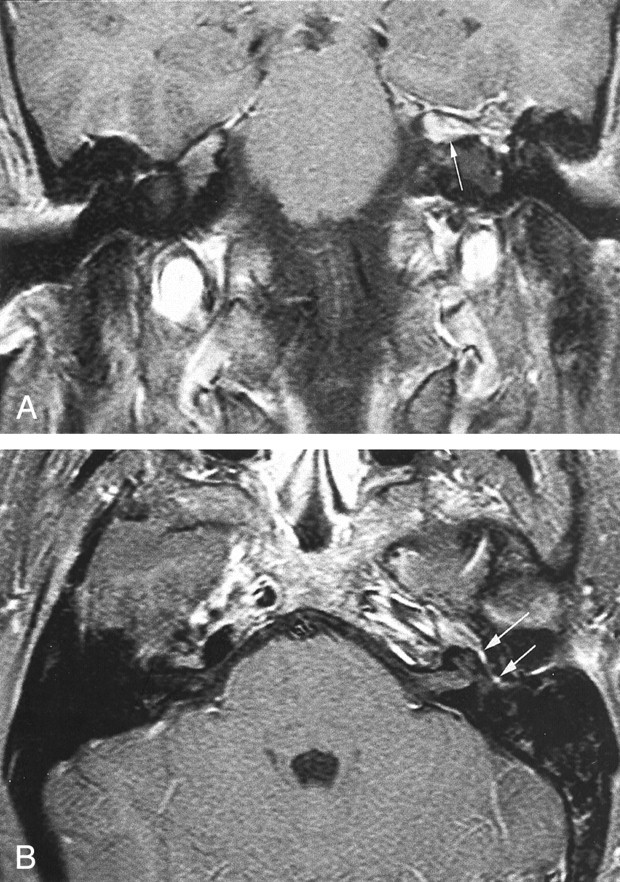
- Fig 1.
Initial MR images (600/10 [TR/TE]) obtained at presentation.
A, Contrast-enhanced fat-suppressed coronal T1-weighted image shows focal area of enhancement along the anterosuperior surface of the left petrous pyramid (arrow).
B, Contrast-enhanced fat-suppressed axial T1-weighted image shows enhancement along the anterosuperior surface of the left petrous pyramid with extension into the geniculate ganglion (long arrow) and the tympanic segment (short arrow) of the left facial nerve. On an adjacent image (not shown), the mass extended into the posterior left cavernous sinus.
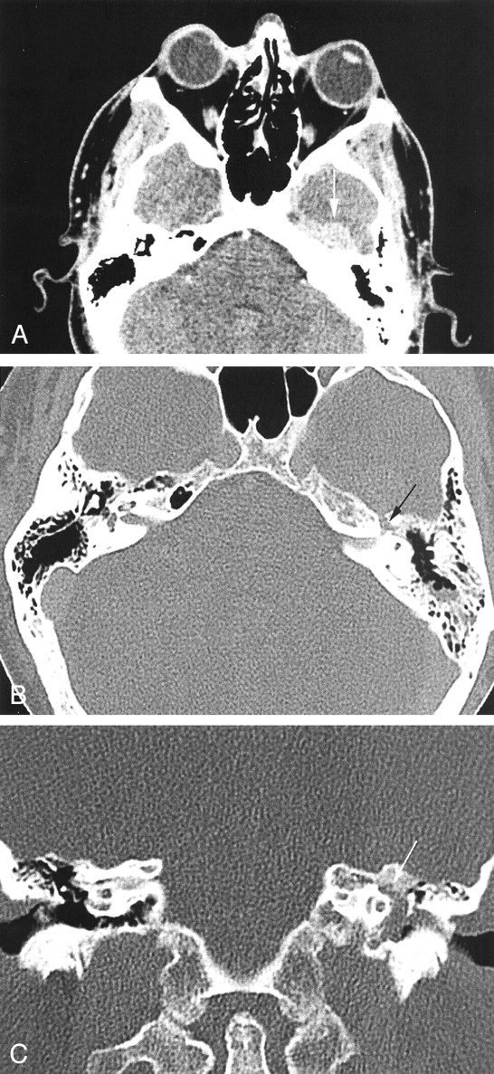
- Fig 2.
CT scans obtained 2 months later.
A, Contrast-enhanced axial CT scan shows a well-defined approximately 2-cm mass (arrow) along the anterosuperior aspect of the left petrous pyramid. Anteriorly, it is in continuity with the cavernous sinus.
B, High-spatial-resolution axial CT scan (bone window) shows widening of the left facial nerve canal in the region of the geniculate ganglion (arrow). Opacification of the mastoid air cells by serous fluid is also noted, probably secondary to blockage of the eustachian tube by the petrous apex mass.
C, Coronal CT scan, obtained at the level of the cochlea, shows extension of the soft tissue mass into the left middle ear cavity. Erosion of the facial nerve canal at the geniculate ganglion (arrow) is seen.
- Fig 3.
CT scans obtained after chemotherapy and resolution of symptoms.
A, Axial CT scan shows resolution of mass, previously noted along the anterosuperior aspect of the left petrous pyramid.
B, Axial CT scan (bone window), obtained at the level of the cochlea, shows no mass within the middle ear cavity and clear mastoid air cells. The anterior portion of the left carotid canal is faintly seen, and this could represent a residual (erosive) change from the previously noted mass.

In this issue


Jump to section
Related Articles
Cited By...
- No citing articles found.