
Article Figures & Data
Figures
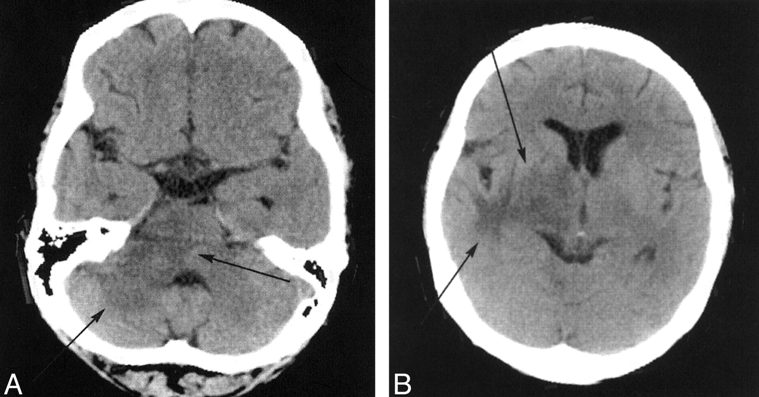
- Fig 1.
Axial nonenhanced CT scans reveal abnormal hypoattenuation in the right subinsular white matter, basal ganglia, and thalamus and within the middle cerebellar peduncle (arrows).
A, Image at the level of the middle cerebellar peduncle.
B, Image at the level of the insular cortex.
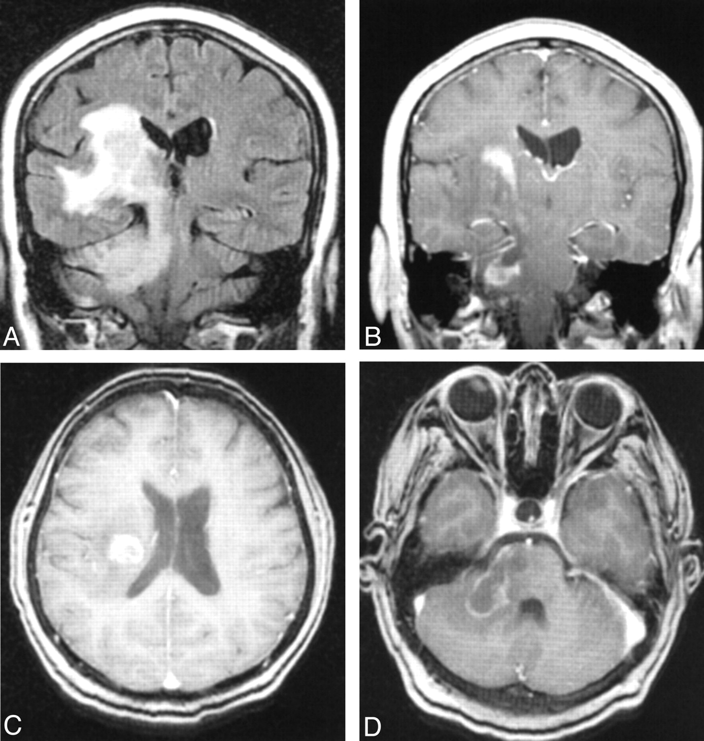
- Fig 2.
Brain MR images obtained at the time of initial clinical presentation show peripheral enhancement of a mass in the right cerebral white matter that extends through the cerebral peduncle and into the right cerebellar hemisphere via the right middle cerebellar peduncle.
A, Coronal fluid-attenuated inversion recovery (TR/TE/TI, 8000/112/2700) image demonstrates marked adjacent vasogenic edema.
B, Coronal spin-echo gadolinium-enhanced T1-weighted image (TR/TE, 600/15).
C and D, Axial spoiled-gradient postcontrast T1-weighted images (9.2/2.0; flip angle, 20° at the level of the lateral ventricles [C] and middle cerebellar peduncle [D]).
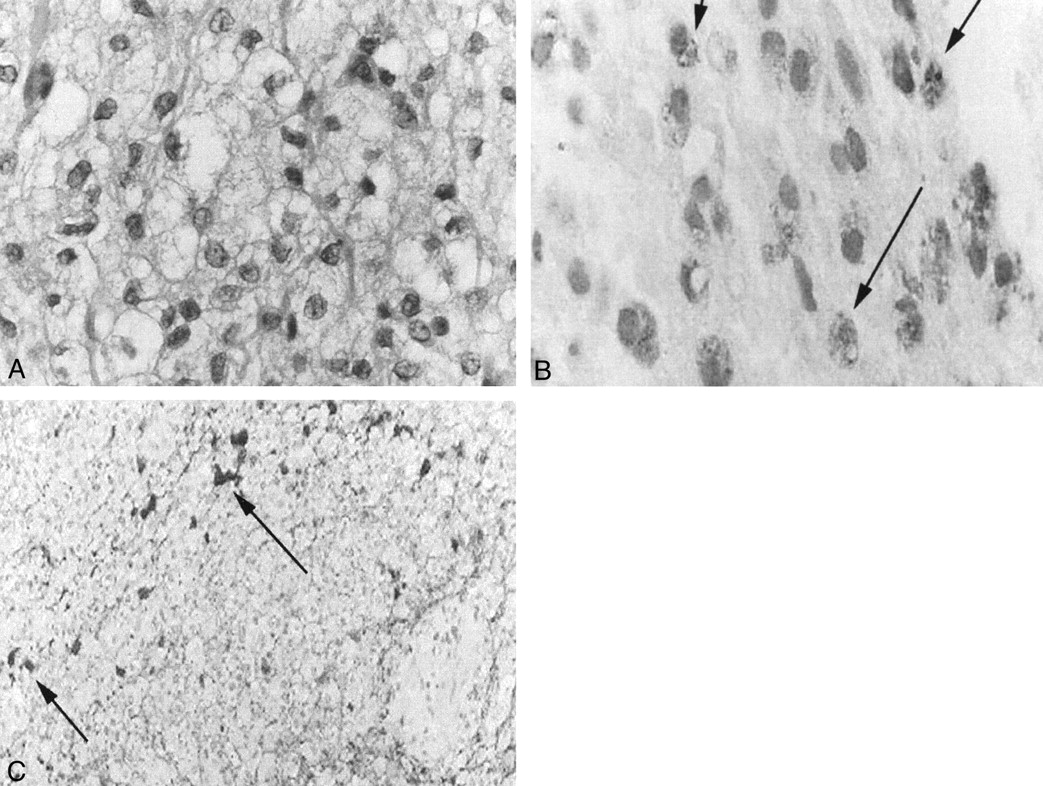
- Fig 3.
Histopathologic findings.
A, Photomicrograph of a routine paraffin section stained with hematoxylin-eosin (from a stereotactic brain biopsy) shows a hypercellular lesion populated by foamy histiocytes with uniform, benign nuclei (original magnification, ×360).
B, Photomicrograph of the brain biopsy stained with immunocytochemical method for CD68 antigen shows granular cytoplasmic positivity (arrows) (original magnification, ×360).
C, The stain for gliofibrillary acidic protein shows the histiocytes that are negative and a few reactive astrocytes and their processes that are darkly stained (arrows) (original magnification, ×120).
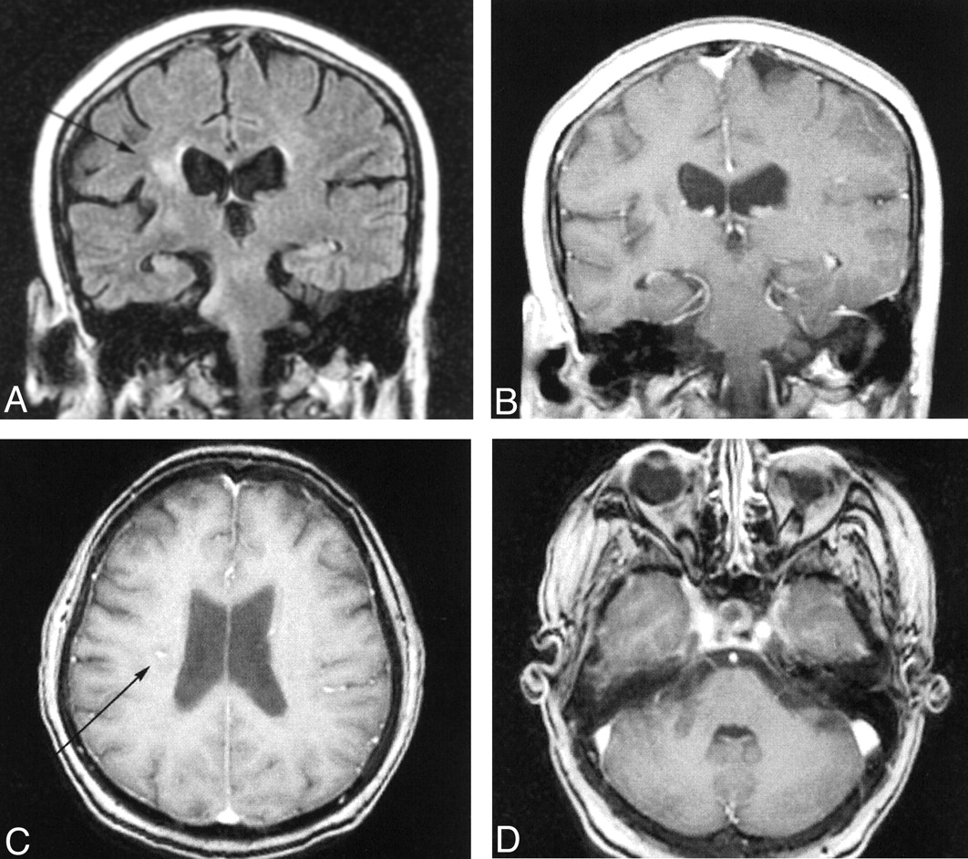
- Fig 4.
Follow-up brain MR images obtained 4 months post diagnosis, after treatment with steroids. The appearance of the brain is markedly improved compared with the appearance at diagnosis (see Fig 2).
A, Coronal fluid-attenuated inversion recovery (8000/112/2700) shows only small amounts of hyperintense signal.
B, Coronal spin-echo gadolinium-enhanced T1-weighted image (600/15).
C and D, Axial spoiled-gradient postcontrast T1-weighted images (9 .2/2.0; flip angle, 20° at the levels of the lateral ventricles [C] and middle cerebellar peduncle [D]) show small amounts of contrast enhancement (arrows).

In this issue


Jump to section
Related Articles
Cited By...
- No citing articles found.