Advertisement

Main menu

  • Home
  • Content
    • Current Issue
    • Accepted Manuscripts
    • Article Preview
    • Past Issue Archive
    • Video Articles
    • AJNR Case Collection
    • Case of the Week Archive
    • Case of the Month Archive
    • Classic Case Archive
  • Special Collections
    • AJNR Awards
    • ASNR Foundation Special Collection
    • Most Impactful AJNR Articles
    • Photon-Counting CT
    • Spinal CSF Leak Articles (Jan 2020-June 2024)
  • Multimedia
    • AJNR Podcasts
    • AJNR SCANtastic
    • Trainee Corner
    • MRI Safety Corner
    • Imaging Protocols
  • For Authors
    • Submit a Manuscript
    • Submit a Video Article
    • Submit an eLetter to the Editor/Response
    • Manuscript Submission Guidelines
    • Statistical Tips
    • Fast Publishing of Accepted Manuscripts
    • Graphical Abstract Preparation
    • Imaging Protocol Submission
    • Author Policies
  • About Us
    • About AJNR
    • Editorial Board
    • Editorial Board Alumni
  • More
    • Become a Reviewer/Academy of Reviewers
    • Subscribers
    • Permissions
    • Alerts
    • Feedback
    • Advertisers
    • ASNR Home

User menu

  • Alerts
  • Log in

Search

  • Advanced search
American Journal of Neuroradiology

American Journal of Neuroradiology

ASHNR American Society of Functional Neuroradiology ASHNR American Society of Pediatric Neuroradiology ASSR
  • Alerts
  • Log in

Advanced Search

  • Home
  • Content
    • Current Issue
    • Accepted Manuscripts
    • Article Preview
    • Past Issue Archive
    • Video Articles
    • AJNR Case Collection
    • Case of the Week Archive
    • Case of the Month Archive
    • Classic Case Archive
  • Special Collections
    • AJNR Awards
    • ASNR Foundation Special Collection
    • Most Impactful AJNR Articles
    • Photon-Counting CT
    • Spinal CSF Leak Articles (Jan 2020-June 2024)
  • Multimedia
    • AJNR Podcasts
    • AJNR SCANtastic
    • Trainee Corner
    • MRI Safety Corner
    • Imaging Protocols
  • For Authors
    • Submit a Manuscript
    • Submit a Video Article
    • Submit an eLetter to the Editor/Response
    • Manuscript Submission Guidelines
    • Statistical Tips
    • Fast Publishing of Accepted Manuscripts
    • Graphical Abstract Preparation
    • Imaging Protocol Submission
    • Author Policies
  • About Us
    • About AJNR
    • Editorial Board
    • Editorial Board Alumni
  • More
    • Become a Reviewer/Academy of Reviewers
    • Subscribers
    • Permissions
    • Alerts
    • Feedback
    • Advertisers
    • ASNR Home
  • Follow AJNR on Twitter
  • Visit AJNR on Facebook
  • Follow AJNR on Instagram
  • Join AJNR on LinkedIn
  • RSS Feeds

AJNR is seeking candidates for the AJNR Podcast Editor. Read the position description.

Research ArticleSPINE

Anterior Spinal Artery and Artery of Adamkiewicz Detected by Using Multi-Detector Row CT

Kohsuke Kudo, Satoshi Terae, Takeshi Asano, Masaki Oka, Kenshi Kaneko, Satoshi Ushikoshi and Kazuo Miyasaka
American Journal of Neuroradiology January 2003, 24 (1) 13-17;
Kohsuke Kudo
  • Find this author on Google Scholar
  • Find this author on PubMed
  • Search for this author on this site
Satoshi Terae
  • Find this author on Google Scholar
  • Find this author on PubMed
  • Search for this author on this site
Takeshi Asano
  • Find this author on Google Scholar
  • Find this author on PubMed
  • Search for this author on this site
Masaki Oka
  • Find this author on Google Scholar
  • Find this author on PubMed
  • Search for this author on this site
Kenshi Kaneko
  • Find this author on Google Scholar
  • Find this author on PubMed
  • Search for this author on this site
Satoshi Ushikoshi
  • Find this author on Google Scholar
  • Find this author on PubMed
  • Search for this author on this site
Kazuo Miyasaka
  • Find this author on Google Scholar
  • Find this author on PubMed
  • Search for this author on this site
  • Article
  • Figures & Data
  • Info & Metrics
  • Responses
  • References
  • PDF
Loading

Article Figures & Data

Figures

  • Tables
  • Fig 1.
    • Download figure
    • Open in new tab
    • Download powerpoint
    Fig 1.

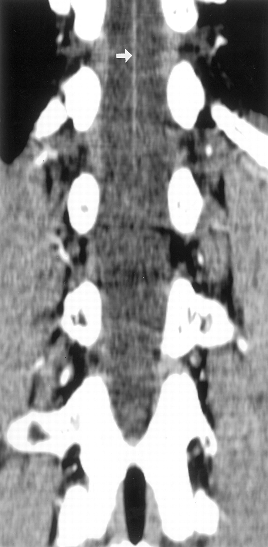
    CT scans obtained from patient 2, a 52-year-old woman.

    A, Coronal multiplanar reconstruction scan shows the ASA along the anterior midsagittal surface of the spinal cord (small white arrows). The large radiculomedullary artery (black arrow), which is defined as the AKA, arises from the left T10 level and joins the ASA. Both the ascending and descending branches of the ASA distal to the junction with the AKA are seen, but the ascending branch is narrower than the descending branch. The spinal branch (curved white arrow) arises from the left 10th intercostal artery (large white arrow).

    B, On the dorsal section of the coronal view, the continuity of the radiculomedullary artery and intercostal artery (large white arrow) via the spinal branch (curved white arrow) is confirmed.

    C and D, Axial scans show the ASA (small white arrows) and AKA (black arrows) in the dural sac. The continuity of the intercostal artery (large white arrow), spinal branch (curved white arrows), and radiculomedullary artery (black arrows) is also seen.

    E, Curved multiplanar reconstruction scan allows the visualization of the continuity of the intercostal artery (large white arrow), spinal branch (curved white arrow), radiculomedullary artery (black arrow), and ASA (small white arrows) on a single section. The change of caliber of these vessels is also seen.

  • Fig 2.
    • Download figure
    • Open in new tab
    • Download powerpoint
    Fig 2.

    CT scans obtained in patient 3, a 54-year-old man.

    A and B, Coronal scans show ASA (small white arrow) and AKA (black arrows). Thorough inspection reveals that the top of the hairpin curve is visualized at the upper edge of the scan. The continuity of the 10th intercostal artery (large white arrow), spinal branch (curved white arrow), and radiculomedullary artery (black arrows) is obscure because of close attachment of these vessels to the bone as a result of narrow intervertebral foramen.

  • Fig 3.
    • Download figure
    • Open in new tab
    • Download powerpoint
    Fig 3.

    CT scans obtained in patient 13, a 52-year-old man.

    A, Coronal multiplanar reconstruction scan shows the radiculomedullary artery (black arrows) arising from the left L1 level and joining the ASA (small white arrows). Both the ascending and descending branches of the ASA are seen.

    B, Because the radiculomedullary artery (black arrow) is slightly tortuous, the continuity of the vessel is confirmed on the section dorsal to that shown in A.

  • Fig 4.
    • Download figure
    • Open in new tab
    • Download powerpoint
    Fig 4.

    CT scan obtained in patient 17, a 25-year-old male patient. Coronal view multiplanar reconstruction scan shows the ASA (small white arrow) as visualized from the top of the scanning range. The radiculomedullary artery is not detected. The upper level of the scanning range is T11 level for this patient. The AKA is expected to arise from a more cranial level.

Tables

  • Figures
    • View popup
    TABLE 1:

    List of Patients and Results

    Patient No.Age (yr)SexBackgroundScanning RangeRange of ASA DetectedLevel of AKA Origin
    Upper LimitLower LimitUpper LimitLower Limit
    165FChronic hepatitisT8L2T10–T11L1–L2Right T11
    252FLiver tumorT8–T9L2–L3UL1–L2Left T10
    354MPost-liver transplantation (donor)T9L2–L3UT12–L1Left T10
    476MHCCT9L2–L3T10–T11T12–L1Left T11
    571MHCCT9L2–L3UT12–L1N.D.
    657FEchinococcosisT9L2UL1–L2N.D.
    755FLiver cirrhosisT9–T10L3–L4ULLeft T10
    851MHCCT9–T10L3UT12–L1Right T11
    973MHCCT9–T10L2–L3UL1–L2N.D.
    1060MHCCT9–T10L2UT11–T12Left T10
    1132MPost-liver transplantation (recipient)T10L3–L4UL12–L1Right T11
    1251MLiver hemangiomaT10L3–L4UL2–L3N.D.
    1352MHCCT10L3ULLeft L1
    1456MHCCT10L2–L3ULRight T12
    1548FPost-liver transplantation (recipient)T10–T11L4–L5T11–T12L1–L2Left L2
    1668MAutoimmune hepatitisT10–T11L3UT12–L1Left T11
    1725MPost-liver transplantation (recipient)T11L4UT12–L1N.D.
    1864MHCCT11L3UT12–L1Left L1
    1958MLiver cirrhosisT11L3UT12–L1N.D.
    • Note.—ASA indicates anterior spinal artery; AKA, artery of Adamkiewicz; F, female; M, male; HCC, hepatocellular carcinoma; U, upper limit of scanning range; L, lower limit of scanning range; N.D., not detected.

PreviousNext
Back to top

In this issue

American Journal of Neuroradiology: 24 (1)
American Journal of Neuroradiology
Vol. 24, Issue 1
1 Jan 2003
  • Table of Contents
  • Index by author
Advertisement
Print
Download PDF
Email Article

Thank you for your interest in spreading the word on American Journal of Neuroradiology.

NOTE: We only request your email address so that the person you are recommending the page to knows that you wanted them to see it, and that it is not junk mail. We do not capture any email address.

Enter multiple addresses on separate lines or separate them with commas.
Anterior Spinal Artery and Artery of Adamkiewicz Detected by Using Multi-Detector Row CT
(Your Name) has sent you a message from American Journal of Neuroradiology
(Your Name) thought you would like to see the American Journal of Neuroradiology web site.
CAPTCHA
This question is for testing whether or not you are a human visitor and to prevent automated spam submissions.
Cite this article
Kohsuke Kudo, Satoshi Terae, Takeshi Asano, Masaki Oka, Kenshi Kaneko, Satoshi Ushikoshi, Kazuo Miyasaka
Anterior Spinal Artery and Artery of Adamkiewicz Detected by Using Multi-Detector Row CT
American Journal of Neuroradiology Jan 2003, 24 (1) 13-17;

Citation Manager Formats

  • BibTeX
  • Bookends
  • EasyBib
  • EndNote (tagged)
  • EndNote 8 (xml)
  • Medlars
  • Mendeley
  • Papers
  • RefWorks Tagged
  • Ref Manager
  • RIS
  • Zotero
0 Responses
Respond to this article
Share
Bookmark this article
Anterior Spinal Artery and Artery of Adamkiewicz Detected by Using Multi-Detector Row CT
Kohsuke Kudo, Satoshi Terae, Takeshi Asano, Masaki Oka, Kenshi Kaneko, Satoshi Ushikoshi, Kazuo Miyasaka
American Journal of Neuroradiology Jan 2003, 24 (1) 13-17;
del.icio.us logo Twitter logo Facebook logo Mendeley logo
  • Tweet Widget
  • Facebook Like
  • Google Plus One

Jump to section

  • Article
    • Abstract
    • Methods
    • Results
    • Discussion
    • Conclusion
    • Footnotes
    • References
  • Figures & Data
  • Info & Metrics
  • Responses
  • References
  • PDF

Related Articles

  • No related articles found.
  • PubMed
  • Google Scholar

Cited By...

  • Assessing Vascularity of Osseous Spinal Metastases with Dual-Energy CT-DSA: A Pilot Study Compared with Catheter Angiography
  • Advantages of 70-kV CT Angiography for the Visualization of the Adamkiewicz Artery: Comparison with 120-kV Imaging
  • Surfers' myelopathy: A case series of 19 novice surfers with nontraumatic myelopathy
  • Crossref
  • Google Scholar

This article has not yet been cited by articles in journals that are participating in Crossref Cited-by Linking.

More in this TOC Section

  • Bern Score Validity for SIH
  • MP2RAGE 7T in MS Lesions of the Cervical Spine
  • Resisted Inspiration for CSF-Venous Fistula
Show more Spine

Similar Articles

Advertisement

Indexed Content

  • Current Issue
  • Accepted Manuscripts
  • Article Preview
  • Past Issues
  • Editorials
  • Editor's Choice
  • Fellows' Journal Club
  • Letters to the Editor
  • Video Articles

Cases

  • Case Collection
  • Archive - Case of the Week
  • Archive - Case of the Month
  • Archive - Classic Case

Special Collections

  • AJNR Awards
  • ASNR Foundation Special Collection
  • Most Impactful AJNR Articles
  • Photon-Counting CT
  • Spinal CSF Leak Articles (Jan 2020-June 2024)

More from AJNR

  • Trainee Corner
  • Imaging Protocols
  • MRI Safety Corner

Multimedia

  • AJNR Podcasts
  • AJNR Scantastics

Resources

  • Turnaround Time
  • Submit a Manuscript
  • Submit a Video Article
  • Submit an eLetter to the Editor/Response
  • Manuscript Submission Guidelines
  • Statistical Tips
  • Fast Publishing of Accepted Manuscripts
  • Graphical Abstract Preparation
  • Imaging Protocol Submission
  • Evidence-Based Medicine Level Guide
  • Publishing Checklists
  • Author Policies
  • Become a Reviewer/Academy of Reviewers
  • News and Updates

About Us

  • About AJNR
  • Editorial Board
  • Editorial Board Alumni
  • Alerts
  • Permissions
  • Not an AJNR Subscriber? Join Now
  • Advertise with Us
  • Librarian Resources
  • Feedback
  • Terms and Conditions
  • AJNR Editorial Board Alumni

American Society of Neuroradiology

  • Not an ASNR Member? Join Now

© 2025 by the American Society of Neuroradiology All rights, including for text and data mining, AI training, and similar technologies, are reserved.
Print ISSN: 0195-6108 Online ISSN: 1936-959X

Powered by HighWire