
Abstract
BACKGROUND AND PURPOSE: Volume changes in subcortical structures and cerebella have been associated with localization-related epilepsy and psychiatric illnesses. This study evaluated the effect of handedness and sex on the volumes of selected subcortical structures and cerebellar hemispheres in normal persons.
METHODS: Volumetric measurements were performed of the caudate heads, thalami, and cerebellar hemispheres in 34 (20 right- and 14 left-handed, 19 female and 15 male) normal persons. Amygdalar and hippocampal volumes were reported previously for these persons. All study participants completed a 10-item handedness questionnaire. The MR imaging sequence was a 3D T1-weighted gradient-echo acquisition of the whole brain (24/6 [TR/TE]; flip angle, 25 degrees). MR images were spatially normalized, and volumes were painted with a 1.0 mm3 resolution cursor on an SGI workstation. The effects of handedness and sex on standardized volumes and right-to-left volume ratios were calculated, and volumes were compared between right and left sides for each structure.
RESULTS: Handedness did not affect standardized volumes or asymmetries of the caudate heads, thalami, or cerebellar hemispheres. The volumes of subcortical structures were bilaterally larger in women than in men. Right-to-left asymmetries were significant for the caudate head and cerebellum but not for the thalamus.
CONCLUSION: These findings show that women have significantly larger subcortical structures than do men after spatial normalization to account for differences in brain size. Sex-specific normal ranges may be needed for evaluating volume changes related to epilepsy or other disease processes.
MR volumetric measurements of subcortical structures have recently gained importance in schizophrenia (1–4) and epilepsy research (5, 6). The structures targeted most commonly by studies assessing newly diagnosed psychosis or medically treated schizophrenia include the thalamus, caudate, putamen, and globus pallidus. Volume reductions of the thalamus were reported in patients with first episode psychosis and neuroleptic naïve schizophrenia (1–3). Patients with medically treated schizophrenia had enlargement of all basal ganglia, including caudate, putamen, and globus pallidus, and thalamus, suggesting that use of neuroleptic medications may lead to hypertrophy of these structures (1). In addition to volume loss of the hippocampus and amygdala ipsilateral to a temporal seizure focus, subcortical structures seem affected bilaterally, including the caudate, putamen, globus pallidus, and thalamus (5, 6). Cerebellar volume changes due to supratentorial lesions or injury are rare, but unilateral cerebral lesions have been associated with atrophy of the contralateral cerebellar hemisphere (7). Bilateral cerebellar atrophy has been described in the setting of temporal lobe epilepsy (8–10).
The analysis of subcortical and cerebellar volumes in normal persons is essential to identify changes related to disease processes. Most of the studies above used normal persons, but none specified handedness of the study participants (1–6, 9, 10). Considering that right-to-left structural asymmetries have been reported for specific brain regions and structures, assessment of potential effects of handedness is important (11–14). Other studies looking at subcortical volumes in normal persons studied only males (15) or did not look at sex-related differences (2–6, 9, 10). Because volumes of subcortical structures and cerebella depend on whole brain volume, it is necessary to control for brain size effects (1, 9, 16). In this study, we standardized the brain volumes by using spatial normalization, whereby 3D MR imaging datasets were reformatted to the dimensions of the Talairach brain (17). The method of spatial normalization implemented in this study standardized volumes of cerebral structures to total cerebral volumes in a proportional manner, whereas the cerebellar volumes were adjusted passively but proportional to total cerebral volumes.
The aim of this study was to establish norms for volumes of the caudate head, thalamus, and cerebellum and to evaluate their interactions with sex and handedness. In conjunction with hippocampal and amygdalar volumetry, these structures were chosen to show structural changes associated with chronic disease processes, specifically those related to localization-related epilepsy.
Methods
Participants
MR images with the appropriate sequences and resolution were found for 14 left-handed participants (eight women, six men) from a database of 170 normal persons (10). MR images of 20 right-handed participants (11 women, nine men) were selected randomly from the same database. The mean age of the participants was 28 years (range, 19–38 years). All participants completed a handedness questionnaire at the time of the MR imaging. The results of the questionnaire were described previously (14). The mean ages of right- and left-handed persons were 27 and 28 years, respectively.
Image Acquisition and Processing
All participants underwent imaging with a 1.9-T MR imaging system (GE/Elscint Prestige, Milwaukee, WI). The MR imaging sequence used for volumetric analysis was a 3D T1-weighted gradient-echo acquisition of the whole brain (24/6 [TR/TE]; flip angle, 25 degrees; field of view, 256 × 256 mm; number of sections, 192). The right side was marked by a 1-cm-diameter plastic tube, filled with water, placed caudally from the participant’s ear and additionally defined in the header file. Pixels were 1.0 × 1.0 mm, and section thickness was 1 mm without an intersection gap. All images of the brain were spatially normalized into registration with the Talairach brain atlas by using the software package “SN,” developed at our center (17). This algorithm uses a nine-parameter fit and interactive denotation of the anterior commissure-posterior commissure line. Images were resectioned into 60 sections by using tri-linear interpolation, with an image matrix size of 60 × 128 × 128 mm3 and with each voxel being 1 × 1 × 1 mm3. All volumes were painted with a 1.0 mm3 resolution cursor on an SGI workstation by using Display (Montréal Neurological Institute, Canada), which allowed simultaneous visualization of the structures in three planes (Fig 1).
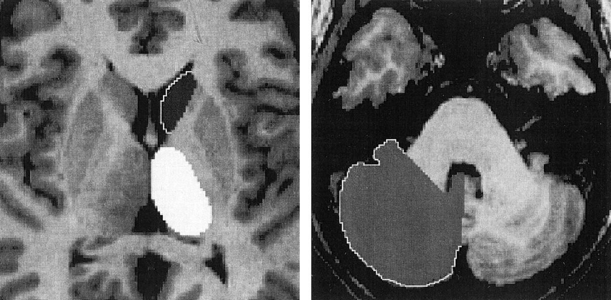
Axial view images of the painted left caudate head and thalamus and right cerebellar hemisphere.
All volumetric measurements were performed by one of two investigators (C.A.S., C.C.), who were blinded to patients’ handedness and sex. Eight pairs of caudate heads and thalami and five pairs of cerebellar hemispheres were chosen randomly for repeated measurements to assess inter-rater inter-trial reliability (Table 1).
Inter-rater, inter-trial correlations
Caudate Head.
The caudate head was delineated posteriorly by the coronal image 1 mm anterior to the anterior commissure. The border between the caudate head and the nucleus accumbens was determined in the sagittal plane by extending the inferior posterior border of the caudate anteriorly. The lateral border was featured by the anterior limb of internal capsule, the superior border by the corpus callosum, and the medial boundary by the lateral ventricles.
Thalamus.
The two thalami were disconnected between two smallest cross-sections through the massa intermedia in the sagittal plane. The anatomic boundary featured the internal capsule laterally, the perithalamic white matter tracts superiorly, and the plane including both the anterior and posterior commissures inferiorly. The posterior border was demarcated as the section showing the fornices in their entire width in the coronal plane. The medial and lateral geniculate bodies were excluded.
Cerebellar Hemisphere.
The cerebellar hemispheres were bisected between the two smallest sections through the medial vermis in the sagittal plane. When this boundary was distorted or curved, adjustments were made by using simultaneous visualization in the axial and coronal planes. Disarticulation from the brain stem was performed by transecting the cerebellar peduncles along the plane of their entrance into the cerebellum, namely at the shortest segment between the posterior recess of the fourth ventricle and the junction between the lateral border of the peduncles and the cerebellar cortex. Using this technique, the deep cerebellar nuclei were retained in the measured volumes whereas the peduncles were largely excluded. The cerebellar hemispheres were painted individually by using thresholds to facilitate the demarcation regarding CSF. Only 30 participants had cerebella completely imaged.
Statistical Analysis.
A wide range of statistical tests was conducted on both standardized volumes and right-to-left volume ratios. Analysis of variance with 2 × 3 factorial design was used to detect significant differences in volume ratios due to handedness and structure (caudate head, thalamus, cerebellar hemisphere). Sex effects on volume ratios were assessed by using a similar 2 × 3 factor analysis of variance. Because the cerebellum was much larger than the thalamus and caudate head, it was excluded from the multivariate comparison of volumes. A 2 × 4 analysis of variance was used to assess the effects of handedness and structures (caudate head, thalamus, amygdala, hippocampus). Hippocampal and amygdalar volumes of the same normal persons were reported previously (14). Cerebellar volumes were compared between right- and left-handed participants and between men and women by using group t tests. For all statistical comparisons of volume ratio data, a logarithmic transform was applied before the comparisons because these data did not conform to a gaussian distribution. Right- and left-sided caudate heads, thalami, and cerebellar hemispheres were compared by group t tests.
Results
Standardized volumes and right-to-left volume ratios were not statistically different between right- and left-handed participants for the caudate head or thalamus. Inter-sex comparison of standardized volumes and right-to-left volume ratios of caudate heads, thalami, and cerebellar hemispheres are listed in Table 2. Comparison of standardized volumes of hippocampi and amygdalae in the same patients is also shown in Table 2. The standardized volumes of subcortical structures, including caudate head, thalamus, hippocampus, and amygdala, were significantly larger in women than in men (F = 11.40, P < .001). However, no interactions between sex and individual structures were found. Furthermore, sex did not affect right-to-left volume ratios for the caudate head or thalamus. No significant differences were observed between cerebellar volumes regarding handedness or sex.
Standardized volumes and ratios of the caudate head, thalamus, hippocampus, amygdala, and cerebellum
Caudate heads (P < .05) and cerebellar hemispheres (P < .005) were larger on the left than on the right for the entire group. No side-to-side difference was noted for the thalamus, yet a left-greater-than-right characteristic was found in 65% of right-handed, compared with 36% of left-handed normal persons.
Discussion
This MR imaging volumetric study assessed caudate heads, thalami, and cerebellar hemispheres of normal persons. Amygdalar and hippocampal volumes were previously reported for these persons (14). Although the study found no significant effect of handedness on the volumes or asymmetries of these structures, sex seemed to affect standardized volumes of subcortical structures. In proportion to total cerebral volume, women had larger subcortical volumes bilaterally than did men. This difference was also evident for amygdala and hippocampus (14). Right-to-left thalamic and caudate ratios were not affected by handedness or sex.
The finding that women seem to have larger subcortical cerebral structures than those of men is consistent with findings of other studies looking at sex differences in development (18, 19). Brain maturation in women during childhood and adolescence resulted in relative increases in gray matter volumes but reduction of white matter volumes (18, 19). The reasons for the sex difference are unclear but may be related to hormonal effects. Developmental effects of hormones in adolescence or changes in neuronal or glial volumes related to fluctuating hormone levels during the menstrual cycle could be responsible for the sex difference.
Asymmetries between right and left sides were noted for the caudate head and cerebellar hemispheres but not for the thalamus. The left-greater-than-right asymmetry of the caudate heads may reflect asymmetries previously reported for putamen and globus pallidus (1, 5, 15). On the other hand, the left-greater-than-right asymmetry of the cerebellar hemispheres differed from the findings of other studies (10, 11). This discrepancy may have been due to the use of different landmarks and techniques. In both cases, however, the asymmetries suggest functional lateralization, which was independent of handedness. It is possible that the asymmetries are related to language lateralization, but further studies correlating language mapping and volumetry are necessary to assess this relationship.
In summary, sex may affect standardized volumes of some subcortical gray matter structures, whereas right-to-left volume ratios were not affected. Although the amygdalar and hippocampal volume ratios seem to be affected by handedness (14), asymmetries of the caudate heads, thalami, and cerebellar hemispheres did not depend on handedness. Nonetheless, the small but significant asymmetries of the caudate heads and cerebellar hemispheres may reflect functional lateralization. More studies need to be conducted to assess the relationship between asymmetries of subcortical structures and cerebellar hemispheres and well-lateralized functions, such as handedness and language. Accurate and stratified control data will help to better define subcortical and cerebellar volume changes occurring in association with several disease processes, including epilepsy and schizophrenia.
Footnotes
Supported by the EJLB Foundation and the National Institute of Mental Health (P20 DA52176) and by an Internal Review Grant through the University of Texas Health Science Center at San Antonio (grant 00–0021).
References
- Received March 18, 2002.
- Accepted after revision October 17, 2002.
- Copyright © American Society of Neuroradiology


