
Article Figures & Data
Figures
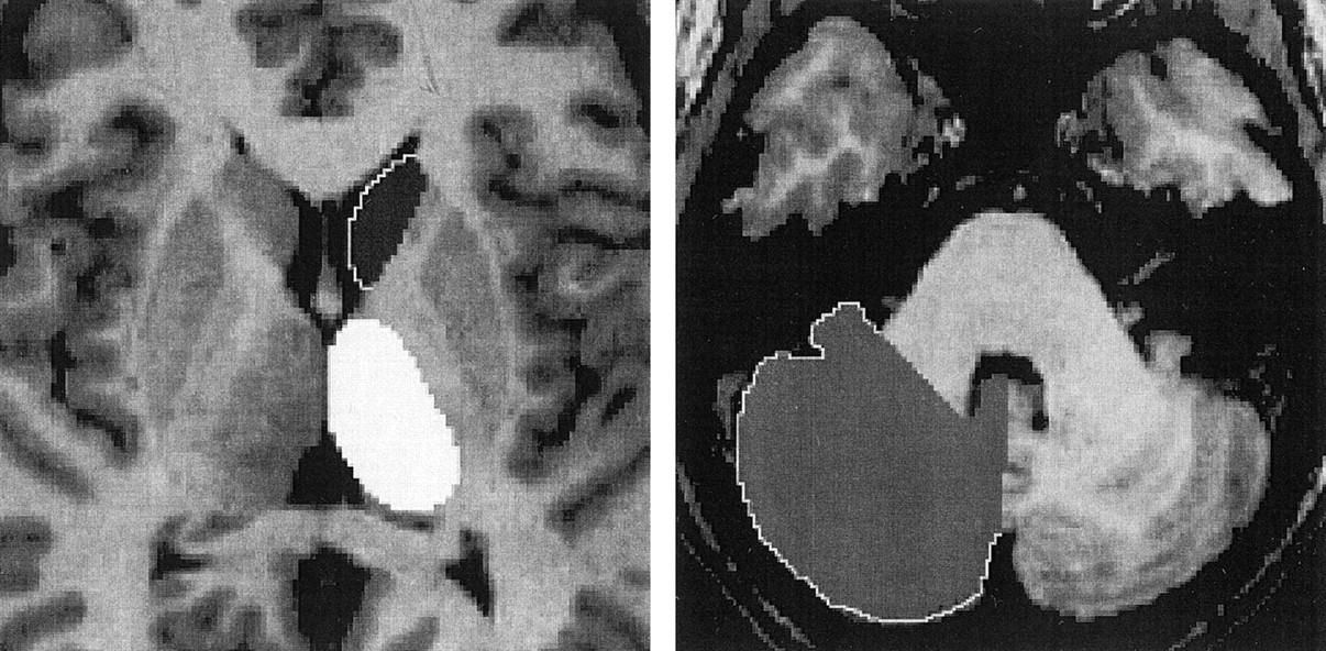
- Fig 1.
Axial view images of the painted left caudate head and thalamus and right cerebellar hemisphere.
Tables
Thalamus Caudate Head Cerebellum Volume R/L VR Volume R/L VR Volume R/L VR Investigator 1 5850 1.03 3314 0.98 73720 0.98 Investigator 2 5950 1.04 3218 1.00 74960 0.98 Statistical analyses P < 0.001* P < 0.05† P < 0.001* P < 0.05† P < 0.0001* P < 0.05† (r = 0.81) (r = 0.82) (r = 0.97) Note.—R/L VR indicates right-to-left volume ratio. Mean volumes are expressed as mm3.
* Pearson correlation tests.
† Two-tailed t tests.
- TABLE 2:
Standardized volumes and ratios of the caudate head, thalamus, hippocampus, amygdala, and cerebellum
Participants Age (yr) Caudate Head Volumes Thalamic Volumes Hippocampal Volumes (14) Amygdalar Volumes (14) Cerebellar Volumes (n = 30) Right Left R/L Right Left R/L Right Left R/L Right Left R/L Right Left R/L All (n = 34) 28 (5) 3170 (398) 3277 (250) 0.97 (0.08) 6193 (507) 6187 (534) 1.00 (0.05) 2986 (402) 2922 (409) 1.02 (0.06) 1608 (150) 1530 (170) 1.06 (0.07) 69768 (8934) 70614 (8763) 0.99 (0.03) Women (n = 19) 27 (5) 3329 (435) 3375 (432) 0.99 (0.05) 6334 (746) 6405 (716) 0.99 (0.07) 3102 (443) 3008 (457) 1.03 (0.06) 1650 (148) 1586 (193) 1.04 (0.07) 70300 (5969) 71550 (5792) 0.98 (0.03) Men (n = 15) 28 (5) 3148 (393) 3257 (218) 0.96 (0.08) 6126 (553) 6155 (529) 1.00 (0.06) 2838 (294) 2813 (320) 1.01 (0.05) 1555 (140) 1459 (164) 1.07 (0.07) 68619 (9404) 69486 (9224) 0.99 (0.02) Note.—R/L indicates right-to-left volume ratio. Numbers and volumes are expressed as mm3. SD are shown in parentheses.


