Advertisement

Main menu

  • Home
  • Content
    • Current Issue
    • Accepted Manuscripts
    • Article Preview
    • Past Issue Archive
    • Video Articles
    • AJNR Case Collection
    • Case of the Week Archive
    • Case of the Month Archive
    • Classic Case Archive
  • Special Collections
    • AJNR Awards
    • ASNR Foundation Special Collection
    • Most Impactful AJNR Articles
    • Photon-Counting CT
    • Spinal CSF Leak Articles (Jan 2020-June 2024)
  • Multimedia
    • AJNR Podcasts
    • AJNR SCANtastic
    • Trainee Corner
    • MRI Safety Corner
    • Imaging Protocols
  • For Authors
    • Submit a Manuscript
    • Submit a Video Article
    • Submit an eLetter to the Editor/Response
    • Manuscript Submission Guidelines
    • Statistical Tips
    • Fast Publishing of Accepted Manuscripts
    • Graphical Abstract Preparation
    • Imaging Protocol Submission
    • Author Policies
  • About Us
    • About AJNR
    • Editorial Board
    • Editorial Board Alumni
  • More
    • Become a Reviewer/Academy of Reviewers
    • Subscribers
    • Permissions
    • Alerts
    • Feedback
    • Advertisers
    • ASNR Home

User menu

  • Alerts
  • Log in

Search

  • Advanced search
American Journal of Neuroradiology

American Journal of Neuroradiology

ASHNR American Society of Functional Neuroradiology ASHNR American Society of Pediatric Neuroradiology ASSR
  • Alerts
  • Log in

Advanced Search

  • Home
  • Content
    • Current Issue
    • Accepted Manuscripts
    • Article Preview
    • Past Issue Archive
    • Video Articles
    • AJNR Case Collection
    • Case of the Week Archive
    • Case of the Month Archive
    • Classic Case Archive
  • Special Collections
    • AJNR Awards
    • ASNR Foundation Special Collection
    • Most Impactful AJNR Articles
    • Photon-Counting CT
    • Spinal CSF Leak Articles (Jan 2020-June 2024)
  • Multimedia
    • AJNR Podcasts
    • AJNR SCANtastic
    • Trainee Corner
    • MRI Safety Corner
    • Imaging Protocols
  • For Authors
    • Submit a Manuscript
    • Submit a Video Article
    • Submit an eLetter to the Editor/Response
    • Manuscript Submission Guidelines
    • Statistical Tips
    • Fast Publishing of Accepted Manuscripts
    • Graphical Abstract Preparation
    • Imaging Protocol Submission
    • Author Policies
  • About Us
    • About AJNR
    • Editorial Board
    • Editorial Board Alumni
  • More
    • Become a Reviewer/Academy of Reviewers
    • Subscribers
    • Permissions
    • Alerts
    • Feedback
    • Advertisers
    • ASNR Home
  • Follow AJNR on Twitter
  • Visit AJNR on Facebook
  • Follow AJNR on Instagram
  • Join AJNR on LinkedIn
  • RSS Feeds

AJNR is seeking candidates for the AJNR Podcast Editor. Read the position description.

Research ArticlePEDIATRICS

Postoperative Evaluation of Moyamoya Disease with Perfusion-Weighted MR Imaging: Initial Experience

Seung-Koo Lee, Dong Ik Kim, Eun-Kee Jeong, Si-Yeon Kim, Sang Heum Kim, Yon Kwon In, Dong-Seok Kim and Joong-Uhn Choi
American Journal of Neuroradiology April 2003, 24 (4) 741-747;
Seung-Koo Lee
  • Find this author on Google Scholar
  • Find this author on PubMed
  • Search for this author on this site
Dong Ik Kim
  • Find this author on Google Scholar
  • Find this author on PubMed
  • Search for this author on this site
Eun-Kee Jeong
  • Find this author on Google Scholar
  • Find this author on PubMed
  • Search for this author on this site
Si-Yeon Kim
  • Find this author on Google Scholar
  • Find this author on PubMed
  • Search for this author on this site
Sang Heum Kim
  • Find this author on Google Scholar
  • Find this author on PubMed
  • Search for this author on this site
Yon Kwon In
  • Find this author on Google Scholar
  • Find this author on PubMed
  • Search for this author on this site
Dong-Seok Kim
  • Find this author on Google Scholar
  • Find this author on PubMed
  • Search for this author on this site
Joong-Uhn Choi
  • Find this author on Google Scholar
  • Find this author on PubMed
  • Search for this author on this site
  • Article
  • Figures & Data
  • Info & Metrics
  • Responses
  • References
  • PDF
Loading

Article Figures & Data

Figures

  • Tables
  • Fig 1.
    • Download figure
    • Open in new tab
    • Download powerpoint
    Fig 1.

    Regions of interest (MCA, PCA, and BG) are drawn on a TTP map. On the rCBV maps (not shown), the relative signal intensity ratios of the MCA and the BG to the PCA territory were obtained, whereas on the TTP maps the time differences in the MCA and BG were calculated and compared with that of the PCA territory.

  • Fig 2.
    • Download figure
    • Open in new tab
    • Download powerpoint
    Fig 2.

    9-year-old boy with moyamoya disease.

    A, Anteroposterior angiogram of the right ICA shows an occlusion of the supraclinoid portion, with development of multiple basal collateral vessels (moyamoya vessels).

    B, Left ICA angiogram depicts mild changes of the luminal caliber in the left MCA, but with preserved patency. Left ACA shows severe narrowing.

    C, Technetium-99m ethyl cysteinate dimer SPECT scan (left scan) reveals decreased perfusion in both hemispheres, more prominent on the right side. After the acetazolamide (Diamox) injection (right scan), there is no evidence of a significant perfusion reduction.

    D, Perfusion MR images show delayed TTP in the right hemisphere before EDAS (arrows in left TTP map) and a signal intensity reduction after EDAS (arrows in right TTP map), which means a restoration of rapid flow at the surgical site. Note, rCBV maps show nothing significant.

  • Fig 3.
    • Download figure
    • Open in new tab
    • Download powerpoint
    Fig 3.

    A and B, Graphs show changes in the rCBV ratios to the posterior circulation of the MCA territory (A) and the basal ganglia (B). The rCBV can be increased or decreased before (Preop) and after (Postop) surgery, which cannot characterize the rCBV patterns of moyamoya disease.

  • Fig 4.
    • Download figure
    • Open in new tab
    • Download powerpoint
    Fig 4.

    A and B, Graphs show changes in the TTP delay to the posterior circulation of the MCA territory (A) and basal ganglia (B). Note the markedly increased TTP in the MCA territory, with a significant reduction after surgery (Postop). There were no specific changes in the TTP values in the basal ganglia.
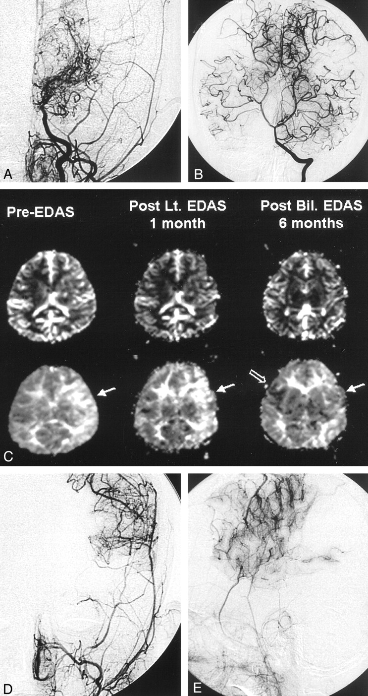
  • Fig 5.
    • Download figure
    • Open in new tab
    • Download powerpoint
    Fig 5.

    7-year-old boy with moyamoya disease.

    A, Preoperative left carotid angiogram shows an occlusion of the supraclinoid ICA with basal collateral vessels. No transdural collateral vessels from the superficial temporal or middle meningeal arteries are seen.

    B, Vertebral artery angiogram shows prominent leptomeningeal collateral vessels supplying the bilateral hemispheres.

    C, Serial perfusion-weighted MR images (rCBV, top row; TTP, bottom row) were obtained because the patient underwent bilateral EDAS procedures with a 1-month interval. One-month follow-up perfusion MR image shows a mild reduction in the TTP delay (arrow in middle TTP map), which means neovascularization is beginning to develop. The last perfusion MR image (right TTP map) shows near-complete normalization of the TTP in the left hemisphere (solid arrow) and a partial TTP shortening on the right side (open arrow).

    D and E, Postoperative 7-month follow-up left ECA angiograms show successful neovascularization from the middle meningeal and superficial temporal arteries.

Tables

  • Figures
  • rCBV Ratios and TTP Values in the Control Group and Patients

    ParameterControl Group (n = 5)Patient Group (n = 13)P Value*
    rCBV ratio
     MCA to PCA1.35 ± 0.141.31 ± 0.66.950
     BG to PCA0.79 ± 0.120.78 ± 0.25.756
    TTP difference (sec)
     MCA to PCA04.37 ± 2.25.0006
     BG to PCA−0.76 ± 0.22−1.08 ± 1.99.765
    • Note.—Data are the mean ± SD.

    • * Two-tailed t test.

PreviousNext
Back to top

In this issue

American Journal of Neuroradiology: 24 (4)
American Journal of Neuroradiology
Vol. 24, Issue 4
1 Apr 2003
  • Table of Contents
  • Index by author
Advertisement
Print
Download PDF
Email Article

Thank you for your interest in spreading the word on American Journal of Neuroradiology.

NOTE: We only request your email address so that the person you are recommending the page to knows that you wanted them to see it, and that it is not junk mail. We do not capture any email address.

Enter multiple addresses on separate lines or separate them with commas.
Postoperative Evaluation of Moyamoya Disease with Perfusion-Weighted MR Imaging: Initial Experience
(Your Name) has sent you a message from American Journal of Neuroradiology
(Your Name) thought you would like to see the American Journal of Neuroradiology web site.
CAPTCHA
This question is for testing whether or not you are a human visitor and to prevent automated spam submissions.
Cite this article
Seung-Koo Lee, Dong Ik Kim, Eun-Kee Jeong, Si-Yeon Kim, Sang Heum Kim, Yon Kwon In, Dong-Seok Kim, Joong-Uhn Choi
Postoperative Evaluation of Moyamoya Disease with Perfusion-Weighted MR Imaging: Initial Experience
American Journal of Neuroradiology Apr 2003, 24 (4) 741-747;

Citation Manager Formats

  • BibTeX
  • Bookends
  • EasyBib
  • EndNote (tagged)
  • EndNote 8 (xml)
  • Medlars
  • Mendeley
  • Papers
  • RefWorks Tagged
  • Ref Manager
  • RIS
  • Zotero
0 Responses
Respond to this article
Share
Bookmark this article
Postoperative Evaluation of Moyamoya Disease with Perfusion-Weighted MR Imaging: Initial Experience
Seung-Koo Lee, Dong Ik Kim, Eun-Kee Jeong, Si-Yeon Kim, Sang Heum Kim, Yon Kwon In, Dong-Seok Kim, Joong-Uhn Choi
American Journal of Neuroradiology Apr 2003, 24 (4) 741-747;
del.icio.us logo Twitter logo Facebook logo Mendeley logo
  • Tweet Widget
  • Facebook Like
  • Google Plus One

Jump to section

  • Article
    • Abstract
    • Methods
    • Results
    • Discussion
    • Conclusion
    • Acknowledgments
    • Footnotes
    • References
  • Figures & Data
  • Info & Metrics
  • Responses
  • References
  • PDF

Related Articles

  • No related articles found.
  • PubMed
  • Google Scholar

Cited By...

  • Understanding external carotid artery collateralisation after cerebral revascularisation in moyamoya disease: insights from quantitative analysis
  • Standardized MR Perfusion Scoring System for Evaluation of Sequential Perfusion Changes and Surgical Outcome of Moyamoya Disease
  • Noninvasive Assessment of Hemodynamic Stress Distribution after Indirect Revascularization for Pediatric Moyamoya Vasculopathy
  • Evaluation of Encephaloduroarteriosynangiosis Efficacy Using Probabilistic Independent Component Analysis Applied to Dynamic Susceptibility Contrast Perfusion MRI
  • Changes in Integrity of Normal-Appearing White Matter in Patients with Moyamoya Disease: A Diffusion Tensor Imaging Study
  • The Acetazolamide Challenge: Techniques and Applications in the Evaluation of Chronic Cerebral Ischemia
  • Changing ischaemic lesion patterns in adult moyamoya disease
  • Crossref
  • Google Scholar

This article has not yet been cited by articles in journals that are participating in Crossref Cited-by Linking.

More in this TOC Section

  • SyMRI & MR Fingerprinting in Brainstem Myelination
  • Comparison of Image Quality and Radiation Dose in Pediatric Temporal Bone CT Using Photon-Counting Detector CT and Energy-Integrating Detector CT
  • Neonatal Hypocalcemia: Cerebral MRI Abnormalities
Show more Pediatrics

Similar Articles

Advertisement

Indexed Content

  • Current Issue
  • Accepted Manuscripts
  • Article Preview
  • Past Issues
  • Editorials
  • Editor's Choice
  • Fellows' Journal Club
  • Letters to the Editor
  • Video Articles

Cases

  • Case Collection
  • Archive - Case of the Week
  • Archive - Case of the Month
  • Archive - Classic Case

Special Collections

  • AJNR Awards
  • ASNR Foundation Special Collection
  • Most Impactful AJNR Articles
  • Photon-Counting CT
  • Spinal CSF Leak Articles (Jan 2020-June 2024)

More from AJNR

  • Trainee Corner
  • Imaging Protocols
  • MRI Safety Corner

Multimedia

  • AJNR Podcasts
  • AJNR Scantastics

Resources

  • Turnaround Time
  • Submit a Manuscript
  • Submit a Video Article
  • Submit an eLetter to the Editor/Response
  • Manuscript Submission Guidelines
  • Statistical Tips
  • Fast Publishing of Accepted Manuscripts
  • Graphical Abstract Preparation
  • Imaging Protocol Submission
  • Evidence-Based Medicine Level Guide
  • Publishing Checklists
  • Author Policies
  • Become a Reviewer/Academy of Reviewers
  • News and Updates

About Us

  • About AJNR
  • Editorial Board
  • Editorial Board Alumni
  • Alerts
  • Permissions
  • Not an AJNR Subscriber? Join Now
  • Advertise with Us
  • Librarian Resources
  • Feedback
  • Terms and Conditions
  • AJNR Editorial Board Alumni

American Society of Neuroradiology

  • Not an ASNR Member? Join Now

© 2025 by the American Society of Neuroradiology All rights, including for text and data mining, AI training, and similar technologies, are reserved.
Print ISSN: 0195-6108 Online ISSN: 1936-959X

Powered by HighWire