
Article Figures & Data
Figures
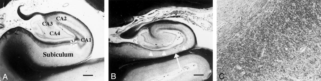
- Fig 1.
Coronal photomicrographs of normal and atrophic hippocampus (Klüver-Barrera stain).
A, Image in a 56-year-old man with dentatorubro-pallidoluysian atrophy shows no atrophy of the left subiculum or hippocampus proper. Image represents a normal hippocampus. The hippocampus proper consists of the CA1, CA2, CA3, and CA4 fields. Bar indicates 1 mm.
B, Image in a 78-year-old man with AD (duration of illness, 15 years) shows severe atrophy of the subiculum (arrow). The superficial medullary lamina (arrowheads) is thinner than that of a normal hippocampus. Bar indicates 1 mm.
C, Magnified (×200) view of the image in A shows numerous nerve fibers running in the anteroposterior direction in the superficial medullary lamina of the hippocampal formation.
- Fig 2.
Schematic representation of the hippocampal formation shows the measurement points for the subiculum and the hippocampus proper. PHG indicates the parahippocampal gyrus.
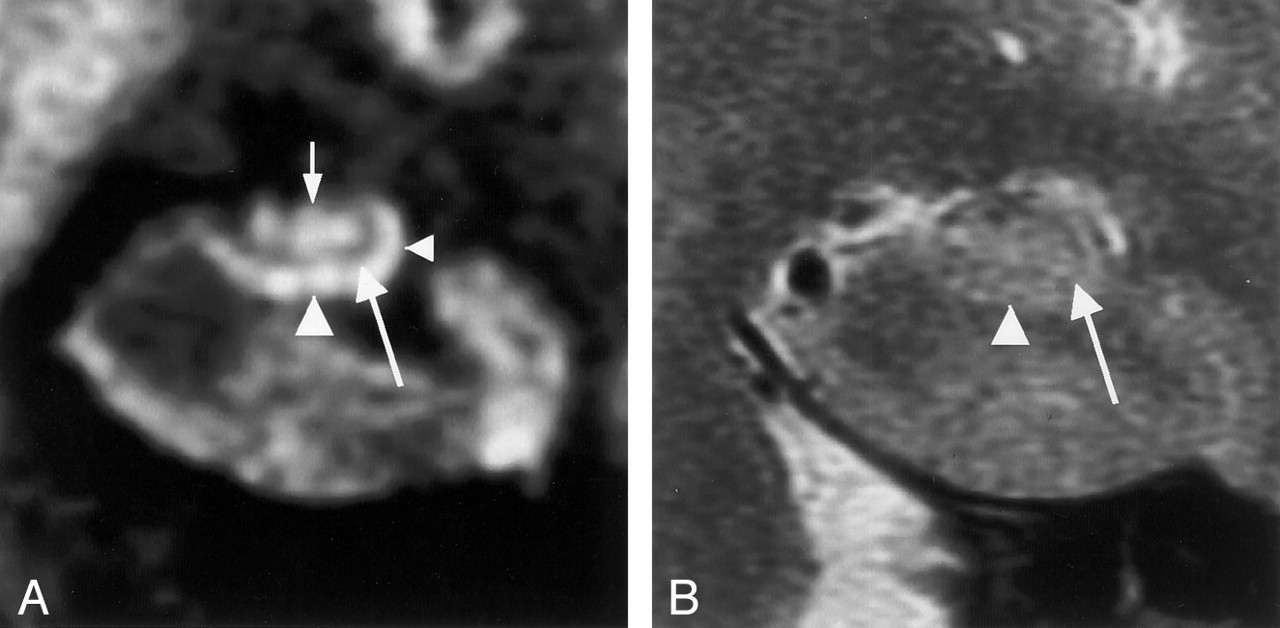
- Fig 3.
Coronal MR images in a 59-year-old man (control subject).
A, Multishot diffusion-weighted image clearly shows the inner structure of the left hippocampus. The subiculum (left arrowhead), CA1 of the hippocampus proper (right arrowhead), and CA3–4 (top arrow) are demonstrated as hyperintense areas, and the superficial medullary lamina (bottom arrow) is shown as a hypointense area.
B, T2-weighted image faintly shows the subiculum (arrowhead), the hippocampus proper, and the superficial medullary lamina (arrow).
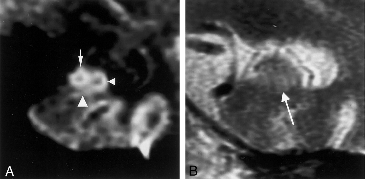
- Fig 4.
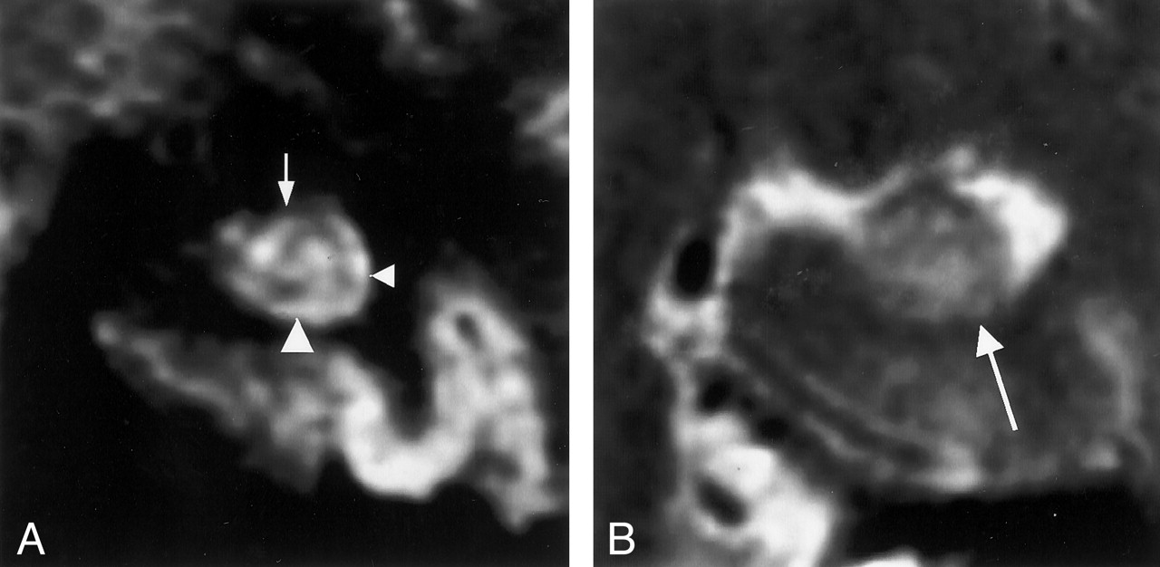
MR images in a 71-year-old woman with mild AD (MMSE score, 27; duration of illness, 2 years).
A, Multishot diffusion-weighted coronal image shows atrophic subiculum (left arrowhead) and CA1 (right arrowhead). However, the CA3–4 (arrow) is spared.
B, T2-weighted image scarcely demonstrates the inner structure of the left hippocampus (arrow).
- Fig 5.
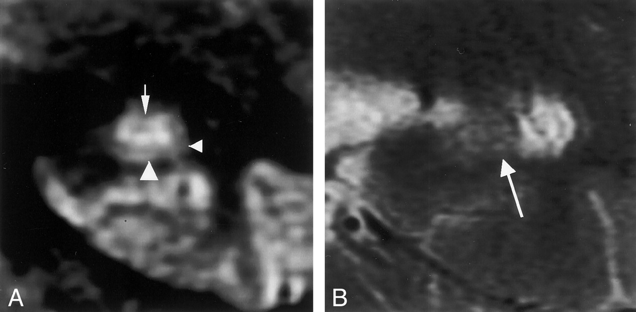
MR images in a 68-year-old man with mild AD (MMSE score, 25; duration of illness, 3 years).
A, Multishot diffusion-weighted coronal image shows an atrophic subiculum (left arrowhead) and CA1 (right arrowhead). CA3–4 (arrow) is spared.
B, T2-weighted image scarcely demonstrates the inner structure of the left hippocampus (arrow).
- Fig 6.
MR images in a 64-year-old woman with moderate AD (MMSE score, 8; duration of illness, 12 years).
A, Multishot diffusion-weighted coronal image shows an atrophic subiculum (left arrowhead). CA1 (right arrowhead) is comparatively spared; however, CA3–4 (arrow) is atrophic.
B, T2-weighted image scarcely demonstrates the inner structure of the left hippocampus (arrow).
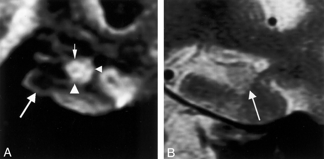
- Fig 7.
MR images in a 71-year-old woman with moderate AD (MMSE score, 6; duration of illness, 11 years).
A, Multishot diffusion-weighted coronal image shows severe atrophy of the subiculum (left arrowhead) and CA3–4 (small arrow). Atrophy of CA1 (right arrowhead) is mild. Image also shows atrophy of the parahippocampal gyrus (large arrow).
B, T2-weighted image barely shows the inner structure of the hippocampus (arrow).
- Fig 8.
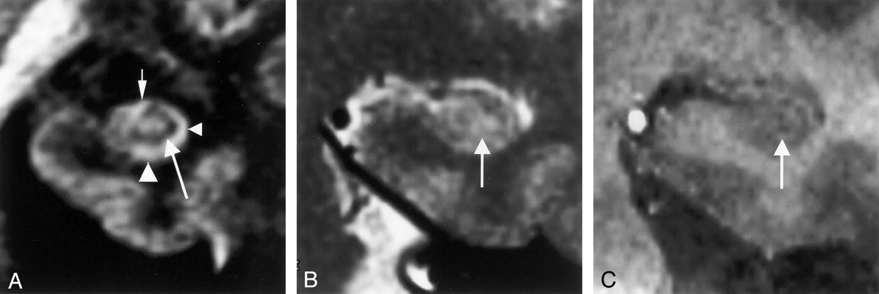
MR images in a 45-year-old man (control subject).
A, Multishot diffusion-weighted coronal image clearly shows the inner structure of the left hippocampus: the subiculum (left arrowhead), CA1 of the hippocampus proper (right arrowhead), CA3–4 (small arrow), and the superficial medullary lamina (large arrow).
B, T2-weighted coronal image faintly shows the superficial medullary lamina (arrow) as a hypointense area.
C, T1-weighted 3D volumetric SPGR image (26/4.3; FOV, 18 cm; matrix, 256 × 256; thickness, 0.8 mm; slab, 64 mm) shows the superficial medullary lamina (arrow) as a hyperintense area and depicts it more faintly than does the T2-weighted image.


Tables
Measurement Site and Group No. of Cases with Measurable Site On Multishot Diffusion-Weighted Images On T2-Weighted Images Width of subiculum Control 14 (100) 1 (7) Mild AD 10 (83) 0 (0) Moderate AD 10 (71) 0 (0) Width of CA1 Control 14 (100) 3 (21) Mild AD 11 (92) 0 (0) Moderate AD 11 (79) 2 (14) Width of CA3–4 Control 14 (100) 1 (7) Mild AD 11 (92) 1 (8) Moderate AD 12 (86) 0 (0) Height of CA3–4 Control 14 (100) 2 (14) Mild AD 10 (83) 0 (0) Moderate AD 11 (79) 0 (0) Note.—Data are the numbers of cases. Data in parentheses are percentages. The control group included 14 participants; the mild AD group, 12; and the moderate AD group, 14.
Measurement Site Intraobserver Variability, % Interobserver Variability % Observer 1 Observer 1 Width of subiculum 9.70 ± 8.89 8.95 ± 6.09 8.34 ± 6.10 Width of CA1 6.01 ± 5.00 6.40 ± 4.27 7.97 ± 5.31 Width of CA3–4 3.91 ± 3.22 4.58 ± 4.05 4.60 ± 4.55 Height of CA3–4 6.23 ± 5.76 5.92 ± 5.56 6.99 ± 4.53 Note.—Data are the mean coefficient of variation ± standard deviation.
Measurement Site Control Group Mild AD Group Moderate AD Group Width of subiculum, mm 2.60 ± 0.53 1.28 ± 0.15 1.14 ± 0.17 Width of CA1, mm 1.90 ± 0.14 1.46 ± 0.14 1.39 ± 0.19 Width of CA3–4, mm 7.18 ± 0.72 6.56 ± 0.85 5.70 ± 0.70 Height of CA3–4, mm 4.32 ± 0.57 4.30 ± 0.58 4.23 ± 0.63 Note.—Data represent the mean dimension ± standard deviation.
Measurement Site and Comparison DM* SEM† P Value 95% CI‡ Width of subiculum Control vs mild AD 1.32 0.15 <.001 0.95, 1.70 Control vs moderate AD 1.46 0.15 <.001 1.09, 1.83 Mild AD vs moderate AD 0.14 0.16 .688 −0.27, 0.54 Width of CA1 Control vs mild AD 0.43 0.06 <.001 0.28, 0.59 Control vs moderate AD 0.51 0.06 <.001 0.35, 0.67 Mild AD vs moderate AD 0.08 0.07 .501 −0.09, 0.24 Width of CA3–4 Control vs mild AD 0.62 0.30 .117 −0.12, 1.37 Control vs moderate AD 1.49 0.30 <.001 0.76, 2.21 Mild AD vs moderate AD 0.86 0.31 .025 0.09, 1.64 Height of CA3–4 Control vs mild AD 0.02 0.24 .995 −0.58, 0.63 Control vs moderate AD 0.10 0.24 .911 −0.49, 0.68 Mild AD vs moderate AD 0.07 0.26 .955 −0.56, 0.71 Note.—Intergroup analysis was performed by using the Tukey honestly significant difference adjustment.
* Difference of the means.
† Standard error of the mean.
‡ 95% confidence interval.
In this issue


Jump to section
Related Articles
Cited By...
- Mesoscopic whole-brain T2*-weighted and associated quantitative MRI in healthy humans at 10.5 T
- A Large-scale Comparison of Cortical and Subcortical Structural Segmentation Methods in Alzheimers Disease: a Statistical Approach
- Human anterolateral entorhinal cortex volumes are associated with preclinical cognitive decline
- Shared Vulnerability of Two Synaptically-Connected Medial Temporal Lobe Areas to Age and Cognitive Decline: A Seven Tesla Magnetic Resonance Imaging Study
- Hippocampal CA1 apical neuropil atrophy in mild Alzheimer disease visualized with 7-T MRI
- Diffusion Tensor Microscopy Indicates the Cytoarchitectural Basis for Diffusion Anisotropy in the Human Hippocampus