
Article Figures & Data
Figures
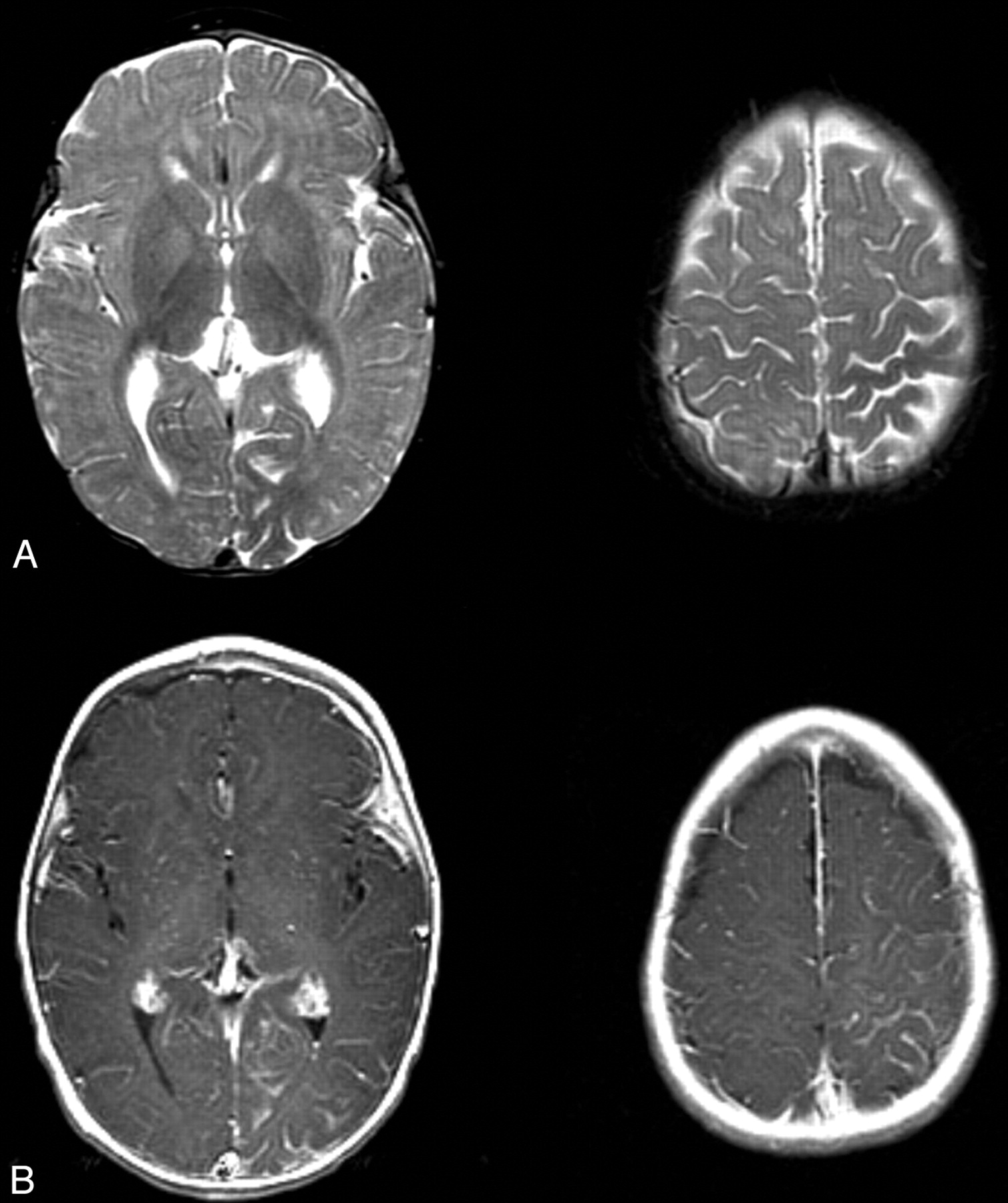
- Fig 1.
T2- and contrast-enhanced T1-weighted images from the case of a 9-month-old male patient with Sturge-Weber syndrome and new onset of seizure.
A, Axial T2-weighted images show mild parenchymal volume loss involving the left parietal and occipital lobes and decreased signal intensity in the subcortical white matter underlying the area of leptomeningeal enhancement seen in B.
B, Axial contrast-enhanced T1-weighted images. Leptomeningeal enhancement is most marked in the left parietal lobe and medial occipital lobe.
- Fig 2.
T2-weighted images, time-series curves, and parameter maps generated from the same contrast-enhanced T2*-weighted perfusion image.
A, Time-series plot from contrast-enhanced T2*-weighted perfusion study shows delayed recovery phase, reflecting impaired venous drainage in the left parietal lobe.
B, Time-series plot shows delayed bolus arrival and protracted recovery, indicating arterial and venous perfusion abnormality more focally in the high left parietal cortex.
C, Symmetric time-series curves in the unaffected frontal lobes.
D, Parameter maps of regional cerebral blood flow, mean transit time, and time-to- peak show decreased flow and increased transit time in the same region. A very mild increase in regional cerebral blood volume is noted.
- Fig 3.
MR spectroscopy at the level of the centrum semiovale shows nearly symmetric and normal levels of N-acetylaspartate and creatine. Mild elevation of choline is shown in the left parietal region, which is best depicted on the metabolic map. No lactate is detected. Asterisk marks broad peaks at 1.2 through 1.4 ppm that most likely reflect lipid contamination.


In this issue


Jump to section
Related Articles
Cited By...
- Alternative Venous Pathways: A Potential Key Imaging Feature for Early Diagnosis of Sturge-Weber Syndrome Type 1
- Leptomeningeal Enhancement in Multiple Sclerosis and Other Neurological Diseases: A Systematic Review and Meta-Analysis
- The Bone Does Not Predict the Brain in Sturge-Weber Syndrome
- Hemodynamic Effects of Developmental Venous Anomalies with and without Cavernous Malformations
- Clinical Correlates of White Matter Blood Flow Perfusion Changes in Sturge-Weber Syndrome: A Dynamic MR Perfusion-Weighted Imaging Study
- Teaching NeuroImages: Sturge-Weber syndrome presenting in a 58-year-old woman with seizures