Advertisement

Main menu

  • Home
  • Content
    • Current Issue
    • Accepted Manuscripts
    • Article Preview
    • Past Issue Archive
    • Video Articles
    • AJNR Case Collection
    • Case of the Week Archive
    • Case of the Month Archive
    • Classic Case Archive
  • Special Collections
    • AJNR Awards
    • ASNR Foundation Special Collection
    • Most Impactful AJNR Articles
    • Photon-Counting CT
    • Spinal CSF Leak Articles (Jan 2020-June 2024)
  • Multimedia
    • AJNR Podcasts
    • AJNR SCANtastic
    • Trainee Corner
    • MRI Safety Corner
    • Imaging Protocols
  • For Authors
    • Submit a Manuscript
    • Submit a Video Article
    • Submit an eLetter to the Editor/Response
    • Manuscript Submission Guidelines
    • Statistical Tips
    • Fast Publishing of Accepted Manuscripts
    • Graphical Abstract Preparation
    • Imaging Protocol Submission
    • Author Policies
  • About Us
    • About AJNR
    • Editorial Board
    • Editorial Board Alumni
  • More
    • Become a Reviewer/Academy of Reviewers
    • Subscribers
    • Permissions
    • Alerts
    • Feedback
    • Advertisers
    • ASNR Home

User menu

  • Alerts
  • Log in

Search

  • Advanced search
American Journal of Neuroradiology

American Journal of Neuroradiology

ASHNR American Society of Functional Neuroradiology ASHNR American Society of Pediatric Neuroradiology ASSR
  • Alerts
  • Log in

Advanced Search

  • Home
  • Content
    • Current Issue
    • Accepted Manuscripts
    • Article Preview
    • Past Issue Archive
    • Video Articles
    • AJNR Case Collection
    • Case of the Week Archive
    • Case of the Month Archive
    • Classic Case Archive
  • Special Collections
    • AJNR Awards
    • ASNR Foundation Special Collection
    • Most Impactful AJNR Articles
    • Photon-Counting CT
    • Spinal CSF Leak Articles (Jan 2020-June 2024)
  • Multimedia
    • AJNR Podcasts
    • AJNR SCANtastic
    • Trainee Corner
    • MRI Safety Corner
    • Imaging Protocols
  • For Authors
    • Submit a Manuscript
    • Submit a Video Article
    • Submit an eLetter to the Editor/Response
    • Manuscript Submission Guidelines
    • Statistical Tips
    • Fast Publishing of Accepted Manuscripts
    • Graphical Abstract Preparation
    • Imaging Protocol Submission
    • Author Policies
  • About Us
    • About AJNR
    • Editorial Board
    • Editorial Board Alumni
  • More
    • Become a Reviewer/Academy of Reviewers
    • Subscribers
    • Permissions
    • Alerts
    • Feedback
    • Advertisers
    • ASNR Home
  • Follow AJNR on Twitter
  • Visit AJNR on Facebook
  • Follow AJNR on Instagram
  • Join AJNR on LinkedIn
  • RSS Feeds

AJNR is seeking candidates for the AJNR Podcast Editor. Read the position description.

Research ArticlePEDIATRICS

MR Imaging Quantification of Cerebellar Growth Following Hypoxic-Ischemic Injury to the Neonatal Brain

Elisabeth Le Strange, Nadeem Saeed, Frances M. Cowan, A. David Edwards and Mary A. Rutherford
American Journal of Neuroradiology March 2004, 25 (3) 463-468;
Elisabeth Le Strange
  • Find this author on Google Scholar
  • Find this author on PubMed
  • Search for this author on this site
Nadeem Saeed
  • Find this author on Google Scholar
  • Find this author on PubMed
  • Search for this author on this site
Frances M. Cowan
  • Find this author on Google Scholar
  • Find this author on PubMed
  • Search for this author on this site
A. David Edwards
  • Find this author on Google Scholar
  • Find this author on PubMed
  • Search for this author on this site
Mary A. Rutherford
  • Find this author on Google Scholar
  • Find this author on PubMed
  • Search for this author on this site
  • Article
  • Figures & Data
  • Info & Metrics
  • Responses
  • References
  • PDF
Loading

Article Figures & Data

Figures

  • Tables
  • Fig 1.
    • Download figure
    • Open in new tab
    • Download powerpoint
    Fig 1.

    T1-weighted MR images (860/20).

    A, Day 5 image shows focal infarction. There is a loss of gray matter–white matter differentiation in the posterior lobe on the left. This is consistent with perinatal infarction within the territory of the left middle cerebral artery.

    B, Day 12 image shows basal ganglia and thalamic lesions. There are persistent, abnormal high-signal-intensity regions in the posterior lentiform nuclei and lateral thalami (arrows). The intervening posterior limb of the internal capsule shows reduced high signal intensity from myelin.

  • Fig 2.
    • Download figure
    • Open in new tab
    • Download powerpoint
    Fig 2.

    Total cerebellar volume versus age in all infants, by group (n = 20).

  • Fig 3.
    • Download figure
    • Open in new tab
    • Download powerpoint
    Fig 3.

    Cerebellar hemispheres in infants with cerebral infarcts. Volume ipsilateral to the infarct versus volume contralateral to the infarct (n = 11). The line is the line of equality.

  • Fig 4.
    • Download figure
    • Open in new tab
    • Download powerpoint
    Fig 4.

    Cerebellar hemispheres in all infants. Volume of the right hemisphere versus volume of the left hemisphere (n = 20). The line is the line of equality.
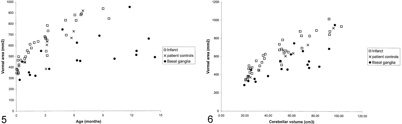
  • Fig 5.
    • Download figure
    • Open in new tab
    • Download powerpoint
    Fig 5.

    Cross-sectional area of the vermis versus age (n = 20).

  • Fig 6.
    • Download figure
    • Open in new tab
    • Download powerpoint
    Fig 6.

    Cross-sectional area of the vermis versus total cerebellar volume (n = 23).

Tables

  • Figures
  • Clinical details and timing of studies

    Patient no.GestationFetal Distress*Delivery†Apgar ScoresBirthweight, kgMR Imaging Findings‡Age at StudiesOutcomeHead Circumference at ≥12 mo
    141Ctg/mslNvd5,83.75Transient bg2 d, 3 d, 4 mNormal1st
    240CtgEmcd1,72.46Normal3 m, 6 mNormal25th
    340CtgEmcd1,53.12Transient bg6 d, 1 m, 3 m, 7 mNormal50th
    441NilNvd7,93.56Infarct, left1 m, 2 mNormal25th
    539NilNvd3,53.35Infarct, left1 m, 3 m, 5 mModerate<0.4th
    640NilNvd9,102.84Infarct, left1 m, 3 m, 8 mNormal75th
    740CtgEmcd2,73.68Infarct, left2 d, 3 d, 9 dNormal9–25th
    841Ctg/mslEmcd5,92.9Infarct, right2 d, 9 d, 1 m, 3 m, 6 mNormal2–9th
    941MslEmcs4,93.12Infarct, right3 m, 5 m, 9 mModerate25th
    1041Ctg/mslForceps5,74.25Infarct, left6 d, 1.5 m, 3 m, 6 mNormal25th
    1142MslVentouse6,83.1Infarct, left6 d, 1 m, 3 mNormal25th
    1239NilNvd7,103.59Infarct, left6 m, 21 mModerate91st
    1339NilNvd5,73.34Infarct, left7 d, 1 m, 3 m, 8 mNormal<25th
    1441Ctg/mslEmcd2,83.22Infarct, left9 d, 16 d, 1.5 m, 3 m, 6 mModerate20th
    1542CtgEmcd0NASevere bg1.3 m, 3.5 m, 12 mSevere<3rd
    1641CtgForceps1,42.8Severe bg, wm1.5 m, 6 mSevere<0.4th
    1742Ctg/mslForceps0,04Severe bg11 m, 14 mSevere<0.4th
    2839Ctg/mslEmcd0,03.08Severe bg11 d, 2 m, 6.5 m, 12 mSevere<0.4th
    1940CtgNvd0,33.13Moderate bg14 d, 4.5 m, 11.5 mSevere45th
    2040CtgNvd5,73.98Severe bg6 m, 13.5 mSevere<0.4th
    • * Ctg indicates cardiotocograph; msl, meconium-stained liquor.

    • † Emcd indicates emergency cesarean delivery; nvd, normal vaginal delivery.

    • ‡ bg indicates basal ganglia lesions; wm, white matter.

PreviousNext
Back to top

In this issue

American Journal of Neuroradiology: 25 (3)
American Journal of Neuroradiology
Vol. 25, Issue 3
1 Mar 2004
  • Table of Contents
  • Index by author
Advertisement
Print
Download PDF
Email Article

Thank you for your interest in spreading the word on American Journal of Neuroradiology.

NOTE: We only request your email address so that the person you are recommending the page to knows that you wanted them to see it, and that it is not junk mail. We do not capture any email address.

Enter multiple addresses on separate lines or separate them with commas.
MR Imaging Quantification of Cerebellar Growth Following Hypoxic-Ischemic Injury to the Neonatal Brain
(Your Name) has sent you a message from American Journal of Neuroradiology
(Your Name) thought you would like to see the American Journal of Neuroradiology web site.
CAPTCHA
This question is for testing whether or not you are a human visitor and to prevent automated spam submissions.
Cite this article
Elisabeth Le Strange, Nadeem Saeed, Frances M. Cowan, A. David Edwards, Mary A. Rutherford
MR Imaging Quantification of Cerebellar Growth Following Hypoxic-Ischemic Injury to the Neonatal Brain
American Journal of Neuroradiology Mar 2004, 25 (3) 463-468;

Citation Manager Formats

  • BibTeX
  • Bookends
  • EasyBib
  • EndNote (tagged)
  • EndNote 8 (xml)
  • Medlars
  • Mendeley
  • Papers
  • RefWorks Tagged
  • Ref Manager
  • RIS
  • Zotero
0 Responses
Respond to this article
Share
Bookmark this article
MR Imaging Quantification of Cerebellar Growth Following Hypoxic-Ischemic Injury to the Neonatal Brain
Elisabeth Le Strange, Nadeem Saeed, Frances M. Cowan, A. David Edwards, Mary A. Rutherford
American Journal of Neuroradiology Mar 2004, 25 (3) 463-468;
del.icio.us logo Twitter logo Facebook logo Mendeley logo
  • Tweet Widget
  • Facebook Like
  • Google Plus One

Jump to section

  • Article
    • Abstract
    • Methods
    • Results
    • Discussion
    • Acknowledgments
    • Footnotes
    • References
  • Figures & Data
  • Info & Metrics
  • Responses
  • References
  • PDF

Related Articles

  • No related articles found.
  • PubMed
  • Google Scholar

Cited By...

  • Evaluation of Posterior Fossa Biometric Measurements on Fetal MRI in the Evaluation of Dandy-Walker Continuum
  • Uneven distribution of Purkinje cell injury in the cerebellar vermis of term neonates with hypoxic-ischemic encephalopathy
  • Injury to the Cerebellum in Term Asphyxiated Newborns Treated with Hypothermia
  • Predicting motor outcome and death in term hypoxic-ischemic encephalopathy
  • Does perinatal asphyxia impair cognitive function without cerebral palsy?
  • Crossref
  • Google Scholar

This article has not yet been cited by articles in journals that are participating in Crossref Cited-by Linking.

More in this TOC Section

  • SyMRI & MR Fingerprinting in Brainstem Myelination
  • Comparison of Image Quality and Radiation Dose in Pediatric Temporal Bone CT Using Photon-Counting Detector CT and Energy-Integrating Detector CT
  • Neonatal Hypocalcemia: Cerebral MRI Abnormalities
Show more Pediatrics

Similar Articles

Advertisement

Indexed Content

  • Current Issue
  • Accepted Manuscripts
  • Article Preview
  • Past Issues
  • Editorials
  • Editor's Choice
  • Fellows' Journal Club
  • Letters to the Editor
  • Video Articles

Cases

  • Case Collection
  • Archive - Case of the Week
  • Archive - Case of the Month
  • Archive - Classic Case

Special Collections

  • AJNR Awards
  • ASNR Foundation Special Collection
  • Most Impactful AJNR Articles
  • Photon-Counting CT
  • Spinal CSF Leak Articles (Jan 2020-June 2024)

More from AJNR

  • Trainee Corner
  • Imaging Protocols
  • MRI Safety Corner

Multimedia

  • AJNR Podcasts
  • AJNR Scantastics

Resources

  • Turnaround Time
  • Submit a Manuscript
  • Submit a Video Article
  • Submit an eLetter to the Editor/Response
  • Manuscript Submission Guidelines
  • Statistical Tips
  • Fast Publishing of Accepted Manuscripts
  • Graphical Abstract Preparation
  • Imaging Protocol Submission
  • Evidence-Based Medicine Level Guide
  • Publishing Checklists
  • Author Policies
  • Become a Reviewer/Academy of Reviewers
  • News and Updates

About Us

  • About AJNR
  • Editorial Board
  • Editorial Board Alumni
  • Alerts
  • Permissions
  • Not an AJNR Subscriber? Join Now
  • Advertise with Us
  • Librarian Resources
  • Feedback
  • Terms and Conditions
  • AJNR Editorial Board Alumni

American Society of Neuroradiology

  • Not an ASNR Member? Join Now

© 2025 by the American Society of Neuroradiology All rights, including for text and data mining, AI training, and similar technologies, are reserved.
Print ISSN: 0195-6108 Online ISSN: 1936-959X

Powered by HighWire