
Article Figures & Data
Figures
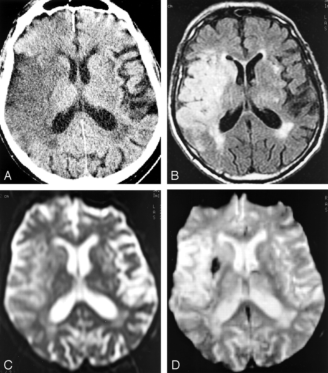
- Fig 1.
Increased conspicuity of HT and upgrade in bleeding categorization at 24 hours by using the different sequences. The patient is a 75-year-old woman admitted 24 hours previously for aphasia, right hemiparesis, and right conjugate ocular deviation who was treated by a combination of 450 mg ASA and low-dose low molecular heparinoids.
A, Axial CT scan, showing an ambiguous isoattenuated area within infarcted left PCA territory, which was ultimately rated as “HI1” after extended debate. The hemorrhagic area displays a similar attenuation as the normal parenchyma raising the hypothesis of spared tissue rather than petechial HT. Consensual interobserver rating of “HI2” was retained.
B, FSE-FLAIR image, showing punctuate hypointense foci confirming petechial hemorrhage and the “HI2” rating.
C, EPI-SE T2-weighted image, demonstrating homogeneous, round hypointense area rated as “PH1” because of the clotlike aspect of the bleeding, without mass effect but with the presence of a surrounding hyperintense ring of ischemic tissue.
D, EPI-GRE T2*-weighted image, demonstrating enlargement of the hemorrhagic area resulting in “PH2” rating since the hypointense artifact had recovered the ring of hyperintense ischemic tissue seen on FSE-FLAIR and EPI-SE T2-weighted images (previous views), thereby filling the theoretical >30% of infarct area criterion for “PH2” rating.
- Fig 2.
EPI-GRE T2*-weighted sequence as the only positive technique for the 24-hour detection of post-thrombolytic petechial hemorrhage in an 86-year-old man admitted 24 hours previously and treated with 58 mg r-tPA IV 110 minutes after the onset of a left hemiplegia (NIH stroke score on admission, 13).
A, CT scan, showing a large acute hypoattenuated area within the MCA territory without sign of fresh blood extravasation.
B, FSE-FLAIR image, showing uniform hyperintensity within the ischemic area without hypointense hemorrhagic focus.
C, EPI-SE T2-weighted image, showing similar findings of positivity for ischemia and negativity for HT as in previous 3B image.
D, EPI-GRE T2*-weighted image, showing hypointense susceptibility artifact within both putamen and globus pallidus, revealing confluent petechiae. The extravasation was rated “HI2.”
- Fig 3.
Discrepancy in CT and MR results between infarcted subareas in a 74-year-old woman admitted 24 hours previously and treated by 67 mg of rt-PA administered intravenously 185 minutes after the sudden onset of a left hemiplegia (NIH stroke score on admission, 14)
A, axial CT scan through the thalamic level shows a round area of isoattenuation relative to normal brain surrounded by ischemic edema within the lenticulate nucleus, which was rated confluent petechial “HI2” after intra- and interobserver controversies (similar to the previous case).
B, EPI-GRE T2*-weighted MR image in the similar location as Fig 2A, confirming the presence of freshly extravasated blood.
C, Axial CT scan through an upper level, showing homogeneous ischemic hypoattenuation without hyperattenuated focus suggesting HT.
D, EPI-GRE T2*-weighted MR image in the similar location as Fig 2C, disclosing unsuspected subependymal hemorrhagic focus.


Tables
Intra- and interobserver consensus for CT and MR sequences
CT FSE-FLAIR EPI-SE T2 EPI-GRE T2* Observer 1: First session 6 9 9 12 Second session 6 9 11 12 Consensus 8 9 9 12 Intraobserver 1 kappa 0.780702 1 0.834437 1 Observer 2: First session 6 9 8 12 Second session 4 9 9 12 Consensus 4 9 8 12 Intraobserver 2 kappa 0.752475 1 0.911032 1 Interobserver consensus 7 9 9 12 Interobserver kappa 0.576271 1 0.911032 1 Note.—Table lists for each pulse sequence the number of patients (roman) considered positive at each individual scoring session, together with the Cohen’s kappa coefficient of agreement (italic) for each session.
In this issue


Jump to section
Related Articles
Cited By...
- Determinants and Clinical Relevance of Iodine Contrast Extravasation after Endovascular Thrombectomy: A Dual-Energy CT Study
- Hemorrhage rates in patients with acute ischemic stroke treated with intravenous alteplase and thrombectomy versus thrombectomy alone
- Type of intracranial hemorrhage after endovascular stroke treatment: association with functional outcome
- Functional Outcome, Recanalization, and Hemorrhage Rates After Large Vessel Occlusion Stroke Treated With Tenecteplase Before Thrombectomy
- Association of initial imaging modality and futile recanalization after thrombectomy
- Dual energy CT after stroke thrombectomy alters assessment of hemorrhagic complications
- Agreement between core laboratory and study investigators for imaging scores in a thrombectomy trial
- Focal Low and Global High Permeability Predict the Possibility, Risk, and Location of Hemorrhagic Transformation following Intra-Arterial Thrombolysis Therapy in Acute Stroke
- Brain Edema Predicts Outcome After Nonlacunar Ischemic Stroke
- Suspicious Neuroimaging Pattern of Thrombotic Microangiopathy
- Substantial Observer Variability in the Differentiation Between Primary Intracerebral Hemorrhage and Hemorrhagic Transformation of Infarction on CT Brain Imaging
- Haemorrhagic transformation in acute ischaemic stroke following thrombolysis therapy: classification, pathogenesis and risk factors
- Angiogenesis Detected After Embolic Stroke in Rat Brain Using Magnetic Resonance T2*WI
- Asymptomatic Hemorrhage After Thrombolysis May Not Be Benign: Prognosis by Hemorrhage Type in the Canadian Alteplase for Stroke Effectiveness Study Registry
- Imaging