
Article Figures & Data
Figures
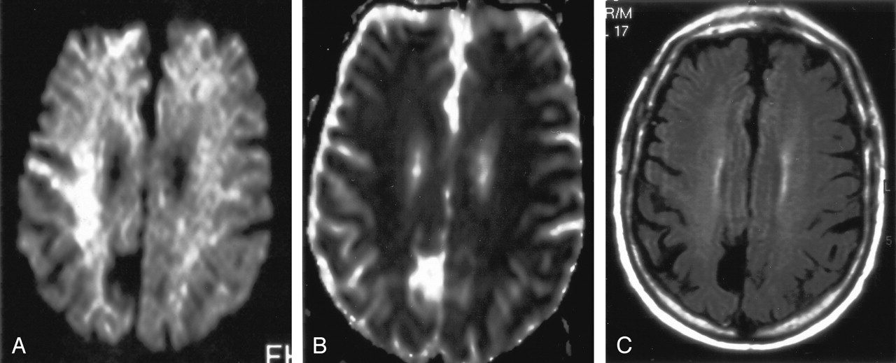
- Fig 1.
Patient with isolated restricted diffusion.
A and B, DW image (A) and ADC map (B) show restricted diffusion in the PLIC.
C and D, Findings on corresponding FLAIR image (C) and contrast-enhanced T1WI (D) are unremarkable.
- Fig 2.
Isolated restricted diffusion in a patient who recovered without residual symptoms.
A, DW image shows asymmetric (right greater than left) high signal intensity in the superior corona radiata and subcortical white matter.
B, Corresponding ADC map shows subtle low signal intensity.
C, No abnormalities are present on the FLAIR image.
- Fig 3.
Images at the level of the basal ganglia and sylvian fissure in a patient with hyperintensity on FLAIR images.
A, Contrast-enhanced FLAIR image shows increased signal intensity in the basal ganglia and PLIC bilaterally, left thalamus, and left periventricular white matter, as well as the temporoparietal and occipital sulci.
B, Contrast-enhanced T1WI shows no corresponding enhancement.
C and D, DW image (C) and ADC map (D) show restricted diffusion is seen in the left periventricular white matter, right basal ganglia, and bilateral PLIC.
E and F, Contiguous FLAIR images show increased signal intensity in the midbrain and medial temporal lobes bilaterally, as well as in the right temporal lobe peripherally. Hyperintensity is again shown in the sulci, signifying WNV meningoencephalitis.
- Fig 4.
Another patient with hyperintense parenchymal abnormalities on FLAIR images.
A, FLAIR image demonstrates increased signal intensity in the bilateral cerebellum and in the left occipital lobe.
B, Contrast-enhanced T1WI shows the same areas of enhancement.
C and D, Corresponding DW image (C) and ADC map (D) show diffusion restriction in the right cerebellum.
- Fig 5.
Patient with meningeal involvement.
A, Axial contrast-enhanced FLAIR image shows increased signal intensity in the sulci compatible with meningitis.
B, Abnormality is not appreciated on the corresponding contrast-enhanced T1WI.
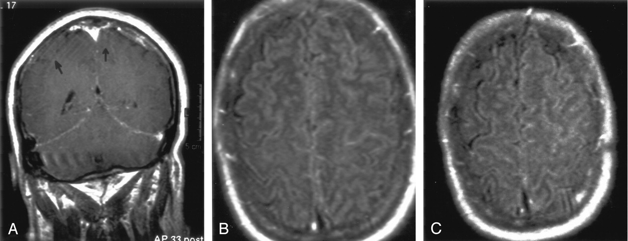
- Fig 6.
Patient 2 with meningeal involvement.
A, Coronal gadolinium-enhanced T1WI shows abnormal enhancement of the tentorial meninges and focal increased meningeal enhancement in the left parietal lobe. Arrows indicate subtle enhancement in the sulci.
B and C, FLAIR images show increased signal intensity in the sulci, more prominent on the left than on the right.
- Fig 7.
Patient with intraspinal abnormalities.
A, Axial contrast-enhanced T1WI through the region of the cauda equina shows marked abnormal enhancement of the nerve roots, which appear as bright dots in the thecal sac.
B, Sagittal gadolinium-enhanced T1WI demonstrates prominently enhancing nerve roots.
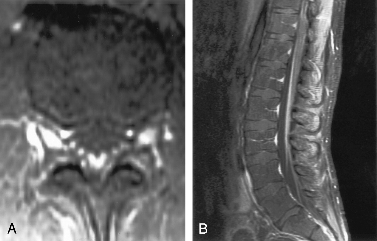
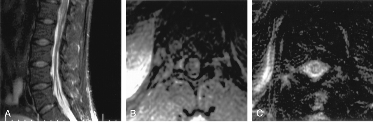
- Fig 8.
Another patient with intraspinal abnormalities.
A, Sagittal T2WI shows abnormal increased signal intensity in the conus medullaris.
B, Axial gadolinium-enhanced T1WI shows abnormal foci of enhancement in the conus medullaris.
C, Axial T2WI shows markedly increased signal intensity in the thoracic cord. This is appreciated despite image degradation due to artifact.
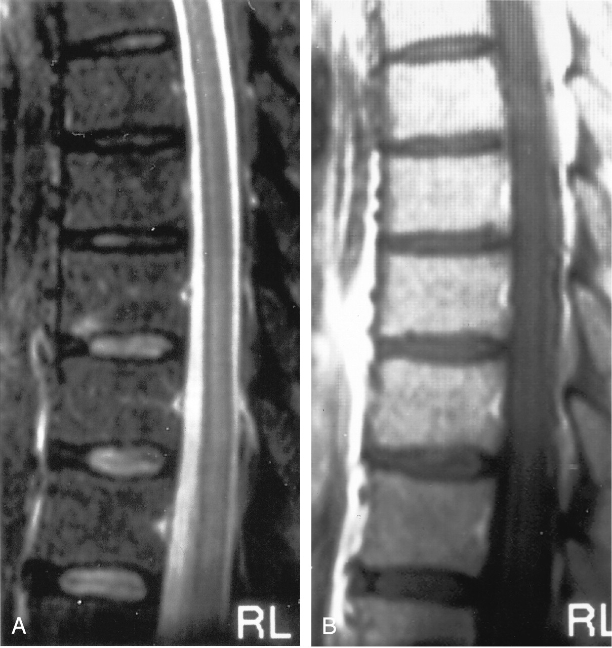
- Fig 9.
Subtle thoracic cord signal abnormalities in a patient with moderate-to-severe neurologic deficits.
A, Sagittal T2WI demonstrates increased signal intensity in the mid-to-lower portion of the central thoracic cord.
B, Sagittal contrast-enhanced T1WI demonstrates patchy areas of subtle enhancement.


Tables
Imaging Finding Mean Age (y) Mean Hospitalization (d) Clinical Outcomes Normal (n = 5) 28.4 (18–43) 6.4 (4–13) Complete recovery (n = 4), mild residual expressive dysphasia (n = 1) Isolated restricted diffusion (n = 4) 75.5 (73–80) 6.5 (4–11) Complete recovery FLAIR and T2WI hyperintensity (n = 3) 63.7 (46–75) 21.3 (11–27) Death (n = 2), severe neurologic deficits (n = 1) Meningeal involvement (n = 2) 60.5 (60–61) 20 (19–21) Severe neurologic deficits Intraspinal abnormalities (n = 3) 36.3 (28–46) 26.6 (10–42) Moderate-to-severe neurologic deficits Note.—Data in parentheses are ranges.
In this issue


Jump to section
Related Articles
Cited By...
- Progressive MRI findings of West Nile virus encephalitis in a patient with diabetes mellitus
- Long-term outcome in neuroZika: When biological diagnosis matters
- Looking out for the blind spot
- Alpha-Synuclein Expression Restricts RNA Viral Infections in the Brain
- Armies of Pestilence: CNS Infections as Potential Weapons of Mass Destruction
- Clinical and Radiological Predictors of Outcome for Murray Valley Encephalitis
- Persistent West Nile Virus Associated with a Neurological Sequela in Hamsters Identified by Motor Unit Number Estimation
- CXCR3 Mediates Region-Specific Antiviral T Cell Trafficking within the Central Nervous System during West Nile Virus Encephalitis
- An 85-year-old man with chronic lymphocytic leukemia and altered mental status