
Article Figures & Data
Figures
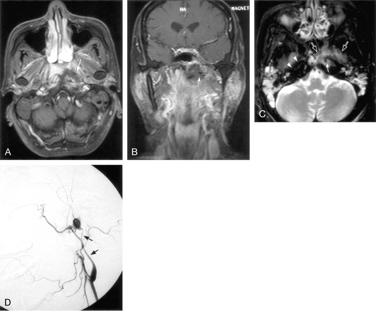
- Fig 1.
Case 1, with nasopharyngeal tissue necrosis and pseudoaneurysm, led to a grave outcome.
A, Axial contrast-enhanced T1-weighted MR image shows the nonenhancing soft tissues, mixed with tiny air bubbles in left-sided nasopharyngeal lateral recess (arrow).
B, Coronal contrast-enhanced T1-weighted image shows a complex mass containing soft tissue and air bubbles (arrow) just beneath skull base.
C, Axial T2-weighted MR image shows an intermediate hyperintense soft tissue mass (open arrows) surrounding the high cervical carotid artery with possible aneurysm formation (arrow). Note the normal right carotid artery for comparison (arrowheads).
D, Left carotid angiogram, lateral projection, shows an aneurysm with rapid contrast medium filling and extravasation in the high cervical internal carotid artery (arrows), after which no antegrade flow beyond the aneuyrsm is identified.
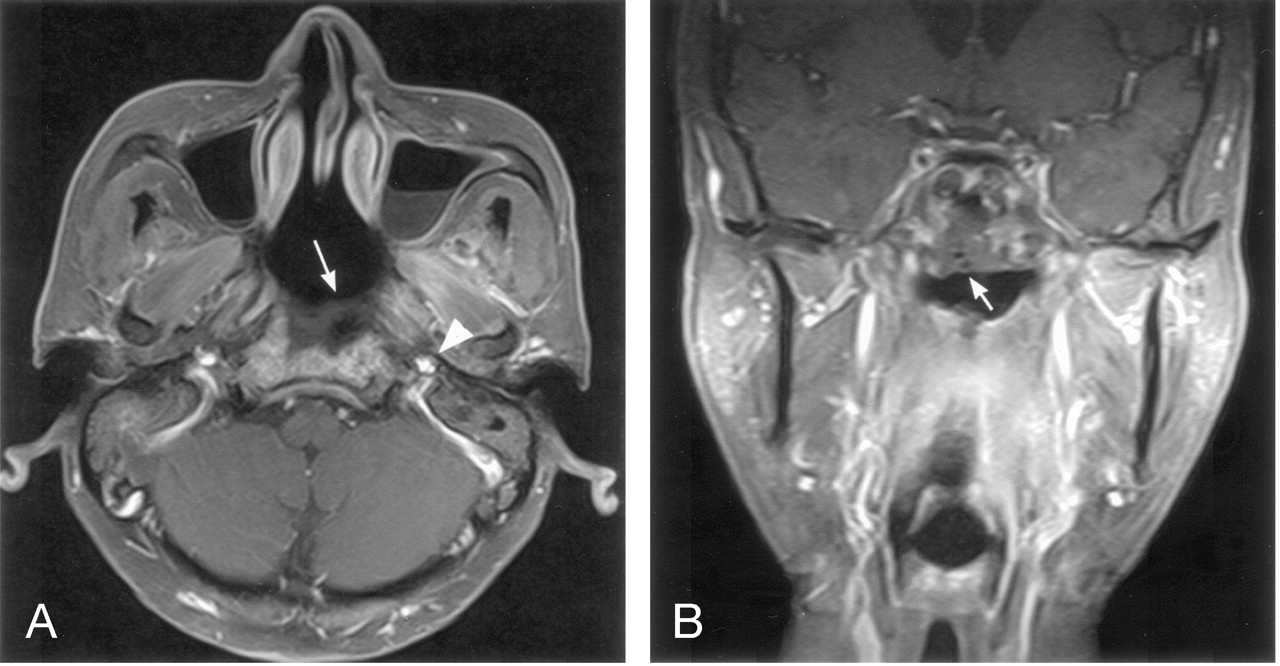
- Fig 2.
Case 3, with similar MR findings to Case 1, had a prompt angiography study and much better prognosis.
A, Axial contrast-enhanced T1-weighted image shows a nonenhancing soft tissue in the right nasopharyngeal lateral recess, extending to the carotid sheath (arrows) and encasing the carotid artery (open arrow). Note the contralateral carotid artery for comparison (arrowhead).
B, Coronal contrast-enhanced T1-weighted MR image shows the necrotic mass with lateral extension into the parapharyngeal space (arrow).


