Advertisement

Main menu

  • Home
  • Content
    • Current Issue
    • Accepted Manuscripts
    • Article Preview
    • Past Issue Archive
    • Video Articles
    • AJNR Case Collection
    • Case of the Week Archive
    • Case of the Month Archive
    • Classic Case Archive
  • Special Collections
    • AJNR Awards
    • ASNR Foundation Special Collection
    • Most Impactful AJNR Articles
    • Photon-Counting CT
    • Spinal CSF Leak Articles (Jan 2020-June 2024)
  • Multimedia
    • AJNR Podcasts
    • AJNR SCANtastic
    • Trainee Corner
    • MRI Safety Corner
    • Imaging Protocols
  • For Authors
    • Submit a Manuscript
    • Submit a Video Article
    • Submit an eLetter to the Editor/Response
    • Manuscript Submission Guidelines
    • Statistical Tips
    • Fast Publishing of Accepted Manuscripts
    • Graphical Abstract Preparation
    • Imaging Protocol Submission
    • Author Policies
  • About Us
    • About AJNR
    • Editorial Board
    • Editorial Board Alumni
  • More
    • Become a Reviewer/Academy of Reviewers
    • Subscribers
    • Permissions
    • Alerts
    • Feedback
    • Advertisers
    • ASNR Home

User menu

  • Alerts
  • Log in

Search

  • Advanced search
American Journal of Neuroradiology

American Journal of Neuroradiology

ASHNR American Society of Functional Neuroradiology ASHNR American Society of Pediatric Neuroradiology ASSR
  • Alerts
  • Log in

Advanced Search

  • Home
  • Content
    • Current Issue
    • Accepted Manuscripts
    • Article Preview
    • Past Issue Archive
    • Video Articles
    • AJNR Case Collection
    • Case of the Week Archive
    • Case of the Month Archive
    • Classic Case Archive
  • Special Collections
    • AJNR Awards
    • ASNR Foundation Special Collection
    • Most Impactful AJNR Articles
    • Photon-Counting CT
    • Spinal CSF Leak Articles (Jan 2020-June 2024)
  • Multimedia
    • AJNR Podcasts
    • AJNR SCANtastic
    • Trainee Corner
    • MRI Safety Corner
    • Imaging Protocols
  • For Authors
    • Submit a Manuscript
    • Submit a Video Article
    • Submit an eLetter to the Editor/Response
    • Manuscript Submission Guidelines
    • Statistical Tips
    • Fast Publishing of Accepted Manuscripts
    • Graphical Abstract Preparation
    • Imaging Protocol Submission
    • Author Policies
  • About Us
    • About AJNR
    • Editorial Board
    • Editorial Board Alumni
  • More
    • Become a Reviewer/Academy of Reviewers
    • Subscribers
    • Permissions
    • Alerts
    • Feedback
    • Advertisers
    • ASNR Home
  • Follow AJNR on Twitter
  • Visit AJNR on Facebook
  • Follow AJNR on Instagram
  • Join AJNR on LinkedIn
  • RSS Feeds

AJNR is seeking candidates for the AJNR Podcast Editor. Read the position description.

OtherPEDIATRICS

Brain Apparent Diffusion Coefficient Decrease During Correction of Severe Hypernatremic Dehydration

Andrea Righini, Luca Ramenghi, Salvatore Zirpoli, Fabio Mosca and Fabio Triulzi
American Journal of Neuroradiology August 2005, 26 (7) 1690-1694;
Andrea Righini
  • Find this author on Google Scholar
  • Find this author on PubMed
  • Search for this author on this site
Luca Ramenghi
  • Find this author on Google Scholar
  • Find this author on PubMed
  • Search for this author on this site
Salvatore Zirpoli
  • Find this author on Google Scholar
  • Find this author on PubMed
  • Search for this author on this site
Fabio Mosca
  • Find this author on Google Scholar
  • Find this author on PubMed
  • Search for this author on this site
Fabio Triulzi
  • Find this author on Google Scholar
  • Find this author on PubMed
  • Search for this author on this site
  • Article
  • Figures & Data
  • Info & Metrics
  • Responses
  • References
  • PDF
Loading

Article Figures & Data

Figures

  • Fig 1.
    • Download figure
    • Open in new tab
    • Download powerpoint
    Fig 1.

    Time course of sodium and chlorum serum concentrations during the neonatal intensive care unit stay. I MRI indicates the first imaging study.

  • Fig 2.
    • Download figure
    • Open in new tab
    • Download powerpoint
    Fig 2.

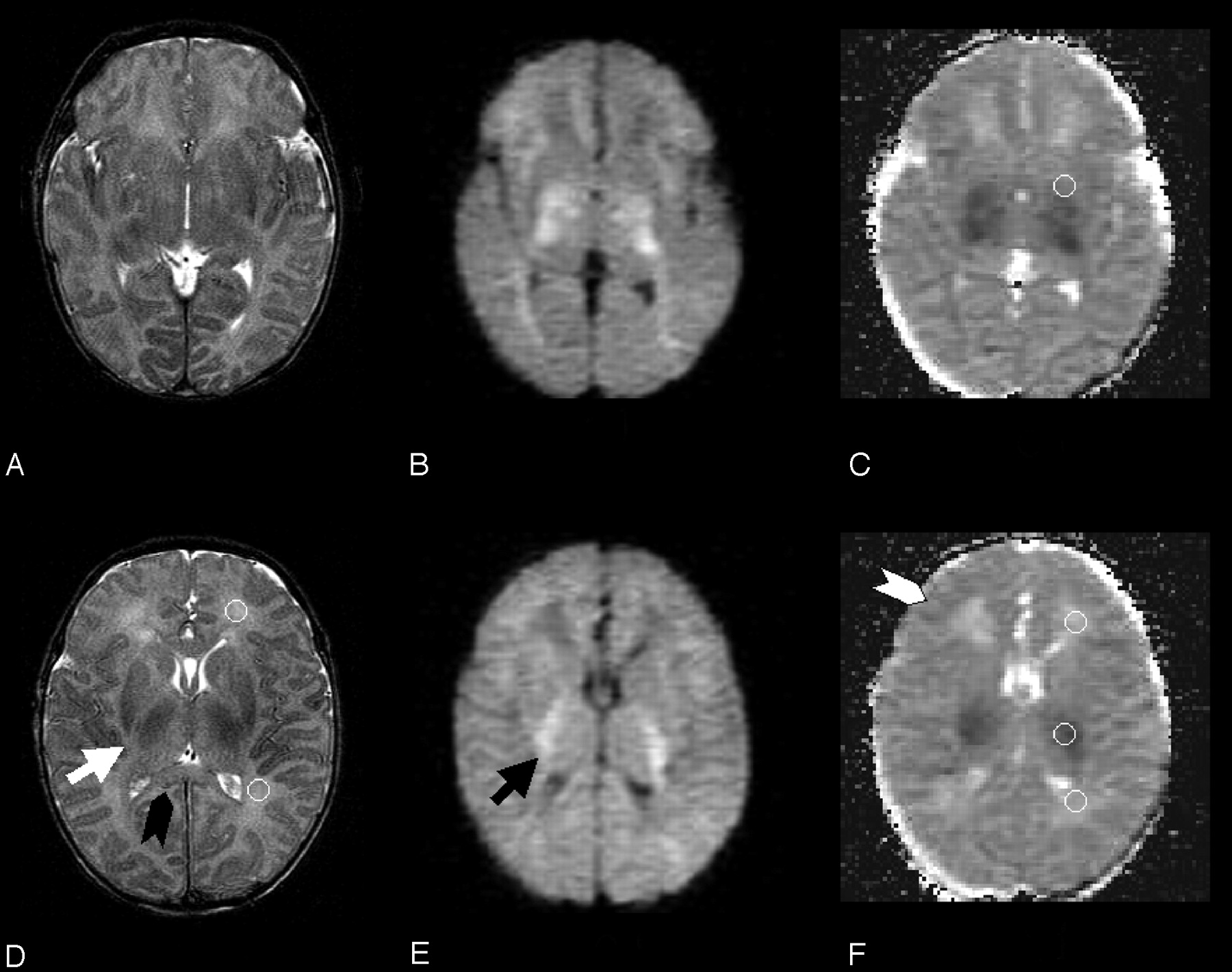
    Two T2-weighted fast spin-echo (A, D), diffusion-weighted (B, E), and trace apparent diffusion coefficient–calculated (C) f-axial sections from the first (time course, 0 hours) MR imaging study are reported. Ventricular and cortical cerebrospinal fluid spaces are clearly reduced. The black arrowhead shows clear swelling of the corpus callosum (especially if compared with Fig 4). The white arrow shows the thickened posterior limb of the internal capsula, and the black arrow shows increased diffusion-weighted signal intensity within the myelinated white matter at the same location. Moderate T2-weighted signal intensity increase is visible within the posterior thalamus bilaterally. The white arrowhead indicates the swollen cortical rim. Circular regions of interest for frontal and occipital unmyelinated white matter are shown for one hemisphere on T2-weighted section (D). Regions of interest for basal ganglia, frontal and occipital unmyelinated white matter, and myelinated white matter of the internal capsula are depicted on apparent diffusion coefficient maps (C and F). These regions of interest were transferred from T2 b = 0 corresponding sections. Cortical gray matter regions of interest could not be traced because of partial volume effect.

  • Fig 3.
    • Download figure
    • Open in new tab
    • Download powerpoint
    Fig 3.

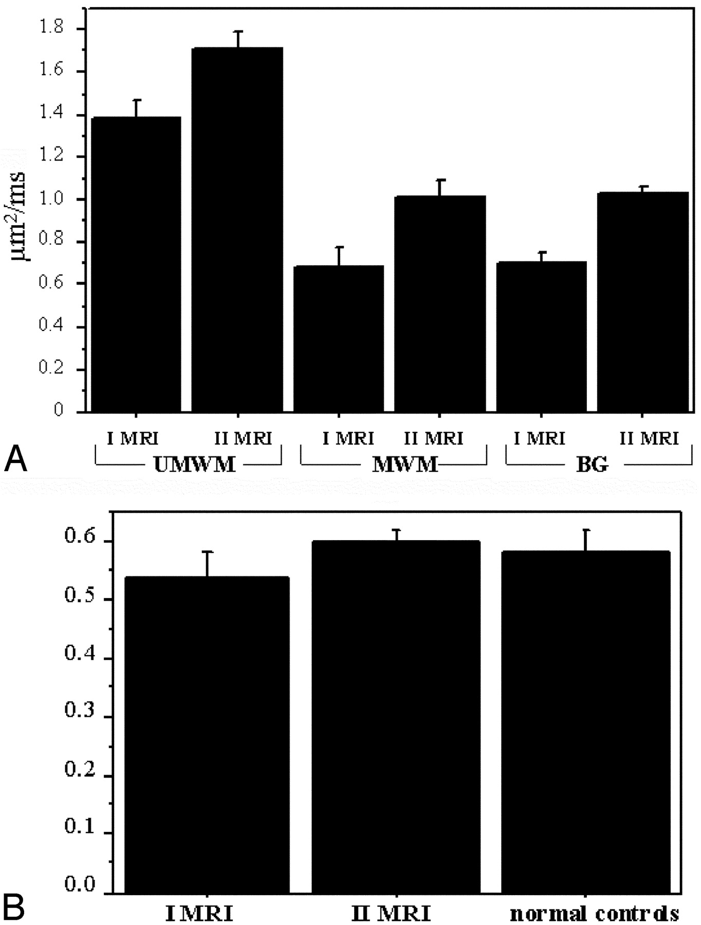
    A, Graph shows plot of the average apparent diffusion coefficient value (with standard deviation of unmyelinated white matter [UMWM]), myelinated white matter (MWM), and basal ganglia (BG) regions of interest in the first (I MRI) and second (II MRI) MR imaging study. B, Graph shows plot of the average value (with standard deviation) of the ratio between T2-weighted signal intensity of frontal-occipital unmyelinated white matter and of cerebrospinal fluid in the first and second MR imaging studies and in normal controls (n = 5).

  • Fig 4.
    • Download figure
    • Open in new tab
    • Download powerpoint
    Fig 4.

    Two T2-weighted (A, D), diffusion-weighted (B, E), and trace apparent diffusion coefficient–calculated (C) f-axial sections from the second (at 200 hours) MR imaging study, obtained at a location similar to that of Fig 2, are reported. Cerebrospinal fluid spaces and brain parenchyma signal intensity are normal. The cortical rim and corpus callosum are no longer swollen. The regions of interest are the same ones as depicted in Fig 2.

PreviousNext
Back to top

In this issue

American Journal of Neuroradiology: 26 (7)
American Journal of Neuroradiology
Vol. 26, Issue 7
1 Aug 2005
  • Table of Contents
  • Index by author
Advertisement
Print
Download PDF
Email Article

Thank you for your interest in spreading the word on American Journal of Neuroradiology.

NOTE: We only request your email address so that the person you are recommending the page to knows that you wanted them to see it, and that it is not junk mail. We do not capture any email address.

Enter multiple addresses on separate lines or separate them with commas.
Brain Apparent Diffusion Coefficient Decrease During Correction of Severe Hypernatremic Dehydration
(Your Name) has sent you a message from American Journal of Neuroradiology
(Your Name) thought you would like to see the American Journal of Neuroradiology web site.
CAPTCHA
This question is for testing whether or not you are a human visitor and to prevent automated spam submissions.
Cite this article
Andrea Righini, Luca Ramenghi, Salvatore Zirpoli, Fabio Mosca, Fabio Triulzi
Brain Apparent Diffusion Coefficient Decrease During Correction of Severe Hypernatremic Dehydration
American Journal of Neuroradiology Aug 2005, 26 (7) 1690-1694;

Citation Manager Formats

  • BibTeX
  • Bookends
  • EasyBib
  • EndNote (tagged)
  • EndNote 8 (xml)
  • Medlars
  • Mendeley
  • Papers
  • RefWorks Tagged
  • Ref Manager
  • RIS
  • Zotero
0 Responses
Respond to this article
Share
Bookmark this article
Brain Apparent Diffusion Coefficient Decrease During Correction of Severe Hypernatremic Dehydration
Andrea Righini, Luca Ramenghi, Salvatore Zirpoli, Fabio Mosca, Fabio Triulzi
American Journal of Neuroradiology Aug 2005, 26 (7) 1690-1694;
del.icio.us logo Twitter logo Facebook logo Mendeley logo
  • Tweet Widget
  • Facebook Like
  • Google Plus One

Jump to section

  • Article
    • Abstract
    • Case Report
    • Discussion
    • References
  • Figures & Data
  • Info & Metrics
  • Responses
  • References
  • PDF

Related Articles

  • No related articles found.
  • PubMed
  • Google Scholar

Cited By...

  • Involvement of bilateral posterior limb of internal capsule in hypernatraemic dehydration
  • Cerebral Metabolic Alterations in Rats With Diabetic Ketoacidosis: Effects of Treatment With Insulin and Intravenous Fluids and Effects of Bumetanide
  • Crossref
  • Google Scholar

This article has not yet been cited by articles in journals that are participating in Crossref Cited-by Linking.

More in this TOC Section

  • SyMRI & MR Fingerprinting in Brainstem Myelination
  • Comparison of Image Quality and Radiation Dose in Pediatric Temporal Bone CT Using Photon-Counting Detector CT and Energy-Integrating Detector CT
  • Neonatal Hypocalcemia: Cerebral MRI Abnormalities
Show more Pediatrics

Similar Articles

Advertisement

Indexed Content

  • Current Issue
  • Accepted Manuscripts
  • Article Preview
  • Past Issues
  • Editorials
  • Editor's Choice
  • Fellows' Journal Club
  • Letters to the Editor
  • Video Articles

Cases

  • Case Collection
  • Archive - Case of the Week
  • Archive - Case of the Month
  • Archive - Classic Case

Special Collections

  • AJNR Awards
  • ASNR Foundation Special Collection
  • Most Impactful AJNR Articles
  • Photon-Counting CT
  • Spinal CSF Leak Articles (Jan 2020-June 2024)

More from AJNR

  • Trainee Corner
  • Imaging Protocols
  • MRI Safety Corner

Multimedia

  • AJNR Podcasts
  • AJNR Scantastics

Resources

  • Turnaround Time
  • Submit a Manuscript
  • Submit a Video Article
  • Submit an eLetter to the Editor/Response
  • Manuscript Submission Guidelines
  • Statistical Tips
  • Fast Publishing of Accepted Manuscripts
  • Graphical Abstract Preparation
  • Imaging Protocol Submission
  • Evidence-Based Medicine Level Guide
  • Publishing Checklists
  • Author Policies
  • Become a Reviewer/Academy of Reviewers
  • News and Updates

About Us

  • About AJNR
  • Editorial Board
  • Editorial Board Alumni
  • Alerts
  • Permissions
  • Not an AJNR Subscriber? Join Now
  • Advertise with Us
  • Librarian Resources
  • Feedback
  • Terms and Conditions
  • AJNR Editorial Board Alumni

American Society of Neuroradiology

  • Not an ASNR Member? Join Now

© 2025 by the American Society of Neuroradiology All rights, including for text and data mining, AI training, and similar technologies, are reserved.
Print ISSN: 0195-6108 Online ISSN: 1936-959X

Powered by HighWire