
Article Figures & Data
Figures
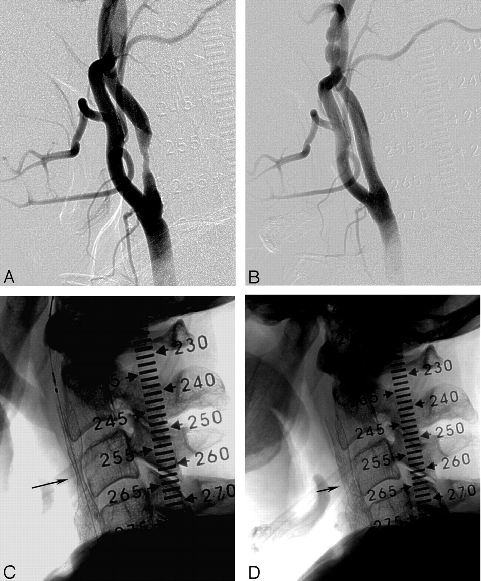
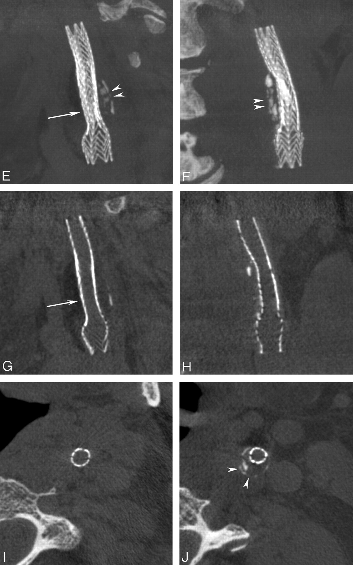
- Fig 1.
Stent placement in an extracranial carotid stenosis.
A, 85% stenosis of the ICA.
B, 6–8 × 40-mm stent (Acculink; Guidant Corporation) is placed.
C, Native image immediately after stenting shows a significant residual stenosis (arrow) requiring postdilation.
D, Satisfactory result and remaining minor narrowing (arrow).
E andF MPRs, Lateral (E) and coronal (F) ACT views (5-mm sections) provide superior visualization of the entire stent, its struts, and the remaining narrowing (arrow) and show the underlying, heavily calcified plaque (arrowheads) responsible for incomplete stent deployment.
G andH, Reducing the section thickness to 0.1 mm allows visualization along a cut plane through the center of the stent, showing the effective remaining narrowing (arrow).
I andJ, On axial views, the different diameters of the recanalized lumen distal to and at the level of the plaque are appreciated. They further reveal the circumferential narrowing by the calcified plaque (arrowheads in J). Note visualization of soft-tissue components in the neck.
- Fig 2.
Stent placement in an 80% stenosis of the intracranial middle cerebral artery.
A, High-grade stenosis (arrow) of the left M1 segment
B, Treatment with a 3 × 8-mm drug-eluting stent (Cypher; Cordis) was successful, completely reestablishing the lumen.
C andD, Nonsubtracted images permit moderate visualization of the deployed stent (arrows).
E andF, Coronal and axial MPRs allow superior visualization of the stent (arrows).
G andH, Orthogonal views (“down the barrel”) through the inner lumen of the stent at its proximal and distal ends show complete and symmetric deployment.
- Fig 3.
Stent placement in a basilar artery stenosis.
A andB, 70% stenosis (arrow) of the midbasilar artery, with poststenotic dilatation.
C andD, Good result after stenting and dilatation. The stent (arrows) was postdilated to better oppose the distal portion to the larger lumen of the basilar artery.
E andF, Nonsubtracted images insufficiently show the stent (arrows). The degree of deployment over its entire length cannot be appreciated with certainty.
G–J, MPRs obtained immediately after stenting (stenting thickness, .1 mm) reveal the asymmetric deployment of the stent with a remaining waist (arrow). Orthogonal projections (I–J) demonstrate the change in caliber (arrows) and show the larger lumen stent distally (J).


In this issue


Jump to section
Related Articles
Cited By...
- Quantitative Analysis of Conebeam CT for Delineating Stents in Stent-Assisted Coil Embolization
- Clinical Impact of Flat Panel Volume CT Angiography in Evaluating the Accurate Intraoperative Deployment of Flow-Diverter Stents
- The Added Value of Volume-of-Interest C-Arm CT Imaging during Endovascular Treatment of Intracranial Aneurysms
- Low-Dose Volume-of-Interest C-Arm CT Imaging of Intracranial Stents and Flow Diverters
- Intravenous C-Arm Conebeam CT Angiography following Long-Term Flow-Diverter Implantation: Technologic Evaluation and Preliminary Results
- Adjunctive value of intra-arterial cone beam CT angiography relative to DSA in the evaluation of cranial and spinal arteriovenous fistulas
- Role of C-Arm VasoCT in the Use of Endovascular WEB Flow Disruption in Intracranial Aneurysm Treatment
- A prospective, multicenter pilot study investigating the utility of flat detector derived parenchymal blood volume maps to estimate cerebral blood volume in stroke patients
- The utility of cone beam volume CT in the evaluation of thrombosed intracranial aneurysms in subarachnoid hemorrhage
- Angiographic CT for Intraprocedural Monitoring of Complex Neuroendovascular Procedures
- The utility of cone beam volume CT in the evaluation of thrombosed intracranial aneurysms in subarachnoid hemorrhage
- Initial experience with a combined multidetector CT and biplane digital subtraction angiography suite with a single interactive table for the diagnosis and treatment of neurovascular disease
- Feasibility of Cerebral Blood Volume Mapping by Flat Panel Detector CT in the Angiography Suite: First Experience in Patients with Acute Middle Cerebral Artery Occlusions
- Contrast-Enhanced Angiographic Cone-Beam CT of Cerebrovascular Stents: Experimental Optimization and Clinical Application
- Angiographic CT after Intravenous Contrast Agent Application: A Noninvasive Follow-Up Tool after Intracranial Angioplasty and Stenting
- Use of Angiographic CT Imaging in the Cardiac Catheterization Laboratory for Congenital Heart Disease
- Feasibility of Angiographic CT in Peri-Interventional Diagnostic Imaging: A Comparative Study with Multidetector CT
- Color-Coded Digital Subtraction Angiography: The End of a Monochromatic Era?
- C-Arm CT Measurement of Cerebral Blood Volume in Ischemic Stroke: An Experimental Study in Canines
- Impact of Intra-Arterial Injection Parameters on Arterial, Capillary, and Venous Time-Concentration Curves in a Canine Model
- C-Arm CT Measurement of Cerebral Blood Volume: An Experimental Study in Canines
- Wall Shear Stress in Intracranial Self-Expanding Stents Studied Using Ultra-High-Resolution 3D Reconstructions
- Large scan field, high spatial resolution flat-panel detector based volumetric CT of the whole human skull base and for maxillofacial imaging
- Stent Conformity in Curved Vascular Models with Simulated Aneurysm Necks Using Flat-Panel CT: An In Vitro Study
- Angiographic Computed Tomography for Imaging of Underdeployed Intracranial Stent