
Article Figures & Data
Figures
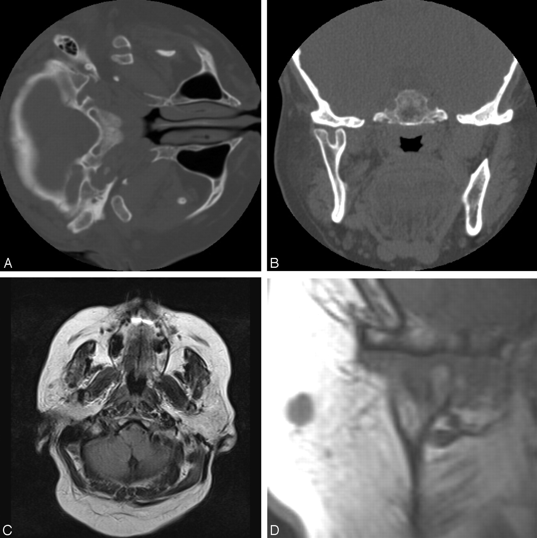
- Fig 1.
Axial (A) and coronal (B) thin-cut CT images through the mandible demonstrate a bifid mandibular condyle on the right side with mediolaterally oriented heads.
C, Axial, T1-weighted MR imaging demonstrates the bifid mandibular condyle on the right side, but also demonstrates subtle fatty atrophy of the muscles of mastication ipsilaterally.
D, Coronal T1-weighted MR imaging images demonstrate the bifid condyle on the right side and also demonstrate that the meniscus has the shape of a T, with a third limb interdigitating between the two mandibular heads.
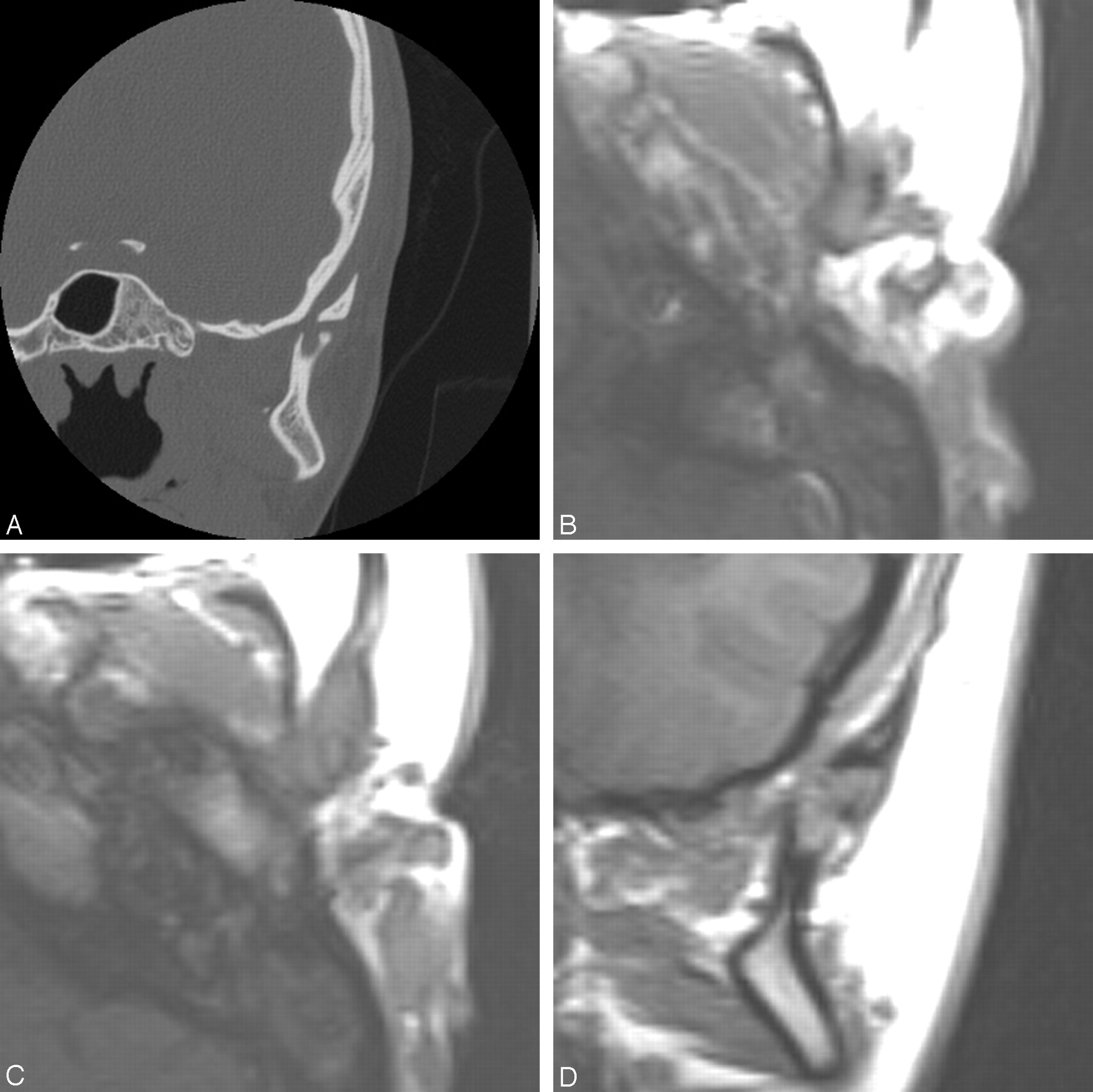
- Fig 2.
A, Coronal thin-cut CT images demonstrate that the left mandibular condyle is bifid, with mediolaterally oriented heads. Although both mandibular heads are small and deformed, the left is much smaller than the right.
B and C, Axial, T1-weighted MR imaging images again demonstrate the bifid mandibular condyle.
D, Coronal images demonstrate the mediolaterally oriented heads and also show the T-shaped meniscus, with the descending limb of the T interdigitating between the mediolaterally oriented heads.

Tables
Cases of bifid condyles reported in literature
Living Cases Cadaveric Cases Hrdlicka (1941) (1) 21 Sicher (1948) (2) 1 Moffett (1966) (3) 1 Stadnicki (1971) (15) 1 1 Lysell and Oberg (1975) (4) 1 Farmand (1981) (5) 1 Forman and Smith (1984) (6) 2 Balciunas (1986) (12) 1 Thomason and Yusuf (1986) (11) 2 Gundlach et al (1987) (7) 4 1 Zohar and Laurian (1987) (9) 1 Sahm and Witt (1989) (16) 1 Szenpetery et al (1990) (8) 7 Phillips and Delzer (1992) (17) 1 Antoniades et al (1993) (18) 1 Fields and Frederiksen (1993) (19) 1 Cowan and Ferguson (1993) (20) 1 Stephanou et al (1998) (21) 4 Garcia-Gonzalez et al (2000) (22) 1 Totals 24 32
In this issue


Jump to section
Related Articles
Cited By...
- Bifid mandibular condyle
- Bifid mandibular condyle: CT and MRI appearance
- Tetrafid mandibular condyle: a unique case report and review of the literature
- Bifid Mandibular Condyle: A Disorder in Its Own Right?
- The frequency of bifid mandibular condyle in a Turkish patient population
- Bifid mandibular condyle with associated temporomandibular joint ankylosis: a computed tomography study of the patterns and morphological variations
- The prevalence of bifid mandibular condyle detected in a Brazilian population