
Article Figures & Data
Figures
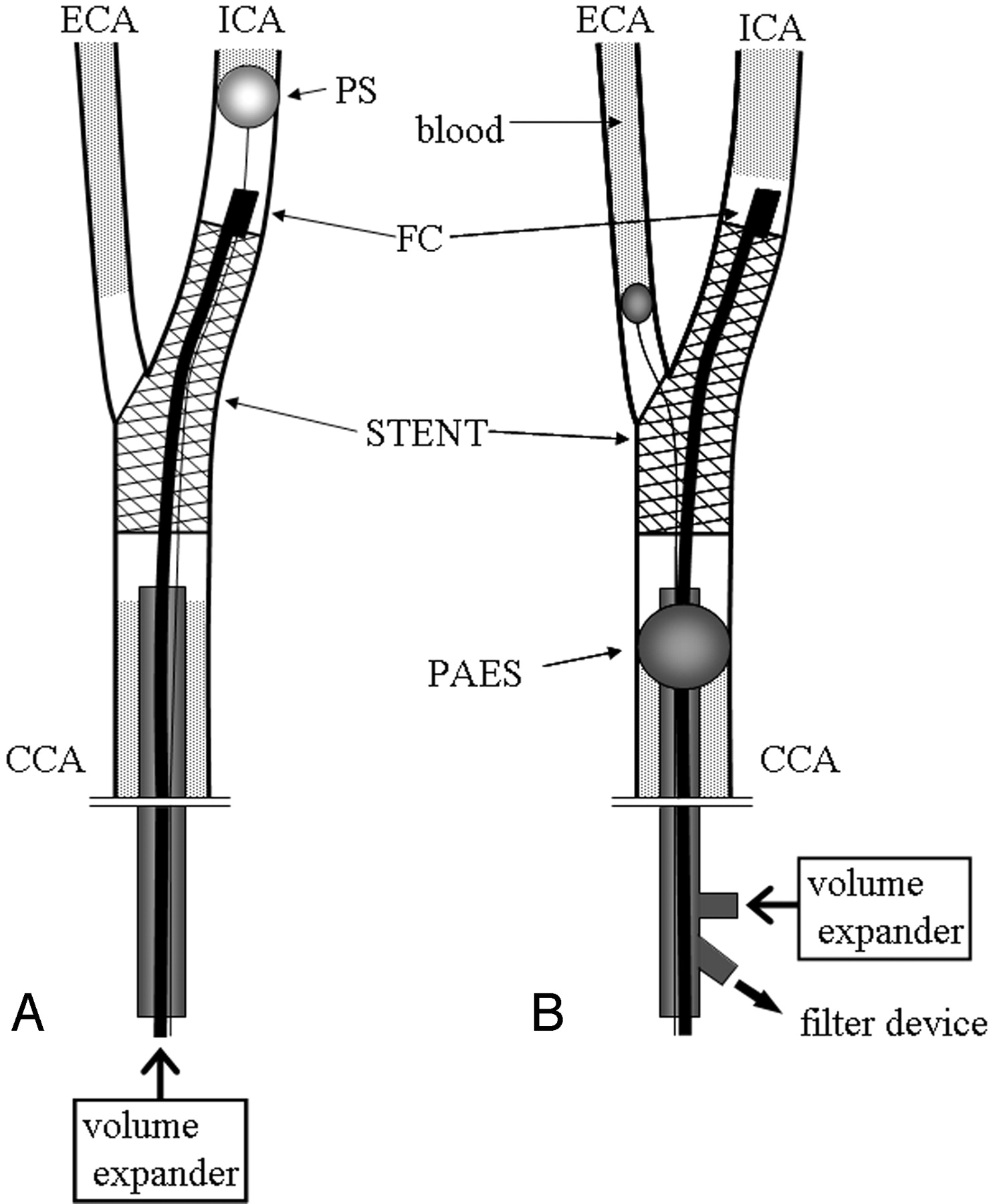
- Fig 1.
Angioscopic observation with distal protection (A) with PercuSurge GuardWire and (B) with the Parodi Antiemboli System. CCA signifies common carotid artery; ICA, internal carotid artery; ECA, external carotid artery; PS, PercuSurge GuardWire; FC, fiber catheter AS-003 with aspiration catheter; PAES, Parodi Antiemboli System.
- Fig 2.
Angioscopic image of the internal carotid artery in case 16 clearly shows stent attachment to the wall.
- Fig 3.
Angioscopic image of the internal carotid artery in case 7. First debris (red arrowheads) detaches from the atherosclerotic carotid wall and then floats to the distal side with time (A–E). Next debris (yellow arrowheads) also detaches from the carotid wall (D–F).
- Fig 4.
Angioscopic image of the internal carotid artery proximal to the stent in case 2 shows the intimal flap (arrows) fluttering from the arterial wall.


Tables
Patient No./Sex/Age (y) Stenosis (%) Symptom Occlusion Time: Total/CAS/AS Protection Device Ischemic Lesion* 1/M/68 70 Minor stroke 19 min 12 s/16 min 12 s/3 min PS Yes 2/M/77 80 Asymptomatic 21 min 34 s/18 min 34 s/3 min PS Yes 3/M/59 80 Asymptomatic 24 min 24 s/20 min 9s/4 min 15 s PS No 4/M/67 70 Asymptomatic 16 min 8 s/10 min 40 s/5 min 28 s PS Yes 5/M/83 95 Minor stroke 25 min/21 min/4 min PS Yes 6/M/70 75 Minor stroke 15 min 47 s/13 min 2 s/2 min 45 s PS Yes 7/M/73 70 Asymptomatic 19 min/14 min 8 s/4 min 52 s PS — 8/M/71 70 Asymptomatic 8 min 56 s/6 min 58 s/1 min 58 s PS, PAES No 9/M/73 70 Asymptomatic 14 min 2 s/9 min 57 s/4 min 5 s PS No 10/M/77 60 Minor stroke 11 min 30 s/11 min/30 s PS Yes 11/M/74 70 Minor stroke 17 min 47 s/14 min 47 s/3 min PS No 12/M/54 70 Asymptomatic 11 min 10 s/9 min 10 s/2 min PS No 13/M/69 90 Asymptomatic 23 min 1 s/16 min 4 s/6 min 57 s PAES No 14/M/76 80 Minor stroke 13 min/10 min/3 min PAES No 15/M/61 90 Asymptomatic 18 min 20 s/13 min 50 s/4 min 30s PAES No 16/M/63 70 Major stroke 15 min 39 s/10 min 24 s/5 min 15 s PAES Yes 17/M/65 70 Asymptomatic 14 min 6 s/8 min 37 s/5 min 29 s PAES Yes 18/M/72 60 Asymptomatic 13 min 47 s/11 min/2 min 47 s PS No Avg 70 74 16 min 48 s/13 min 5 s/3 min 43s Note.—CAS indicates carotid angioplasty with stent; AS, angioscopy; PS, PercuSurge; PAES, Parodi antiemboli system.
* Refers to asymptomatic ischemic lesions detected by diffusion-weighted MRI after the procedure.
Patient No. Angioscopic Findings 1 Stent attachment to the wall 2 Intimal flap 3 Plaque protrusion 4 Stent attachment to the wall 5 Unclear view 6 Unclear view 7 Plaque protrusion, debris detaching form the plaque 8 Stent attachment to the wall 9 Stent attachment to the wall 10 Unclear view 11 Stent attachment to the wall 12 Unclear view 13 Stent attachment to the wall 14 Unclear view 15 Yellowish surface of carotid artery 16 Stent attachment to the wall, plaque protrusion 17 Unclear view 18 Stent attachment to the wall


