Advertisement

Main menu

  • Home
  • Content
    • Current Issue
    • Accepted Manuscripts
    • Article Preview
    • Past Issue Archive
    • Video Articles
    • AJNR Case Collection
    • Case of the Week Archive
    • Case of the Month Archive
    • Classic Case Archive
  • Special Collections
    • AJNR Awards
    • ASNR Foundation Special Collection
    • Most Impactful AJNR Articles
    • Photon-Counting CT
    • Spinal CSF Leak Articles (Jan 2020-June 2024)
  • Multimedia
    • AJNR Podcasts
    • AJNR SCANtastic
    • Trainee Corner
    • MRI Safety Corner
    • Imaging Protocols
  • For Authors
    • Submit a Manuscript
    • Submit a Video Article
    • Submit an eLetter to the Editor/Response
    • Manuscript Submission Guidelines
    • Statistical Tips
    • Fast Publishing of Accepted Manuscripts
    • Graphical Abstract Preparation
    • Imaging Protocol Submission
    • Author Policies
  • About Us
    • About AJNR
    • Editorial Board
    • Editorial Board Alumni
  • More
    • Become a Reviewer/Academy of Reviewers
    • Subscribers
    • Permissions
    • Alerts
    • Feedback
    • Advertisers
    • ASNR Home

User menu

  • Alerts
  • Log in

Search

  • Advanced search
American Journal of Neuroradiology

American Journal of Neuroradiology

ASHNR American Society of Functional Neuroradiology ASHNR American Society of Pediatric Neuroradiology ASSR
  • Alerts
  • Log in

Advanced Search

  • Home
  • Content
    • Current Issue
    • Accepted Manuscripts
    • Article Preview
    • Past Issue Archive
    • Video Articles
    • AJNR Case Collection
    • Case of the Week Archive
    • Case of the Month Archive
    • Classic Case Archive
  • Special Collections
    • AJNR Awards
    • ASNR Foundation Special Collection
    • Most Impactful AJNR Articles
    • Photon-Counting CT
    • Spinal CSF Leak Articles (Jan 2020-June 2024)
  • Multimedia
    • AJNR Podcasts
    • AJNR SCANtastic
    • Trainee Corner
    • MRI Safety Corner
    • Imaging Protocols
  • For Authors
    • Submit a Manuscript
    • Submit a Video Article
    • Submit an eLetter to the Editor/Response
    • Manuscript Submission Guidelines
    • Statistical Tips
    • Fast Publishing of Accepted Manuscripts
    • Graphical Abstract Preparation
    • Imaging Protocol Submission
    • Author Policies
  • About Us
    • About AJNR
    • Editorial Board
    • Editorial Board Alumni
  • More
    • Become a Reviewer/Academy of Reviewers
    • Subscribers
    • Permissions
    • Alerts
    • Feedback
    • Advertisers
    • ASNR Home
  • Follow AJNR on Twitter
  • Visit AJNR on Facebook
  • Follow AJNR on Instagram
  • Join AJNR on LinkedIn
  • RSS Feeds

AJNR is seeking candidates for the AJNR Podcast Editor. Read the position description.

OtherPEDIATRICS

Pantothenate Kinase-Associated Neurodegeneration with Increased Lentiform Nuclei Cerebral Blood Flow

M. Koyama and A. Yagishita
American Journal of Neuroradiology January 2006, 27 (1) 212-213;
M. Koyama
  • Find this author on Google Scholar
  • Find this author on PubMed
  • Search for this author on this site
A. Yagishita
  • Find this author on Google Scholar
  • Find this author on PubMed
  • Search for this author on this site
  • Article
  • Figures & Data
  • Info & Metrics
  • Responses
  • References
  • PDF
Loading

Abstract

Summary: We report a case of pantothenate kinase-associated neurodegeneration with increased regional cerebral blood flow (rCBF) in bilateral lentiform nuclei on technetium Tc-99m ethyl cysteinate dimer single-photon emission CT (ECD-SPECT). A 6-year-old boy presented with opisthotonus. T2*-weighted MR images revealed areas of marked hypointensity with a hyperintense focus in bilateral globus pallidus, creating the characteristic eye-of-the-tiger appearance. ECD-SPECT showed increased rCBF in bilateral lentiform nuclei.

Pantothenate kinase-associated neurodegeneration (PKAN,previously known as Hallervorden-Spatz syndrome) represents an autosomal recessive disorder characterized by dystonia, Parkinsonism, and iron accumulation in the brain. Findings on MR imaging have been described elsewhere,1-4 but no previous reports have described brain perfusion studies on single photon-emission CT (SPECT) or positron-emission tomography. We describe the case of a 6-year-old boy for whom brain perfusion SPECT was performed.

Case Report

A 6-year-old boy presented with opisthotonus. His mother had a history of threatened premature delivery, but he was born by normal vaginal delivery. Development by 1 year of age had been normal. He tended to fall down while walking, and fell backward even when sitting. Verbal development was delayed. Involuntary movements of the right upper extremity began at 4 years of age, and was unable to walk at 5.25 years of age. He was subsequently fed by nasogastric tube. Opisthotonus began at 6.5 years of age.

Emaciation was noted on admission, along with urinary and fecal incontinence. On CT images, calcification was noted in bilateral globus pallidus, and the brain was slightly atrophic.

On T2-weighted MR imaging, areas of marked hypointensity with a focus of signal intensity hyperintensity were evident in bilateral globus pallidus, creating the characteristic eye-of-the-tiger appearance. The lesion showed more prominent hypointensity without hyperintensity foci on T2*-weighted images (Fig 1). Cerebral and cerebellar atrophy were also noted.

Fig 1.
  • Download figure
  • Open in new tab
  • Download powerpoint
Fig 1.

T2*-weighted MR imaging shows an area of marked hypointensity in the globus pallidus.

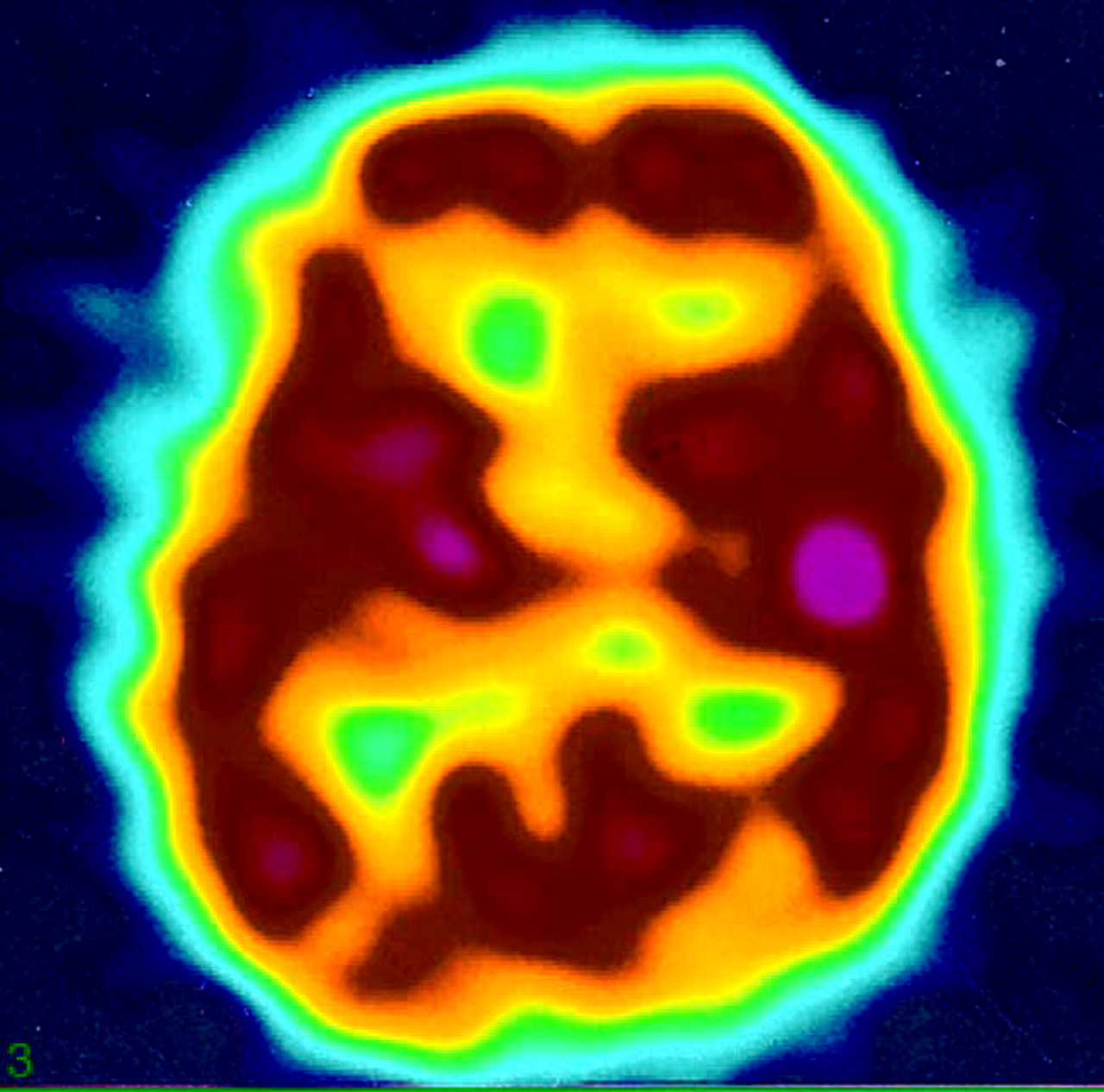
Technetium Tc-99m ethyl cysteinate dimer SPECT (ECD-SPECT) revealed prominent accumulation on the medial side of bilateral lentiform nuclei (Fig 2).

Fig 2.
  • Download figure
  • Open in new tab
  • Download powerpoint
Fig 2.

Transaxial (A) and coronal (B) SPECT images with Tc-99m ECD-SPECT show prominent accumulation in bilateral basal ganglia.

Deep brain stimulation (DBS) of the globus pallidus internus was performed bilaterally. Opisthotonus was markedly decreased after DBS. SPECT images 9 weeks after DBS revealed slightly decreased accumulation in this area. After 19 weeks, hypoperfusion was noted in the right frontotemporal region and basal ganglia, predominantly on the right (Fig. 3).

Fig 3.
  • Download figure
  • Open in new tab
  • Download powerpoint
Fig 3.

SPECT image 19 weeks after DBS. Perfusion is decreased in the right globus pallidus and frontotemporal region. Color setting differs from the initial study due to replacement of the gamma camera and related equipment.

Discussion

Many cases of Hallervorden-Spatz syndrome have recently been found to result from mutations in a gene located on chromosome 20p13. All patients with classic Hallervorden-Spatz syndrome and one third of those with atypical disease display PKAN2 mutations. In all patients with PKAN, whether classic or atypical, T2-weighted MR imaging of the brain displays a specific pattern of hyperintensity within the hypointense medial globus pallidus. This pattern is not seen in patients without mutations.1

On the basis of clinical assessment, early-onset childhood-type (1a) PKAN with rapid progression was diagnosed in this case.5

Prominent accumulation at the medial side of bilateral lentiform nuclei was noted on ECD-SPECT. In patients with epilepsy or convulsions, perfusion in the basal ganglia on ECD-SPECT is reportedly higher in patients younger than 1 year than in older patients.6 Perfusion in the basal ganglia is stable after 1 year of age. Because this patient was 6 years old, hyperperfusion in the lentiform nuclei seems significant.

In epilepsy patients, hyperemia occurs at the epileptogenic focus during ictal status, but perfusion decreases during interictal status at the focus. In this case, SPECT was performed during interictal status. The medial sides of bilateral lentiform nuclei might have acted as continuous epileptogenic foci.

Neurochemical abnormalities have been found in PKAN, with markedly elevated levels of cysteine and glutathione cysteine mixed disulphide in the globus pallidus associated with reduced activity of cysteine dioxygenase.7 Accumulated cysteine may act as a chelating agent and might be responsible for the accumulation of iron. Radiopharmaceuticals such as technetium Tc-99m ECD may also be chelated by cysteine in the globus pallidus.

Recently, DBS of the globus pallidus has emerged as a significant therapeutic alternative in intractable dystonia, including PKAN.8 DBS of the globus pallidus internus seems effective for dystonia, because the process removes the source of inhibitory input to the thalamus and normalizes thalamic neuronal activity. This patient showed decreased perfusion in the right globus pallidus and frontotemporal region after DBS. Decreased perfusion in the right front-temporal region may be due to remote effects.

Acknowledgments

We thank Dr Ryouichi Okiyama, neurologist, for the clinical advice.

References

  1. ↵
    Hayflick SJ, Westaway SK, Levinson B, et al. Genetic, clinical and radiographic delineation of Hallervorden-Spatz syndrome. N Engl J Med 2003;348:33–40
    CrossRefPubMed
  2. Sener RN. Pantothenate kinase-associated neurodegeneration: MR imaging, proton MR spectroscopy, and diffusion MR imaging findings. AJNR Am J Neuroradiol 2003;24:1690–93
    Abstract/FREE Full Text
  3. Guillerman RP. The Eye-of-the-tiger sign. Radiology 2000;217:895–96
    PubMed
  4. ↵
    Savoiardo M, Halliday WC, Nardocci N, et al. Hallervorden-Spatz disease: MR and pathologic findings. AJNR Am J Neuroradiol 1993;14:155–62
    Abstract/FREE Full Text
  5. ↵
    Swaiman KF. Hallervorden-Spatz syndrome. Pediatr Neurol 2001;25:102–108
    CrossRefPubMed
  6. ↵
    Kuji I, Sumiya H, Niida Y, et al. Age-related changes in the cerebral distribution of 99mTc-ECD from infancy to adulthood. J Nucl Med 1999;40:1818–23
    Abstract/FREE Full Text
  7. ↵
    Perry TL, Norman MG, Yong VM, et al. Hallervorden-Spatz disease: cysteine accumulation and cysteine dioxygenase deficiency in the globus pallidus. Ann Neurol 1985;18:482–89
    CrossRefPubMed
  8. ↵
    Umemura A, Jagii JL, Dolinskas CA, et al. Pallidal deep brain stimulation for longstanding severe generalized dystonia in Hallervorden-Spatz syndrome. J Neurosurg 2004;100:706–709
    CrossRefPubMed
  • Received November 7, 2004.
  • Accepted after revision February 11, 2005.
  • Copyright © American Society of Neuroradiology
View Abstract
PreviousNext
Back to top

In this issue

American Journal of Neuroradiology: 27 (1)
American Journal of Neuroradiology
Vol. 27, Issue 1
January, 2006
  • Table of Contents
  • Index by author
Advertisement
Print
Download PDF
Email Article

Thank you for your interest in spreading the word on American Journal of Neuroradiology.

NOTE: We only request your email address so that the person you are recommending the page to knows that you wanted them to see it, and that it is not junk mail. We do not capture any email address.

Enter multiple addresses on separate lines or separate them with commas.
Pantothenate Kinase-Associated Neurodegeneration with Increased Lentiform Nuclei Cerebral Blood Flow
(Your Name) has sent you a message from American Journal of Neuroradiology
(Your Name) thought you would like to see the American Journal of Neuroradiology web site.
CAPTCHA
This question is for testing whether or not you are a human visitor and to prevent automated spam submissions.
Cite this article
M. Koyama, A. Yagishita
Pantothenate Kinase-Associated Neurodegeneration with Increased Lentiform Nuclei Cerebral Blood Flow
American Journal of Neuroradiology Jan 2006, 27 (1) 212-213;

Citation Manager Formats

  • BibTeX
  • Bookends
  • EasyBib
  • EndNote (tagged)
  • EndNote 8 (xml)
  • Medlars
  • Mendeley
  • Papers
  • RefWorks Tagged
  • Ref Manager
  • RIS
  • Zotero
0 Responses
Respond to this article
Share
Bookmark this article
Pantothenate Kinase-Associated Neurodegeneration with Increased Lentiform Nuclei Cerebral Blood Flow
M. Koyama, A. Yagishita
American Journal of Neuroradiology Jan 2006, 27 (1) 212-213;
del.icio.us logo Twitter logo Facebook logo Mendeley logo
  • Tweet Widget
  • Facebook Like
  • Google Plus One

Jump to section

  • Article
    • Abstract
    • Case Report
    • Discussion
    • Acknowledgments
    • References
  • Figures & Data
  • Info & Metrics
  • Responses
  • References
  • PDF

Related Articles

  • No related articles found.
  • PubMed
  • Google Scholar

Cited By...

  • No citing articles found.
  • Crossref
  • Google Scholar

This article has not yet been cited by articles in journals that are participating in Crossref Cited-by Linking.

More in this TOC Section

  • SyMRI & MR Fingerprinting in Brainstem Myelination
  • Comparison of Image Quality and Radiation Dose in Pediatric Temporal Bone CT Using Photon-Counting Detector CT and Energy-Integrating Detector CT
  • Neonatal Hypocalcemia: Cerebral MRI Abnormalities
Show more Pediatrics

Similar Articles

Advertisement

Indexed Content

  • Current Issue
  • Accepted Manuscripts
  • Article Preview
  • Past Issues
  • Editorials
  • Editor's Choice
  • Fellows' Journal Club
  • Letters to the Editor
  • Video Articles

Cases

  • Case Collection
  • Archive - Case of the Week
  • Archive - Case of the Month
  • Archive - Classic Case

Special Collections

  • AJNR Awards
  • ASNR Foundation Special Collection
  • Most Impactful AJNR Articles
  • Photon-Counting CT
  • Spinal CSF Leak Articles (Jan 2020-June 2024)

More from AJNR

  • Trainee Corner
  • Imaging Protocols
  • MRI Safety Corner

Multimedia

  • AJNR Podcasts
  • AJNR Scantastics

Resources

  • Turnaround Time
  • Submit a Manuscript
  • Submit a Video Article
  • Submit an eLetter to the Editor/Response
  • Manuscript Submission Guidelines
  • Statistical Tips
  • Fast Publishing of Accepted Manuscripts
  • Graphical Abstract Preparation
  • Imaging Protocol Submission
  • Evidence-Based Medicine Level Guide
  • Publishing Checklists
  • Author Policies
  • Become a Reviewer/Academy of Reviewers
  • News and Updates

About Us

  • About AJNR
  • Editorial Board
  • Editorial Board Alumni
  • Alerts
  • Permissions
  • Not an AJNR Subscriber? Join Now
  • Advertise with Us
  • Librarian Resources
  • Feedback
  • Terms and Conditions
  • AJNR Editorial Board Alumni

American Society of Neuroradiology

  • Not an ASNR Member? Join Now

© 2025 by the American Society of Neuroradiology All rights, including for text and data mining, AI training, and similar technologies, are reserved.
Print ISSN: 0195-6108 Online ISSN: 1936-959X

Powered by HighWire