
Abstract
SUMMARY: We present 2 patients with giant cell reparative granuloma (GCRG) of the sphenoid bone. The first patient is an 8-year-old boy with involvement of the greater wing, and the second is a 53- year-old man with a lateral pterygoid plate mass. Both patients presented with rapid expansion of lytic bone lesions, which had solid and cystic components and lacked matrix calcification. Biopsies were indeterminate for definitive diagnoses. The radiologic appearance, location, and incidence of the lesions, and the patient’s age and medical history are helpful aids in narrowing the differential diagnosis of sphenoid bone lesions. However, the imaging and, occasionally, even the histologic findings may not suggest the specific diagnosis of GCRG, which must be added into the differential diagnosis of rapidly enlarging cystic bone lesions of the sphenoid bone.
Giant cell reparative granuloma (GCRG) is a reactive inflammatory process related to trauma and intraosseous hemorrhage.1,2 GCRG and giant cell tumor (GCT) are 2 of the many giant cell–containing bone lesions.3 GCRG has frequently been misdiagnosed as GCT.4-6 GCT is a neoplastic process with a potential to metastasize even though it is histologically benign.1,7,8 GCRG arises from the periosteal connective tissue, whereas GCT originates in the connective tissue of the bone marrow.5 Radiologic appearances of GCT and GCRG are indistinguishable. Histologically, abundant multinucleated giant cells are evenly distributed in GCT,1,3 whereas they are unevenly distributed and contain fewer nuclei in GCRG.5,6 On CT, both usually appear as a nonspecific expansile lytic lesion. These lesions present with low signal intensity on T1- and T2-weighted MR imaging because of the hemosiderin and/or fibrous tissue. Both tumors enhance with contrast administration; the degree of enhancement ranges from slight to strong.5 GCRG typically affects the mandible in children and young adults, with extragnathic lesions having a second peak between the fifth and seventh decades of life.6 GCT is mostly observed at the skull base in patients aged 20–40 years.1,9
Case Descriptions
Case 1
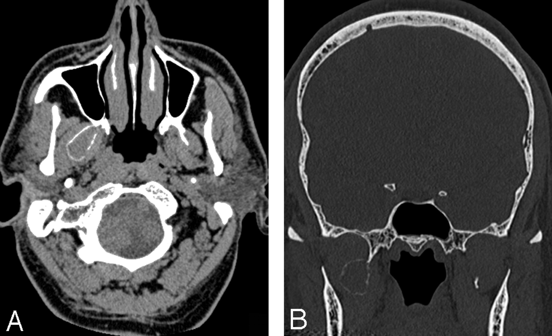
An 8-year-old boy had rapid development of exophthalmos, pain, and diplopia, with normal visual acuity. There was no history of ocular surgery or injury. CT and MR imaging revealed an expansile and lytic bone lesion with solid and cystic components (Figs 1 and 2). The solid component of the mass showed strong enhancement with contrast material. The mass extended into the orbit and middle cranial fossa. Open biopsy suggested a GCT. Thereafter, the patient underwent surgery, with complete resection of the mass and a final histologic diagnosis of GCRG. One year later, MR imaging was repeated with no evidence of recurrence.

Axial CT scan shows a lytic expansile bone lesion of the greater wing of the sphenoid bone protruding into middle cranial fossa and, to a lesser degree, into the orbit. No discontinuity of the cortex is appreciated. Thickening of extraocular muscles is noted.

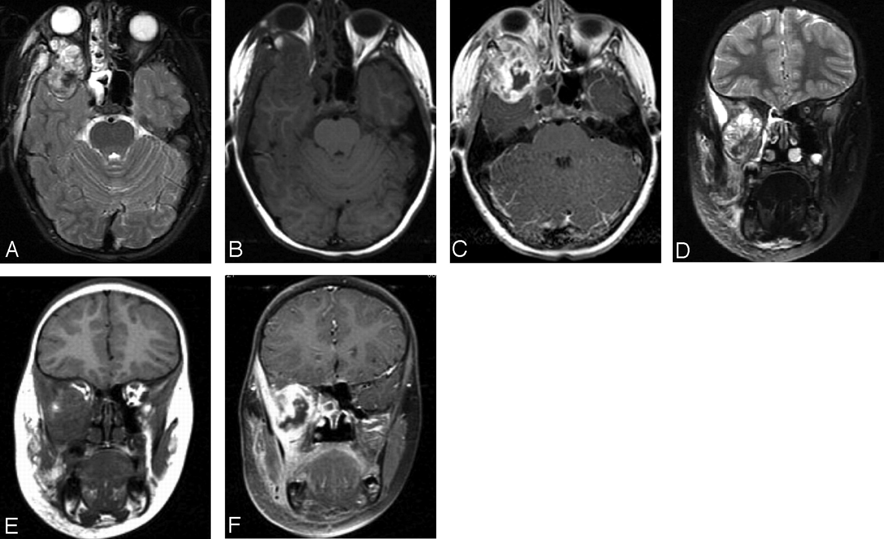
Axial and coronal T2-weighted (A, D), T1-weighted (B, E), and gadolinium-enhanced T1-weighted (C, F) images show a heterogeneously enhancing lesion with extension into the orbit, masticator space, and middle cranial fossa and proptosis of the right eye. Enhancement and edema are also noted within the masticator muscles and surrounding soft tissue. Nonenhancing parts of the lesion with low T2 signal intensity probably represent dense fibrosis identified on the histologic examination.
Case 2
A 53-year-old man with a history of a recent dental procedure and a suggestion of dental abscess underwent CT examination of the neck, demonstrating a lytic and expansile bone lesion in the right lateral pterygoid plate of the sphenoid bone. A comparison with a sinus CT examination performed 3 months previously revealed a similar but much smaller mass in the same region. The patient’s medical history was remarkable for an electrical burn to the forehead 20 years previously that damaged the frontal bone and required a frontal bone augmentation with rib graft as well as obliteration of the frontal sinus with fat. He had chronic frontal sinusitis with acute exacerbations and underwent repeat sinus procedures (Figs 3–5).
Noncontrast axial (A) and coronal (B) CT images show an expansile bone lesion arising from the lateral plate of the right pterygoid process.
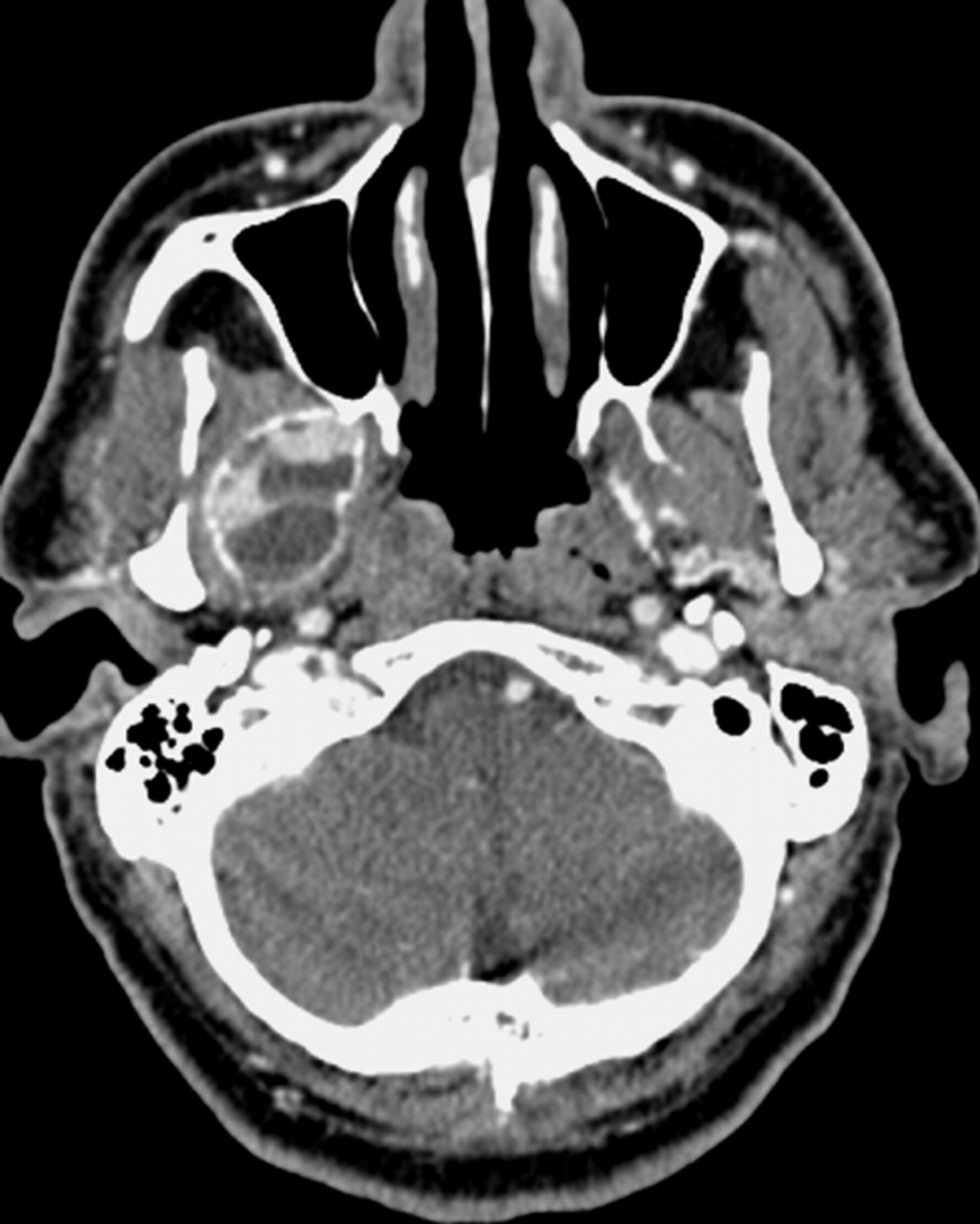
Contrast-enhanced axial CT scan obtained 3 months after Fig 3 shows interval rapid enlargement of the lesion with enhancing soft-tissue component and fluid-fluid levels containing cystic parts.

Axial T1-weighted (A), T2-weighted (B), and postcontrast T1-weighted (C) images demonstrate fluid-fluid levels and an enhancing soft-tissue component. Note that the low T2 signal intensity-enhancing, the enhancing part, which is on the lateral wall of the lesion, likely represents a dense fibrous component rather than hemosiderin.
A CT-guided tru-cut (wide) needle biopsy of the mass showed a giant cell containing bony lesion (Fig 6). Thereafter, the patient underwent surgery involving complete resection of the mass, with the final histologic diagnosis of GCRG (Fig 7).

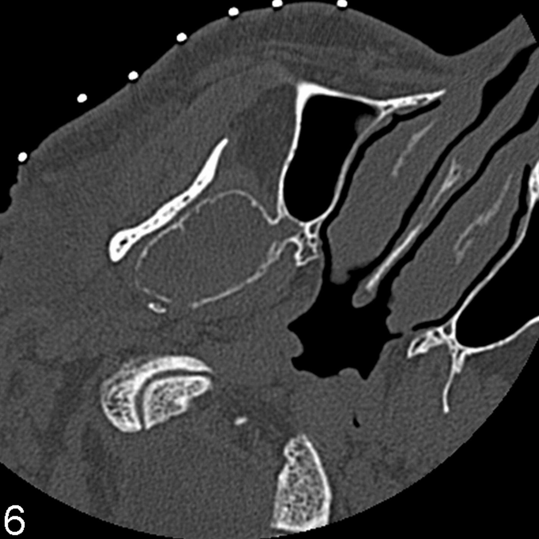
Axial noncontrast image, obtained 1 month after Fig 5 as a part of the imaging-guided biopsy, shows noticeable enlargement of the lesion in the interim. A small cortical breakthrough is seen posteromedially.

Surgical specimen shows multinucleated giant cells interspersed among stromal mononuclear cells in a hemorrhagic background (H&E).
Discussion
GCRG is a reactive inflammatory process secondary to trauma and intraosseous hemorrhage and predominantly affects children and young women.1,6 GCRG occurs in many bones,6 with the mandible, maxilla, and the small bones of the hand and feet being the most common sites.1,6 Extragnathic GCRG has another peak of occurrence in the 50- to 80-year range, with the most common site in the craniofacial region being the temporal bone.1,6 In the English-language literature, to the best of our knowledge, GCRG of the pterygoid plate has not been reported. GCRG is characterized by a non-neoplastic fibrous stroma, with unevenly distributed multinucleated giant cells, particularly appearing around foci of hemorrhage. Reactive osteoid formation is almost always present. GCRG is frequently misdiagnosed as GCT,4-6 which is a neoplastic process.4 In GCRG, giant cells contain fewer nuclei than those of GCT.5,6
Radiologically, GCT and GCRG are indistinguishable. On CT, both usually appear as nonspecific lytic lesions. On MR imaging, most lesions show areas of low signal intensity on T1- and T2-weighted imaging, corresponding to the areas of fibrosis and/or hemosiderin. Both tumors enhance, with the degree of enhancement ranging from slight to strong.5 The primary difference between the 2 is the prognosis.5 GCT has a higher incidence of recurrence than GCRG and may undergo malignant transformation and metastasize.5 GCRG does not undergo malignant transformation,6 and metastasis has not been reported.5
GCT is a histologically benign but locally aggressive bone lesion1,8 and mostly observed between the ages of 20 and 40 years and rarely seen before puberty.1,9 GCT most commonly involves the epiphyses of major long bones, occasionally the bones of the hands, feet, sacrum,1,7,8 and, rarely, the vertebrae above the sacrum.8 Less than 2% of GCTs present in the craniofacial region.1,3 Within the skull, the sphenoid and temporal bones are the most commonly affected.1,3,8 These 2 bones show endochondral ossification like that of the long bones.1 The infrequency of GCT in the other bones of the skull may be related to their intramembranous bone formation.1 Histologic cell types are osteoclast-like multinucleated giant cells, round mononuclear cells resembling monocytes, and a spindle-shaped fibroblast-like stromal cell.1,8 Osteoid or bone formation may occur.1
Giant cells are found in many bone tumors, such as osteosarcoma, chondroblastoma, osteoblastoma, aneurysmal bone cyst, malignant fibrous histiocytoma, chondromyxoid fibroma, fibrous dysplasia, GCRG, eosinophilic granuloma, and the brown tumor of hyperparathyroidism.3 Evenly distributed abundant and uniform multinucleated giant cells and stromal cells are essential for the diagnosis of GCT.1,3
GCT can have a clinically malignant behavior due to its aggressive growth and potential to metastasize and recur.7,8 Radiologically, GCT is seen as a radiolucent lesion without sclerotic borders.8 CT demonstrates cortical expansion or penetration, trabeculations, absence of matrix mineralization, and pathologic fracture. MR imaging shows cystic and solid parts and better delineates the soft-tissue extension, which occurs in more than one third of cases.1,7 GCT is usually well defined and marginated by a rim of low signal intensity, either because of reactive osteosclerosis or a fibrous pseudocapsule. The signal intensity characteristics of solid tissue can be nonspecific, but most GCTs reveal moderate-to-marked hypointensity on T1- and T2-weighted images, which is attributed to hemosiderin deposits and/or high collagen content.10 This feature is useful to exclude chondroid tumors, which have high signal intensity on T2-weighted images. Cystic parts are the result of hemorrhage or formation of a secondary aneurysmal bone cyst.7
GCRG must be also differentiated from the brown tumor, aneurysmal bone cyst, chondroblastoma, fibrous dysplasia, synovial sarcoma, and osteosarcoma.5
Rapid growth, fluid-fluid levels, and an enhancing solid part are suggestive of GCT, GCRG, secondary aneurysmal bone cyst, or telangiectatic osteosarcoma.11 Fluid-fluid levels can be seen in other bony lesions, such as fibrous dysplasia, solitary bone cyst, chondroblastoma, osteosarcoma, ostemyelitis, osteoblastoma, malignant fibrous histiocytoma, synovial sarcomas, and bone metastasis, which can be differentiated from GCT and GCRG with relative ease.12,13 Aneurysmal bone cyst may mimic GCT and GCRG more closely than the others mentioned previously, but it is rarely seen at the skull base.7,11,14 Aneurysmal bone cyst can be a primary lesion or associated with or preceded by other bone lesions, such as GCT, fibrous dysplasia, osteoblastoma, chondroblastoma, unicameral bone cyst, nonossifying fibroma, and fibromyxoma. Presence of an enhancing soft-tissue component without matrix calcification makes the diagnosis of GCT and GCRG more likely.5,7,14
GCRG is more likely than GCT when benign giant cell lesions occur in locations other than the ends of long tubular bones and in skeletally immature patients.4 Accordingly, the most plausible diagnoses in the first case, even though there was no history of injury, were GCRG and secondary aneurysmal bone cyst, and in the second case, GCRG and GCT with or without aneurysmal bone cyst. Of all the lesions of the sphenoid bone, metastases are the most common.11
References
- Received May 2, 2005.
- Accepted after revision October 5, 2005.
- Copyright © American Society of Neuroradiology


