
Article Figures & Data
Figures
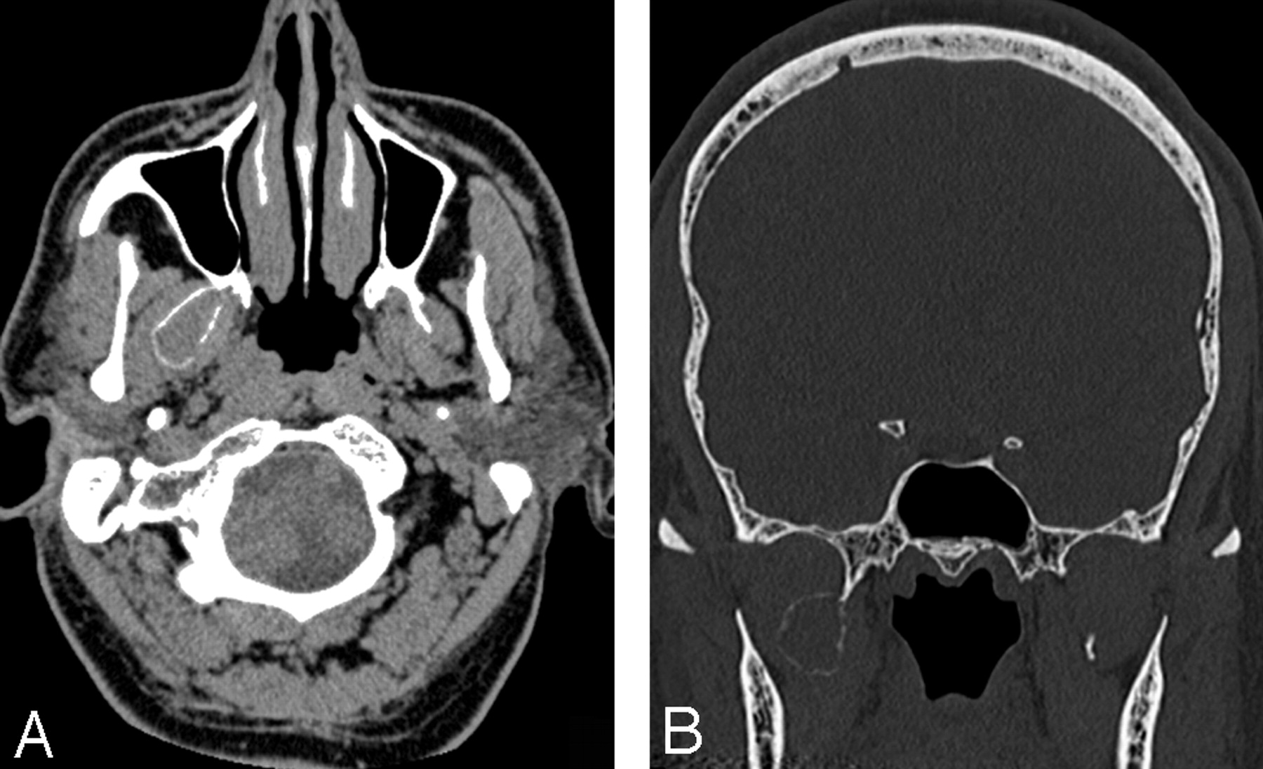
- Fig. 1.
Axial CT scan shows a lytic expansile bone lesion of the greater wing of the sphenoid bone protruding into middle cranial fossa and, to a lesser degree, into the orbit. No discontinuity of the cortex is appreciated. Thickening of extraocular muscles is noted.
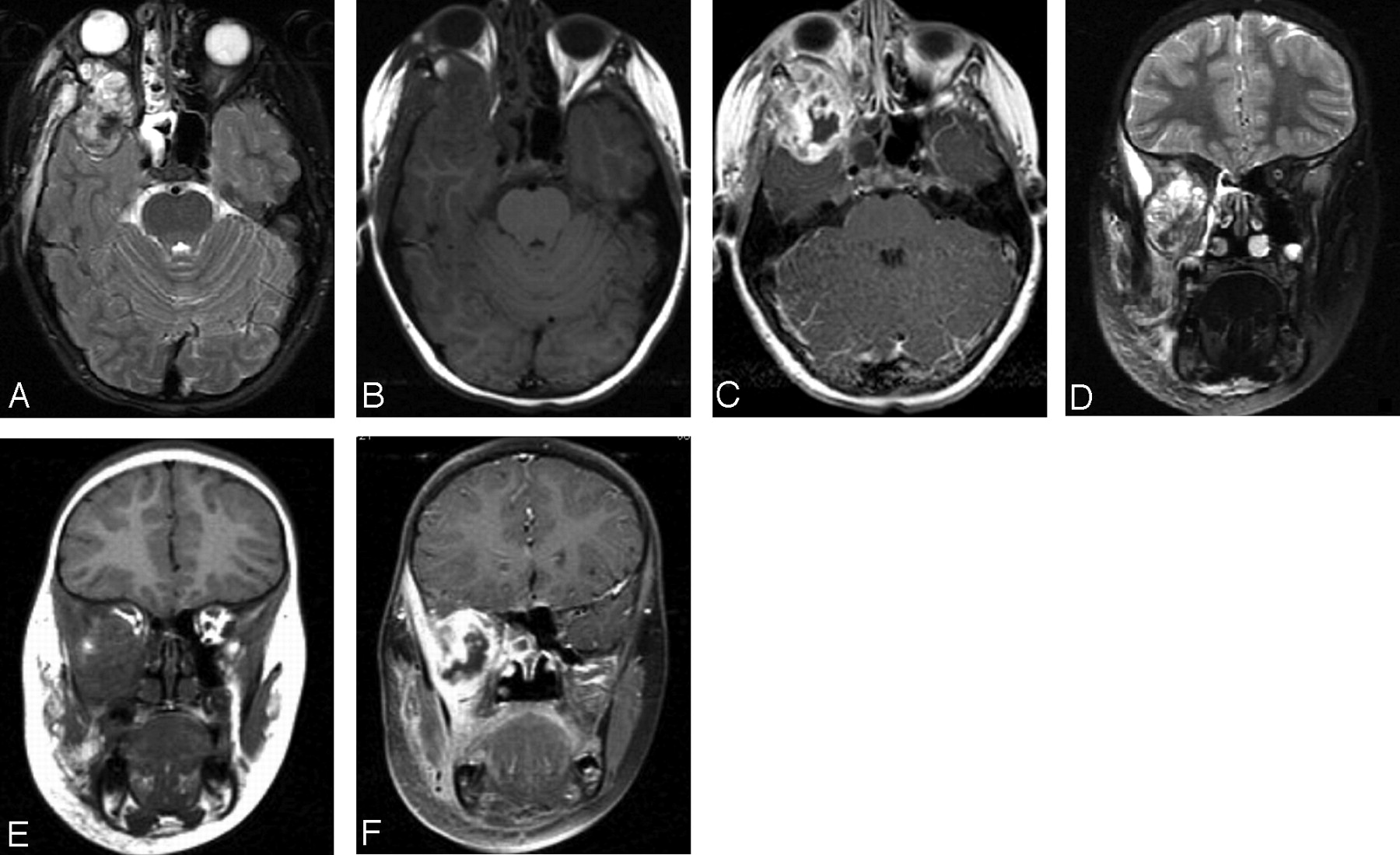
- Fig. 2.
Axial and coronal T2-weighted (A, D), T1-weighted (B, E), and gadolinium-enhanced T1-weighted (C, F) images show a heterogeneously enhancing lesion with extension into the orbit, masticator space, and middle cranial fossa and proptosis of the right eye. Enhancement and edema are also noted within the masticator muscles and surrounding soft tissue. Nonenhancing parts of the lesion with low T2 signal intensity probably represent dense fibrosis identified on the histologic examination.
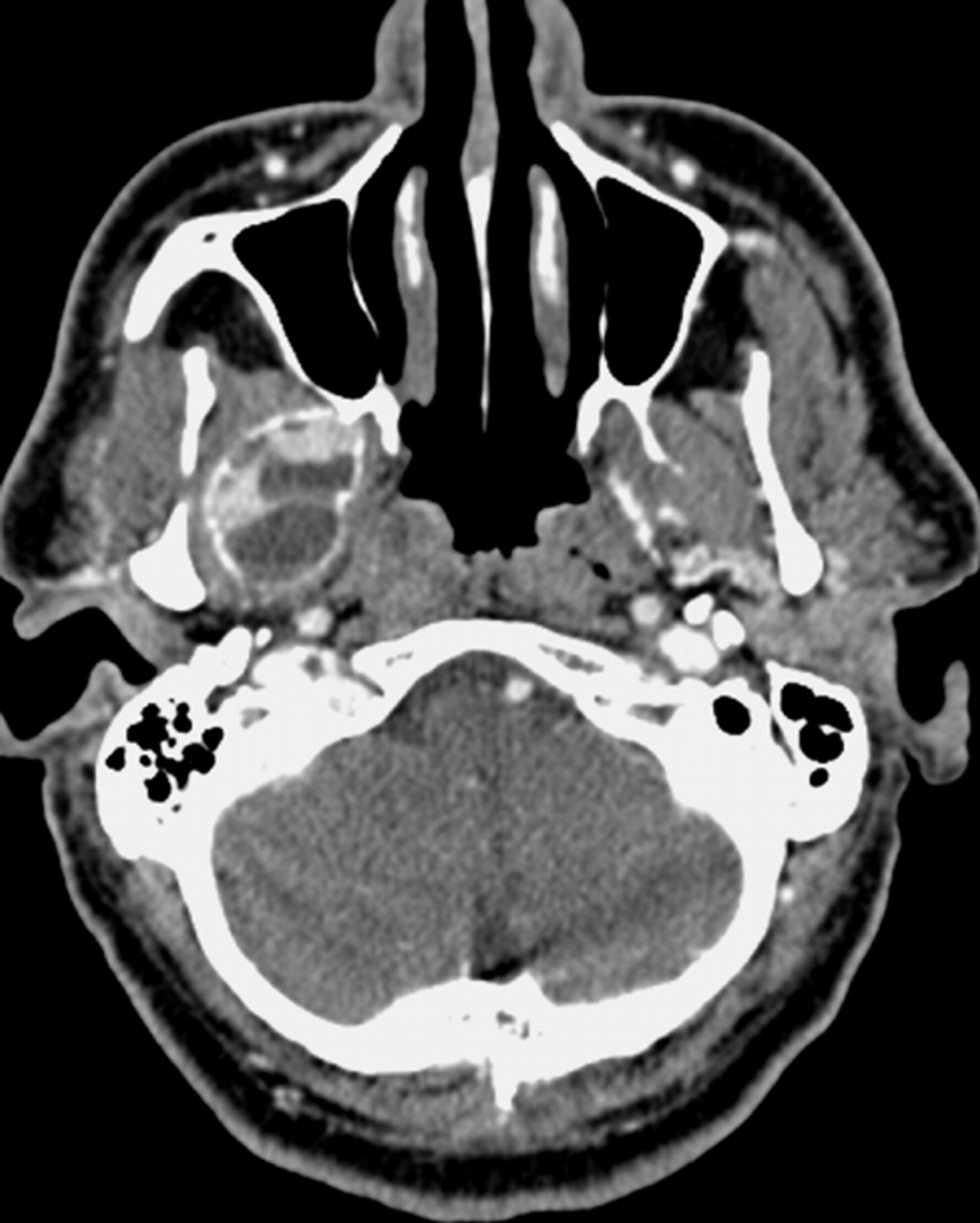
- Fig. 3.
Noncontrast axial (A) and coronal (B) CT images show an expansile bone lesion arising from the lateral plate of the right pterygoid process.
- Fig. 4.
Contrast-enhanced axial CT scan obtained 3 months after Fig 3 shows interval rapid enlargement of the lesion with enhancing soft-tissue component and fluid-fluid levels containing cystic parts.
- Fig. 5.
Axial T1-weighted (A), T2-weighted (B), and postcontrast T1-weighted (C) images demonstrate fluid-fluid levels and an enhancing soft-tissue component. Note that the low T2 signal intensity-enhancing, the enhancing part, which is on the lateral wall of the lesion, likely represents a dense fibrous component rather than hemosiderin.
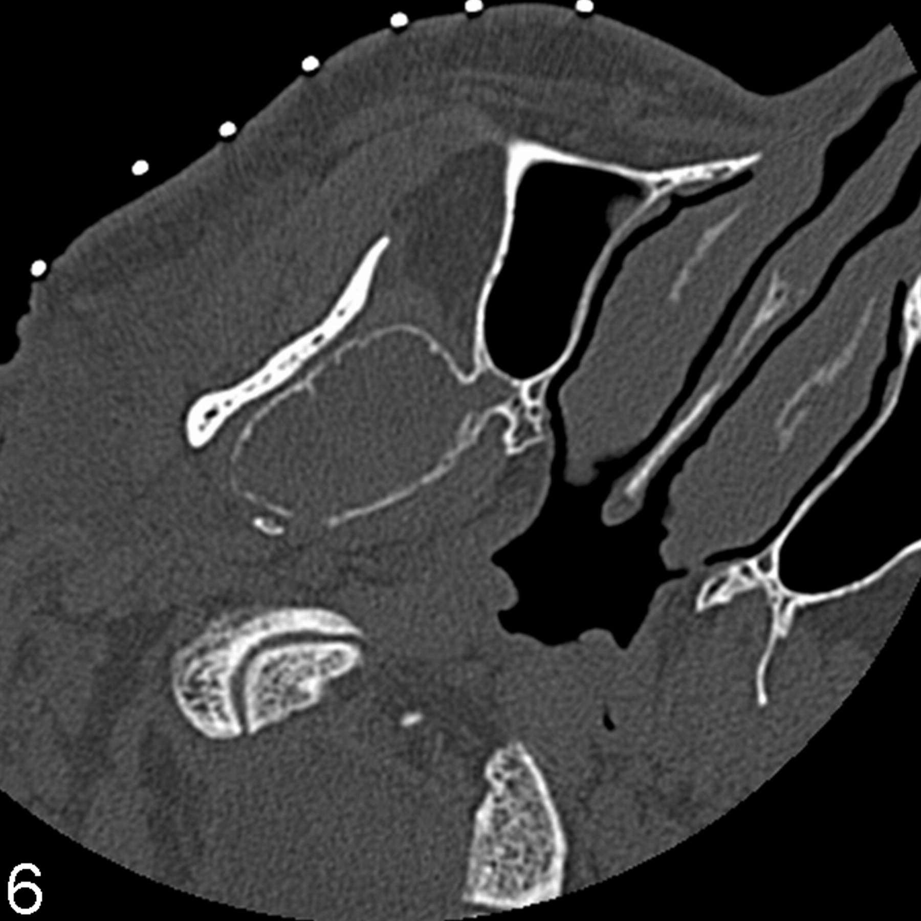
- Fig. 6.
Axial noncontrast image, obtained 1 month after Fig 5 as a part of the imaging-guided biopsy, shows noticeable enlargement of the lesion in the interim. A small cortical breakthrough is seen posteromedially.
- Fig. 7.
Surgical specimen shows multinucleated giant cells interspersed among stromal mononuclear cells in a hemorrhagic background (H&E).


