Advertisement

Main menu

  • Home
  • Content
    • Current Issue
    • Accepted Manuscripts
    • Article Preview
    • Past Issue Archive
    • Video Articles
    • AJNR Case Collection
    • Case of the Week Archive
    • Case of the Month Archive
    • Classic Case Archive
  • Special Collections
    • AJNR Awards
    • ASNR Foundation Special Collection
    • Most Impactful AJNR Articles
    • Photon-Counting CT
    • Spinal CSF Leak Articles (Jan 2020-June 2024)
  • Multimedia
    • AJNR Podcasts
    • AJNR SCANtastic
    • Trainee Corner
    • MRI Safety Corner
    • Imaging Protocols
  • For Authors
    • Submit a Manuscript
    • Submit a Video Article
    • Submit an eLetter to the Editor/Response
    • Manuscript Submission Guidelines
    • Statistical Tips
    • Fast Publishing of Accepted Manuscripts
    • Graphical Abstract Preparation
    • Imaging Protocol Submission
    • Author Policies
  • About Us
    • About AJNR
    • Editorial Board
    • Editorial Board Alumni
  • More
    • Become a Reviewer/Academy of Reviewers
    • Subscribers
    • Permissions
    • Alerts
    • Feedback
    • Advertisers
    • ASNR Home

User menu

  • Alerts
  • Log in

Search

  • Advanced search
American Journal of Neuroradiology

American Journal of Neuroradiology

ASHNR American Society of Functional Neuroradiology ASHNR American Society of Pediatric Neuroradiology ASSR
  • Alerts
  • Log in

Advanced Search

  • Home
  • Content
    • Current Issue
    • Accepted Manuscripts
    • Article Preview
    • Past Issue Archive
    • Video Articles
    • AJNR Case Collection
    • Case of the Week Archive
    • Case of the Month Archive
    • Classic Case Archive
  • Special Collections
    • AJNR Awards
    • ASNR Foundation Special Collection
    • Most Impactful AJNR Articles
    • Photon-Counting CT
    • Spinal CSF Leak Articles (Jan 2020-June 2024)
  • Multimedia
    • AJNR Podcasts
    • AJNR SCANtastic
    • Trainee Corner
    • MRI Safety Corner
    • Imaging Protocols
  • For Authors
    • Submit a Manuscript
    • Submit a Video Article
    • Submit an eLetter to the Editor/Response
    • Manuscript Submission Guidelines
    • Statistical Tips
    • Fast Publishing of Accepted Manuscripts
    • Graphical Abstract Preparation
    • Imaging Protocol Submission
    • Author Policies
  • About Us
    • About AJNR
    • Editorial Board
    • Editorial Board Alumni
  • More
    • Become a Reviewer/Academy of Reviewers
    • Subscribers
    • Permissions
    • Alerts
    • Feedback
    • Advertisers
    • ASNR Home
  • Follow AJNR on Twitter
  • Visit AJNR on Facebook
  • Follow AJNR on Instagram
  • Join AJNR on LinkedIn
  • RSS Feeds

AJNR is seeking candidates for the AJNR Podcast Editor. Read the position description.

Research ArticlePEDIATRICS

Cranial Ultrasound in Metabolic Disorders Presenting in the Neonatal Period: Characteristic Features and Comparison with MR Imaging

L.M. Leijser, L.S. de Vries, M.A. Rutherford, A.Y. Manzur, F. Groenendaal, T.J. de Koning, M. van der Heide-Jalving and F.M. Cowan
American Journal of Neuroradiology August 2007, 28 (7) 1223-1231; DOI: https://doi.org/10.3174/ajnr.A0553
L.M. Leijser
  • Find this author on Google Scholar
  • Find this author on PubMed
  • Search for this author on this site
L.S. de Vries
  • Find this author on Google Scholar
  • Find this author on PubMed
  • Search for this author on this site
M.A. Rutherford
  • Find this author on Google Scholar
  • Find this author on PubMed
  • Search for this author on this site
A.Y. Manzur
  • Find this author on Google Scholar
  • Find this author on PubMed
  • Search for this author on this site
F. Groenendaal
  • Find this author on Google Scholar
  • Find this author on PubMed
  • Search for this author on this site
T.J. de Koning
  • Find this author on Google Scholar
  • Find this author on PubMed
  • Search for this author on this site
M. van der Heide-Jalving
  • Find this author on Google Scholar
  • Find this author on PubMed
  • Search for this author on this site
F.M. Cowan
  • Find this author on Google Scholar
  • Find this author on PubMed
  • Search for this author on this site
  • Article
  • Figures & Data
  • Info & Metrics
  • Responses
  • References
  • PDF
Loading

Article Figures & Data

Figures

  • Tables
  • Fig 1.
    • Download figure
    • Open in new tab
    • Download powerpoint
    Fig 1.

    Ultrasonography images from 1 of 2 siblings with an oxidative phosphorylation disorder.

    A and B, Coronal views showing (A) bilateral marked echogenicity in the lower basal ganglia typical of LSV (arrows) and (B) bilateral increased echogenicity in the white matter (arrows). No MR imaging was obtained in this child.

  • Fig 2.
    • Download figure
    • Open in new tab
    • Download powerpoint
    Fig 2.

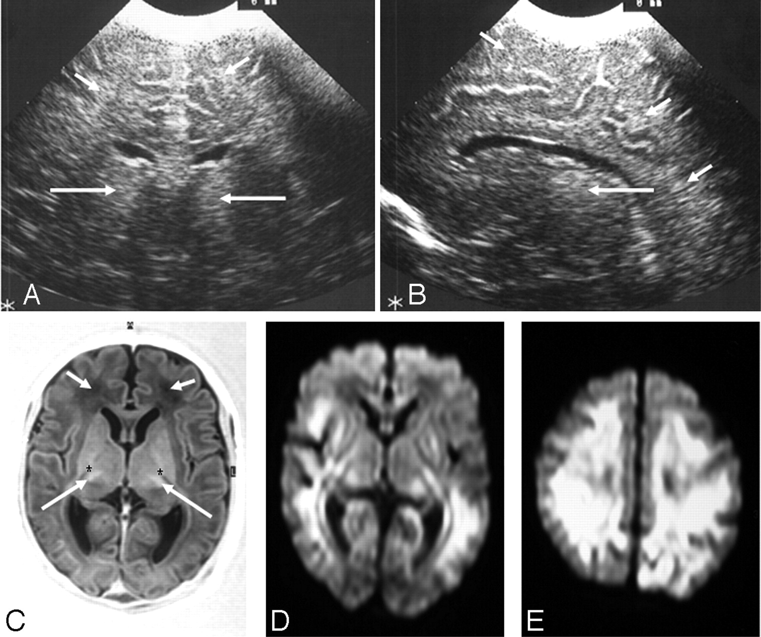
    Ultrasonography (A and B) and MR imaging (C–E) (time interval, 3 days) from an infant with a complex III disorder.

    A, Coronal view showing bilateral echogenicity in the thalami (long arrows) and diffusely increased echogenicity in the subcortical and periventricular white matter (short arrows). B, Parasagittal view showing an echogenic thalamus (long arrow) and diffusely increased echogenicity in the periventricular and subcortical white matter (short arrows).

    C, Axial inversion recovery MR image showing abnormal signal intensity in the thalami (long arrows), absence of normal signal intensity from the internal capsule (*), and abnormal low signal intensity in the white matter (short arrows).

    D and E, Axial diffusion-weighted MR images showing widespread abnormal signal intensity in the periventricular and subcortical white matter and in the lateral basal ganglia and posterior thalami.

  • Fig 3.
    • Download figure
    • Open in new tab
    • Download powerpoint
    Fig 3.

    Ultrasonography (A–C) and MR imaging (D–F) (time interval, 1 day) from an infant with a peroxisomal biogenesis disorder with a Zellweger phenotype.

    A, Coronal view showing GLCs (short arrows), a large cavum septum pellucidum (long arrow), and increased echogenicity in the white matter.

    B, Parasagittal view showing GLCs, cysts, in the choroid plexus (short arrow) and an abnormal appearance to the insula (long arrow).

    C, Extreme parasagittal view showing abnormal development of the Sylvian fissure (arrow) and increased echogenicity in the white matter.

    D and E, Axial inversion recovery and T2-weighted MR images showing a large cavum septum pellucidum, abnormal signal intensity in the frontal white matter (short arrows), and polymicrogyria of the Sylvian fissures (long arrows).

    F, Axial inversion recovery image showing a lack of myelin in the posterior limb of the internal capsule (arrows).

  • Fig 4.
    • Download figure
    • Open in new tab
    • Download powerpoint
    Fig 4.

    Ultrasonography (A–C) and MR imaging (D and E) (time interval, 7 days) from an infant with NKHG.

    A–C, Sagittal views showing (A) a hypoplastic corpus callosum (arrow) and increased echogenicity in the white matter, (B) increased echogenicity in the white matter (arrows), and (C) exaggerated contrast between the white matter and the cortex (arrows).

    D and E, Axial T1- and T2-weighted MR images showing a lack of myelin in the posterior limb of the internal capsule (arrows) and increased T1 and T2 throughout the white matter.

  • Fig 5.
    • Download figure
    • Open in new tab
    • Download powerpoint
    Fig 5.

    Ultrasonography images from an infant with ornithine transcarbamylase deficiency.

    A and B, Coronal views showing straight gyri off the interhemispheric fissure (short arrow), loss of gray/white matter differentiation, and loss of tissue definition in patchy echogenic white matter (long arrow). This child was too unstable to transfer for MR imaging.

  • Fig 6.
    • Download figure
    • Open in new tab
    • Download powerpoint
    Fig 6.

    Ultrasonography (A–C) and MR imaging (D–F) (time interval, 35 days) from an infant with argininosuccinic acid lyase deficiency.

    A, Coronal view showing a thin corpus callosum (long arrow) and widened extracerebral space and interhemispheric fissure (short arrows).

    B, Sagittal view showing LSV (long arrow), increased echogenicity in the white matter most obvious at the trigone (medium arrow), and widened extracerebral space (short arrow).

    C, Sagittal view showing a cyst in the choroid plexus (long arrow) and slightly increased echogenicity in the subcortical white matter (short arrows).

    D, Coronal T1-weighted MR image showing a thin corpus callosum (long arrow) and low signal intensity in the peripheral white matter (short arrow).

    E, Parasagittal T2-weighted MR image showing widespread abnormal signal intensity in the white matter but not the LSV seen on cUS.

    F, Midsagittal T1-weighted MR image showing the small cyst (arrow) also seen on cUS.

  • Fig 7.
    • Download figure
    • Open in new tab
    • Download powerpoint
    Fig 7.

    Ultrasonography (A and B) and MR imaging (C and D) (time interval, 2 days) from an infant with methylmalonic acidaemia.

    A, Coronal view showing straight sulci coming off the interhemispheric fissure (short arrows), bilateral GLCs (medium arrows), LSV (dotted arrow), and slightly widened interhemispheric fissure (long arrow).

    B, Posterior coronal view showing straight sulci (short arrows), slightly widened interhemispheric fissure and extracerebral space (long arrow), and increased echogenicity in the white matter of the trigone (medium arrows).

    C and D, Reconstructed coronal T2-weighted MR images showing features similar to the ultrasonography images except for the LSV (only seen sonographically) and white matter change also seen subcortically on the MR images.

  • Fig 8.
    • Download figure
    • Open in new tab
    • Download powerpoint
    Fig 8.

    Ultrasonography image from an infant with CDG. Sagittal view showing an apparently small cerebellum on visual assessment (arrow). No MR imaging was obtained for this child.

Tables

  • Figures
  • Clinical and cUS and MRI findings in different groups of metabolic disorders

    DiagnosisClinical findingsCharacteristic cUS findingsFindings added by MRI
    Poor responsivenessHypotoniaEarly seizuresPoor feeding
    Oxidative phosphorylation disorders (n = 21; MRI, n = 12)18 (86%)14 (67%)10 (48%)15 (71%)VD (n = 11; 52%)Delayed myelination (n = 3, 25%)
    GLCs; abnormal WM (n = 7; 33%)Abnormal brainstem (n = 2, 17%)
    LSV; widened ECS/IHF (n = 5, 24%)Loss of grey/WM differentiation; large subarachnoid space; intraventricular haemorrhage; abnormal SI in WM; decreased WM volume; abnormal SI in BGT; abnormal hippocampus; abnormal SI in choroid plexus (n = 1, 8%)
    Abnormal cortical folding; small cerebellum; abnormal BGT (n = 4, 19%)
    Thin corpus callosum (n = 3, 14%)
    Peroxisomal biogenesis disorders (n = 13; MRI, n = 9)12 (92%)12 (92%)10 (77%)8 (62%)GLCs; VD (n = 10, 77%)Abnormal/delayed myelination (n = 6, 67%)
    Abnormal cortical folding; LSV (n = 8, 62%)Abnormal SI in WM (n = 3, 33%)
    Abnormal BGT (n = 4, 31%)Migrational disorder; decreased WM volume (n = 1, 11%)
    Absent hin corpus callosum; patent CSP (n = 3, 23%)
    Amino acid metabolism and urea cycle disorders (n = 14; MRI, n = 11)11 (79%)14 (100%)13 (93%)13 (93%)Abnormal cortical folding (n = 9, 64%)Delayed myelination (n = 5, 45%)
    Abnormal WM (n = 8, 57%)Cortical highlighting (n = 3, 27%)
    Abnormal corpus callosum (n = 7, 50%)Punctate WM haemorrhage; abnormal brainstem (n = 2, 18%)
    Unusually shaped lateral ventricles (n = 5, 36%)Abnormal SI in cerebellum; subdural haemorrhage (n = 1, 9%)
    Abnormal BGT (n = 4, 29%)
    Small cerebellum; widened ECS/IHF (n = 2, 14%)
    Organic acid disorders (n = 4; MRI, n = 2)4 (100%)3 (75%)1 (25%)2 (50%)Echogenic periventricular WM (n = 2, 50%)Delayed myelination; abnormal SI in globus pallidus (n = 1, 50%)
    Other disorders (n = 3; MRI, n = 1)3 (100%)1 (33%)0 (0%)0 (0%)Lateral VD (n = 2, 67%)None
    • Note:—Clinical findings are given in number of infants (percentage in group). n indicates number of examinations; VD, ventricular dilatation; GLC, germinolytic cyst; WM, white matter; LSV, lenticulostriate vasculopathy; ECS, extracerebral space; IHF, interhemispheric fissure; BGT, basal ganglia and thalami; SI, signal intensity; CSP, cavum septum pellucidum.

PreviousNext
Back to top

In this issue

American Journal of Neuroradiology: 28 (7)
American Journal of Neuroradiology
Vol. 28, Issue 7
August 2007
  • Table of Contents
  • Index by author
Advertisement
Print
Download PDF
Email Article

Thank you for your interest in spreading the word on American Journal of Neuroradiology.

NOTE: We only request your email address so that the person you are recommending the page to knows that you wanted them to see it, and that it is not junk mail. We do not capture any email address.

Enter multiple addresses on separate lines or separate them with commas.
Cranial Ultrasound in Metabolic Disorders Presenting in the Neonatal Period: Characteristic Features and Comparison with MR Imaging
(Your Name) has sent you a message from American Journal of Neuroradiology
(Your Name) thought you would like to see the American Journal of Neuroradiology web site.
CAPTCHA
This question is for testing whether or not you are a human visitor and to prevent automated spam submissions.
Cite this article
L.M. Leijser, L.S. de Vries, M.A. Rutherford, A.Y. Manzur, F. Groenendaal, T.J. de Koning, M. van der Heide-Jalving, F.M. Cowan
Cranial Ultrasound in Metabolic Disorders Presenting in the Neonatal Period: Characteristic Features and Comparison with MR Imaging
American Journal of Neuroradiology Aug 2007, 28 (7) 1223-1231; DOI: 10.3174/ajnr.A0553

Citation Manager Formats

  • BibTeX
  • Bookends
  • EasyBib
  • EndNote (tagged)
  • EndNote 8 (xml)
  • Medlars
  • Mendeley
  • Papers
  • RefWorks Tagged
  • Ref Manager
  • RIS
  • Zotero
0 Responses
Respond to this article
Share
Bookmark this article
Cranial Ultrasound in Metabolic Disorders Presenting in the Neonatal Period: Characteristic Features and Comparison with MR Imaging
L.M. Leijser, L.S. de Vries, M.A. Rutherford, A.Y. Manzur, F. Groenendaal, T.J. de Koning, M. van der Heide-Jalving, F.M. Cowan
American Journal of Neuroradiology Aug 2007, 28 (7) 1223-1231; DOI: 10.3174/ajnr.A0553
del.icio.us logo Twitter logo Facebook logo Mendeley logo
  • Tweet Widget
  • Facebook Like
  • Google Plus One
Purchase

Jump to section

  • Article
    • Abstract
    • Materials and Methods
    • Results
    • Discussion
    • Conclusion
    • Acknowledgments
    • Footnotes
    • References
  • Figures & Data
  • Info & Metrics
  • Responses
  • References
  • PDF

Related Articles

  • No related articles found.
  • PubMed
  • Google Scholar

Cited By...

  • Concerns about a New Preterm MR Imaging Scoring System
  • Cranial Ultrasonography in Infantile Encephalitic Beriberi: A Useful First-Line Imaging Tool for Screening and Diagnosis in Suspected Cases
  • Is lenticulostriated vasculopathy a sign of central nervous system insult in infants with congenital CMV infection?
  • Lenticulostriate vasculopathy in very preterm infants
  • Patterns of brain injury and outcome in term neonates presenting with postnatal collapse
  • Crossref (52)
  • Google Scholar

This article has been cited by the following articles in journals that are participating in Crossref Cited-by Linking.

  • Patterns of neonatal hypoxic–ischaemic brain injury
    Linda S. de Vries, Floris Groenendaal
    Neuroradiology 2010 52 6
  • Cranial Ultrasonography in Neonates: Role and Limitations
    Gerda van Wezel-Meijler, Sylke J. Steggerda, Lara M. Leijser
    Seminars in Perinatology 2010 34 1
  • Serial brain MRI and ultrasound findings: Relation to gestational age, bilirubin level, neonatal neurologic status and neurodevelopmental outcome in infants at risk of kernicterus
    Konstantina Gkoltsiou, Meropi Tzoufi, Serena Counsell, Mary Rutherford, Frances Cowan
    Early Human Development 2008 84 12
  • Brain imaging findings in very preterm infants throughout the neonatal period: Part I. Incidences and evolution of lesions, comparison between ultrasound and MRI
    Lara M. Leijser, Francisca T. de Bruïne, Sylke J. Steggerda, Jeroen van der Grond, Frans J. Walther, Gerda van Wezel-Meijler
    Early Human Development 2009 85 2
  • Evolving Understanding of Hypoxic-Ischemic Encephalopathy in the Term Infant
    Linda S. de Vries, Frances M. Cowan
    Seminars in Pediatric Neurology 2009 16 4
  • Neonatal neuroimaging findings in inborn errors of metabolism
    Andrea Poretti, Susan I. Blaser, Maarten H. Lequin, Ali Fatemi, Avner Meoded, Frances J. Northington, Eugen Boltshauser, Thierry A.G.M. Huisman
    Journal of Magnetic Resonance Imaging 2013 37 2
  • The aetiology of neonatal seizures and the diagnostic contribution of neonatal cerebral magnetic resonance imaging
    Lauren C Weeke, Floris Groenendaal, Mona C Toet, Manon J N L Benders, Rutger A J Nievelstein, Linda G M van Rooij, Linda S de Vries
    Developmental Medicine & Child Neurology 2015 57 3
  • Neuroimaging Findings of Organic Acidemias and Aminoacidopathies
    Nihaal Reddy, Sonia F. Calloni, Hilary J. Vernon, Eugen Boltshauser, Thierry A. G. M. Huisman, Bruno P. Soares
    RadioGraphics 2018 38 3
  • Imaging the term neonatal brain
    S Todd Sorokan, Ann L Jefferies, Steven P Miller
    Paediatrics & Child Health 2018 23 5
  • Neonatal pyruvate dehydrogenase deficiency due to a R302H mutation in the PDHA1 gene: MRI findings
    João P. Soares-Fernandes, Roseli Teixeira-Gomes, Romeu Cruz, Manuel Ribeiro, Zita Magalhães, Jaime F. Rocha, Lara M. Leijser
    Pediatric Radiology 2008 38 5

More in this TOC Section

  • SyMRI & MR Fingerprinting in Brainstem Myelination
  • Comparison of Image Quality and Radiation Dose in Pediatric Temporal Bone CT Using Photon-Counting Detector CT and Energy-Integrating Detector CT
  • Neonatal Hypocalcemia: Cerebral MRI Abnormalities
Show more Pediatrics

Similar Articles

Advertisement

Indexed Content

  • Current Issue
  • Accepted Manuscripts
  • Article Preview
  • Past Issues
  • Editorials
  • Editor's Choice
  • Fellows' Journal Club
  • Letters to the Editor
  • Video Articles

Cases

  • Case Collection
  • Archive - Case of the Week
  • Archive - Case of the Month
  • Archive - Classic Case

Special Collections

  • AJNR Awards
  • ASNR Foundation Special Collection
  • Most Impactful AJNR Articles
  • Photon-Counting CT
  • Spinal CSF Leak Articles (Jan 2020-June 2024)

More from AJNR

  • Trainee Corner
  • Imaging Protocols
  • MRI Safety Corner

Multimedia

  • AJNR Podcasts
  • AJNR Scantastics

Resources

  • Turnaround Time
  • Submit a Manuscript
  • Submit a Video Article
  • Submit an eLetter to the Editor/Response
  • Manuscript Submission Guidelines
  • Statistical Tips
  • Fast Publishing of Accepted Manuscripts
  • Graphical Abstract Preparation
  • Imaging Protocol Submission
  • Evidence-Based Medicine Level Guide
  • Publishing Checklists
  • Author Policies
  • Become a Reviewer/Academy of Reviewers
  • News and Updates

About Us

  • About AJNR
  • Editorial Board
  • Editorial Board Alumni
  • Alerts
  • Permissions
  • Not an AJNR Subscriber? Join Now
  • Advertise with Us
  • Librarian Resources
  • Feedback
  • Terms and Conditions
  • AJNR Editorial Board Alumni

American Society of Neuroradiology

  • Not an ASNR Member? Join Now

© 2025 by the American Society of Neuroradiology All rights, including for text and data mining, AI training, and similar technologies, are reserved.
Print ISSN: 0195-6108 Online ISSN: 1936-959X

Powered by HighWire