Advertisement

Main menu

  • Home
  • Content
    • Current Issue
    • Accepted Manuscripts
    • Article Preview
    • Past Issue Archive
    • Video Articles
    • AJNR Case Collection
    • Case of the Week Archive
    • Case of the Month Archive
    • Classic Case Archive
  • Special Collections
    • AJNR Awards
    • ASNR Foundation Special Collection
    • Most Impactful AJNR Articles
    • Photon-Counting CT
    • Spinal CSF Leak Articles (Jan 2020-June 2024)
  • Multimedia
    • AJNR Podcasts
    • AJNR SCANtastic
    • Trainee Corner
    • MRI Safety Corner
    • Imaging Protocols
  • For Authors
    • Submit a Manuscript
    • Submit a Video Article
    • Submit an eLetter to the Editor/Response
    • Manuscript Submission Guidelines
    • Statistical Tips
    • Fast Publishing of Accepted Manuscripts
    • Graphical Abstract Preparation
    • Imaging Protocol Submission
    • Author Policies
  • About Us
    • About AJNR
    • Editorial Board
    • Editorial Board Alumni
  • More
    • Become a Reviewer/Academy of Reviewers
    • Subscribers
    • Permissions
    • Alerts
    • Feedback
    • Advertisers
    • ASNR Home

User menu

  • Alerts
  • Log in

Search

  • Advanced search
American Journal of Neuroradiology

American Journal of Neuroradiology

ASHNR American Society of Functional Neuroradiology ASHNR American Society of Pediatric Neuroradiology ASSR
  • Alerts
  • Log in

Advanced Search

  • Home
  • Content
    • Current Issue
    • Accepted Manuscripts
    • Article Preview
    • Past Issue Archive
    • Video Articles
    • AJNR Case Collection
    • Case of the Week Archive
    • Case of the Month Archive
    • Classic Case Archive
  • Special Collections
    • AJNR Awards
    • ASNR Foundation Special Collection
    • Most Impactful AJNR Articles
    • Photon-Counting CT
    • Spinal CSF Leak Articles (Jan 2020-June 2024)
  • Multimedia
    • AJNR Podcasts
    • AJNR SCANtastic
    • Trainee Corner
    • MRI Safety Corner
    • Imaging Protocols
  • For Authors
    • Submit a Manuscript
    • Submit a Video Article
    • Submit an eLetter to the Editor/Response
    • Manuscript Submission Guidelines
    • Statistical Tips
    • Fast Publishing of Accepted Manuscripts
    • Graphical Abstract Preparation
    • Imaging Protocol Submission
    • Author Policies
  • About Us
    • About AJNR
    • Editorial Board
    • Editorial Board Alumni
  • More
    • Become a Reviewer/Academy of Reviewers
    • Subscribers
    • Permissions
    • Alerts
    • Feedback
    • Advertisers
    • ASNR Home
  • Follow AJNR on Twitter
  • Visit AJNR on Facebook
  • Follow AJNR on Instagram
  • Join AJNR on LinkedIn
  • RSS Feeds

AJNR is seeking candidates for the AJNR Podcast Editor. Read the position description.

Research ArticleSPINE

Normal Thickness and Appearance of the Prevertebral Soft Tissues on Multidetector CT

C.A. Rojas, D. Vermess, J.C. Bertozzi, J. Whitlow, C. Guidi and C.R. Martinez
American Journal of Neuroradiology January 2009, 30 (1) 136-141; DOI: https://doi.org/10.3174/ajnr.A1307
C.A. Rojas
  • Find this author on Google Scholar
  • Find this author on PubMed
  • Search for this author on this site
D. Vermess
  • Find this author on Google Scholar
  • Find this author on PubMed
  • Search for this author on this site
J.C. Bertozzi
  • Find this author on Google Scholar
  • Find this author on PubMed
  • Search for this author on this site
J. Whitlow
  • Find this author on Google Scholar
  • Find this author on PubMed
  • Search for this author on this site
C. Guidi
  • Find this author on Google Scholar
  • Find this author on PubMed
  • Search for this author on this site
C.R. Martinez
  • Find this author on Google Scholar
  • Find this author on PubMed
  • Search for this author on this site
  • Article
  • Figures & Data
  • Info & Metrics
  • Responses
  • References
  • PDF
Loading

Article Figures & Data

Figures

  • Tables
  • Fig 1.
    • Download figure
    • Open in new tab
    • Download powerpoint
    Fig 1.

    Midsagittal MDCT image of the cervical spine illustrating the method used to measure the thickness of the PVST.

  • Fig 2.
    • Download figure
    • Open in new tab
    • Download powerpoint
    Fig 2.

    A 24-year-old patient after a motor vehicle crash with closed head injury; no fracture was identified in the cervical spine or craniocervical junction. A, Midsagittal MDCT image of the cervical spine demonstrates abnormal PVST thickening at C1 and C2 (asterisks). B, Correlation with midsagittal short τ inversion recovery MR image obtained the next day confirms the presence of extensive PVST edema and/or hematoma in this region (arrowheads).
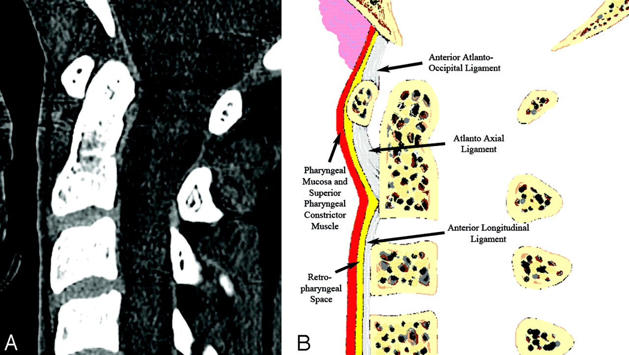
  • Fig 3.
    • Download figure
    • Open in new tab
    • Download powerpoint
    Fig 3.

    Normal appearance of the upper PVST on the midsagittal plane. A, MDCT image demonstrates 2 high-attenuation stripes separated by a central stripe of fat attenuation corresponding to the retropharyngeal/retroesophageal spaces. B, Diagram helps depict the normal anatomic planes of the PVST.
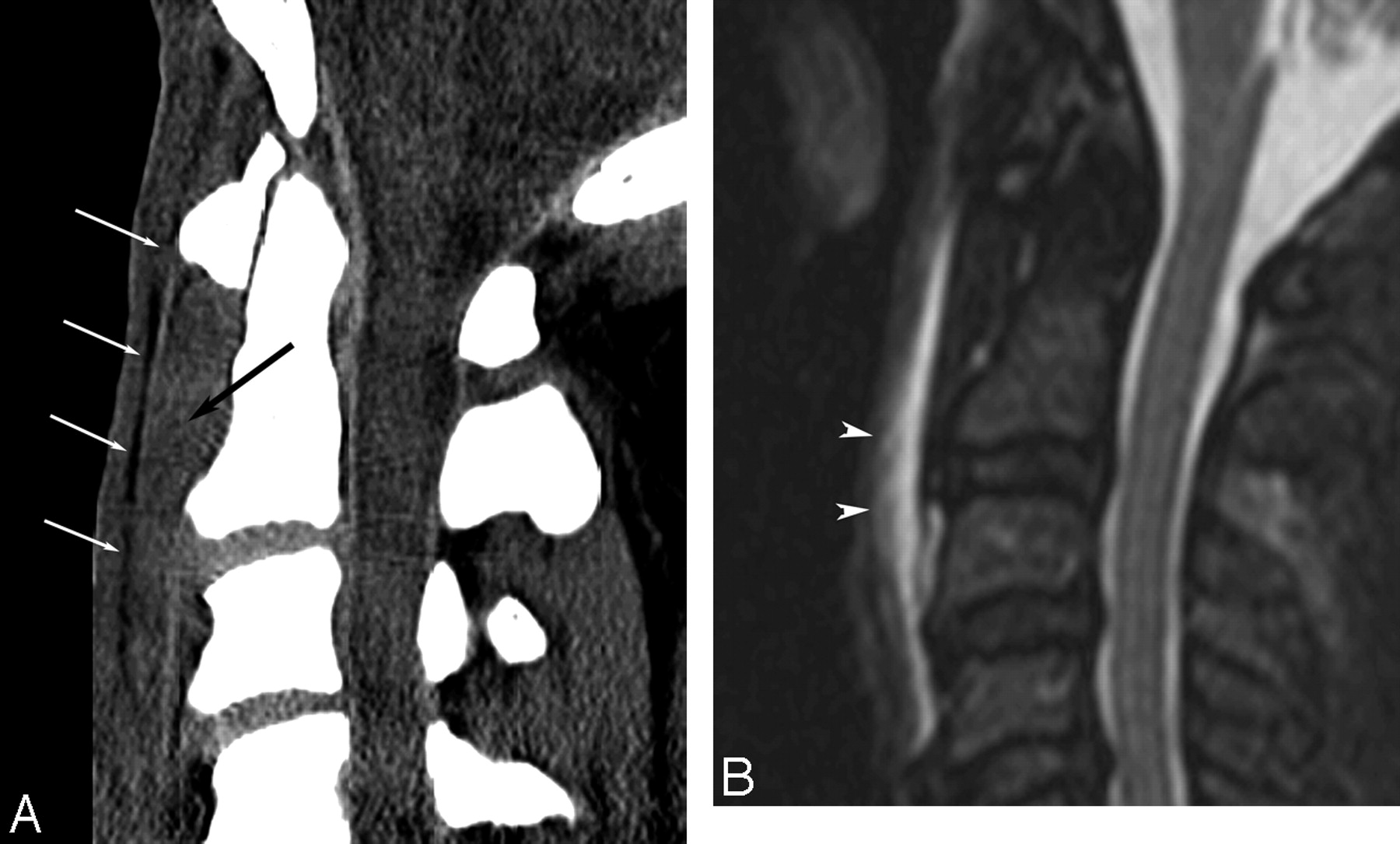
  • Fig 4.
    • Download figure
    • Open in new tab
    • Download powerpoint
    Fig 4.

    A 43- year-old patient after a motor vehicle crash with no osseous injury. A, Midsagittal MDCT image of the cervical spine demonstrates abnormal high attenuation (black arrow) anteriorly displacing the retropharyngeal fat plane (white arrows). B, Short τ inversion recovery MR image obtained the same day shows extensive PVST edema and/or hematoma in this region (arrowheads).

  • Fig 5.
    • Download figure
    • Open in new tab
    • Download powerpoint
    Fig 5.

    A 27-year-old patient after a motor vehicle crash with no osseous injury. A, Midsagittal MDCT image demonstrates normal PVST thickness by using normal values based on MDCT. B, Evaluation of the appearance of the PVST reveals subtle loss of the normal PVST fat plane above the inferior endplate of C3 (arrowhead). C, Correlation with short τ inversion recovery MR image obtained the same day confirms the presence of edema and/or hematoma of the PVST accounting for the MDCT image findings.

Tables

  • Figures
  • Results for mean, range, and upper limits of normal PVST thickness on MDCT*

    Mean PVST Thickness (range) (mm)Upper Limit of Normal PVST Thickness (mm)
    C14.4 (1.5–11)8.5
    C23.7 (1.5–8.5)6
    C34.2 (2–9.5)7
    C47 (2.5–16)N/C
    C512.4 (5–20)N/C
    C613 (5.5–24)18
    C711.6 (2.5–21)18
    • Note:—N/C indicates that upper limits of normal at C4 and C5 were not calculated due to the inconsistent position of the esophagus and larynx among the patient population: PVST, prevertebral soft tissue; MDCT, multidetector CT.

    • * Upper limit of normal calculated as 2 SDs above the mean maximum value for 97.5% of the population.

PreviousNext
Back to top

In this issue

American Journal of Neuroradiology: 30 (1)
American Journal of Neuroradiology
Vol. 30, Issue 1
January 2009
  • Table of Contents
  • Index by author
Advertisement
Print
Download PDF
Email Article

Thank you for your interest in spreading the word on American Journal of Neuroradiology.

NOTE: We only request your email address so that the person you are recommending the page to knows that you wanted them to see it, and that it is not junk mail. We do not capture any email address.

Enter multiple addresses on separate lines or separate them with commas.
Normal Thickness and Appearance of the Prevertebral Soft Tissues on Multidetector CT
(Your Name) has sent you a message from American Journal of Neuroradiology
(Your Name) thought you would like to see the American Journal of Neuroradiology web site.
CAPTCHA
This question is for testing whether or not you are a human visitor and to prevent automated spam submissions.
Cite this article
C.A. Rojas, D. Vermess, J.C. Bertozzi, J. Whitlow, C. Guidi, C.R. Martinez
Normal Thickness and Appearance of the Prevertebral Soft Tissues on Multidetector CT
American Journal of Neuroradiology Jan 2009, 30 (1) 136-141; DOI: 10.3174/ajnr.A1307

Citation Manager Formats

  • BibTeX
  • Bookends
  • EasyBib
  • EndNote (tagged)
  • EndNote 8 (xml)
  • Medlars
  • Mendeley
  • Papers
  • RefWorks Tagged
  • Ref Manager
  • RIS
  • Zotero
0 Responses
Respond to this article
Share
Bookmark this article
Normal Thickness and Appearance of the Prevertebral Soft Tissues on Multidetector CT
C.A. Rojas, D. Vermess, J.C. Bertozzi, J. Whitlow, C. Guidi, C.R. Martinez
American Journal of Neuroradiology Jan 2009, 30 (1) 136-141; DOI: 10.3174/ajnr.A1307
del.icio.us logo Twitter logo Facebook logo Mendeley logo
  • Tweet Widget
  • Facebook Like
  • Google Plus One
Purchase

Jump to section

  • Article
    • Abstract
    • Materials and Methods
    • Results
    • Discussion
    • Conclusions
    • Acknowledgments
    • Footnotes
    • References
  • Figures & Data
  • Info & Metrics
  • Responses
  • References
  • PDF

Related Articles

  • No related articles found.
  • PubMed
  • Google Scholar

Cited By...

  • A 43-year-old man with neck pain and fever
  • Spontaneous retropharyngeal haematoma with direct oral anticoagulant medication
  • Unusual case of acute neck pain: acute calcific longus colli tendinitis
  • Evaluation of the Intervertebral Disk Angle for the Assessment of Anterior Cervical Diskoligamentous Injury
  • Crossref (46)
  • Google Scholar

This article has been cited by the following articles in journals that are participating in Crossref Cited-by Linking.

  • Effect of Retropharyngeal Steroid on Prevertebral Soft Tissue Swelling Following Anterior Cervical Discectomy and Fusion
    Sang-Hun Lee, Ki-Tack Kim, Kyung-Soo Suk, Kyoung-Jun Park, Kyung-Il Oh
    Spine 2011 36 26
  • Predicting Cervical Hyperextension Injury: A Covariance Guided Sine Cosine Support Vector Machine
    Guomin Liu, Wenyuan Jia, Mingjing Wang, Ali Asghar Heidari, Huiling Chen, Yungang Luo, Chengye Li
    IEEE Access 2020 8
  • Airway management in patients with deep neck infections
    Soo Young Cho, Jae Hee Woo, Yoon Jin Kim, Eun Hee Chun, Jong In Han, Dong Yeon Kim, Hee Jung Baik, Rack Kyung Chung
    Medicine 2016 95 27
  • Risk factors for laryngeal penetration-aspiration in patients with acute traumatic cervical spinal cord injury
    Tiina Ihalainen, Irina Rinta-Kiikka, Teemu M. Luoto, Tuomo Thesleff, Mika Helminen, Anna-Maija Korpijaakko-Huuhka, Antti Ronkainen
    The Spine Journal 2018 18 1
  • Normal Pediatric Prevertebral Soft-Tissue Thickness on MDCT
    David Vermess, Carlos A. Rojas, Fawad Shaheen, Pinakpani Roy, Carlos R. Martinez
    American Journal of Roentgenology 2012 199 1
  • Evaluation of CT Changes in the Head and Neck After Cancer Treatment: Development of a Measurement Tool
    Joseph M. Aulino, Elizabeth M. Wulff-Burchfield, Mary S. Dietrich, Sheila H. Ridner, Kenneth J. Niermann, Jie Deng, Bethany A. Rhoten, Jennifer K. Doersam, Lee Ann Jarrett, Kyle Mannion, Barbara A. Murphy
    Lymphatic Research and Biology 2018 16 1
  • Prevertebral Space Invasion in Head and Neck Cancer
    Abdulkadir Imre, Ercan Pinar, Nezahat Erdoğan, Ahmet Ata Ece, Yuksel Olgun, Ibrahim Aladag, Sedat Ozturkcan
    Annals of Otology, Rhinology & Laryngology 2015 124 5
  • Mechanism of Dysphagia after Acute Traumatic Cervical Spinal Cord Injury
    Tetsuo Hayashi, Yuichi Fujiwara, Yuto Ariji, Hiroaki Sakai, Kensuke Kubota, Osamu Kawano, Muneaki Masuda, Yuichiro Morishita, Takeshi Maeda
    Journal of Neurotrauma 2020 37 21
  • Surgical Outcomes of Dysphagia Provoked by Diffuse Idiopathic Skeletal Hyperostosis in the Cervical Spine
    Young Soo Chung, Ho Yeol Zhang, Yoon Ha, Jeong-Yoon Park
    Yonsei Medical Journal 2020 61 4
  • Diagnostic Accuracy of Dual-Layer Spectral CT Using Electron Density Images to Detect Post-Traumatic Prevertebral Hematoma of the Cervical Spine
    Sam Sedaghat, Patrick Langguth, Naomi Larsen, Graeme Campbell, Marcus Both, Olav Jansen
    RöFo - Fortschritte auf dem Gebiet der Röntgenstrahlen und der bildgebenden Verfahren 2021 193 12

More in this TOC Section

  • Bern Score Validity for SIH
  • MP2RAGE 7T in MS Lesions of the Cervical Spine
  • Deep Learning for STIR Spine MRI Quality
Show more Spine

Similar Articles

Advertisement

Indexed Content

  • Current Issue
  • Accepted Manuscripts
  • Article Preview
  • Past Issues
  • Editorials
  • Editor's Choice
  • Fellows' Journal Club
  • Letters to the Editor
  • Video Articles

Cases

  • Case Collection
  • Archive - Case of the Week
  • Archive - Case of the Month
  • Archive - Classic Case

Special Collections

  • AJNR Awards
  • ASNR Foundation Special Collection
  • Most Impactful AJNR Articles
  • Photon-Counting CT
  • Spinal CSF Leak Articles (Jan 2020-June 2024)

More from AJNR

  • Trainee Corner
  • Imaging Protocols
  • MRI Safety Corner

Multimedia

  • AJNR Podcasts
  • AJNR Scantastics

Resources

  • Turnaround Time
  • Submit a Manuscript
  • Submit a Video Article
  • Submit an eLetter to the Editor/Response
  • Manuscript Submission Guidelines
  • Statistical Tips
  • Fast Publishing of Accepted Manuscripts
  • Graphical Abstract Preparation
  • Imaging Protocol Submission
  • Evidence-Based Medicine Level Guide
  • Publishing Checklists
  • Author Policies
  • Become a Reviewer/Academy of Reviewers
  • News and Updates

About Us

  • About AJNR
  • Editorial Board
  • Editorial Board Alumni
  • Alerts
  • Permissions
  • Not an AJNR Subscriber? Join Now
  • Advertise with Us
  • Librarian Resources
  • Feedback
  • Terms and Conditions
  • AJNR Editorial Board Alumni

American Society of Neuroradiology

  • Not an ASNR Member? Join Now

© 2025 by the American Society of Neuroradiology All rights, including for text and data mining, AI training, and similar technologies, are reserved.
Print ISSN: 0195-6108 Online ISSN: 1936-959X

Powered by HighWire