Advertisement

Main menu

  • Home
  • Content
    • Current Issue
    • Accepted Manuscripts
    • Article Preview
    • Past Issue Archive
    • Video Articles
    • AJNR Case Collection
    • Case of the Week Archive
    • Case of the Month Archive
    • Classic Case Archive
  • Special Collections
    • AJNR Awards
    • ASNR Foundation Special Collection
    • Most Impactful AJNR Articles
    • Photon-Counting CT
    • Spinal CSF Leak Articles (Jan 2020-June 2024)
  • Multimedia
    • AJNR Podcasts
    • AJNR SCANtastic
    • Trainee Corner
    • MRI Safety Corner
    • Imaging Protocols
  • For Authors
    • Submit a Manuscript
    • Submit a Video Article
    • Submit an eLetter to the Editor/Response
    • Manuscript Submission Guidelines
    • Statistical Tips
    • Fast Publishing of Accepted Manuscripts
    • Graphical Abstract Preparation
    • Imaging Protocol Submission
    • Author Policies
  • About Us
    • About AJNR
    • Editorial Board
    • Editorial Board Alumni
  • More
    • Become a Reviewer/Academy of Reviewers
    • Subscribers
    • Permissions
    • Alerts
    • Feedback
    • Advertisers
    • ASNR Home

User menu

  • Alerts
  • Log in

Search

  • Advanced search
American Journal of Neuroradiology

American Journal of Neuroradiology

ASHNR American Society of Functional Neuroradiology ASHNR American Society of Pediatric Neuroradiology ASSR
  • Alerts
  • Log in

Advanced Search

  • Home
  • Content
    • Current Issue
    • Accepted Manuscripts
    • Article Preview
    • Past Issue Archive
    • Video Articles
    • AJNR Case Collection
    • Case of the Week Archive
    • Case of the Month Archive
    • Classic Case Archive
  • Special Collections
    • AJNR Awards
    • ASNR Foundation Special Collection
    • Most Impactful AJNR Articles
    • Photon-Counting CT
    • Spinal CSF Leak Articles (Jan 2020-June 2024)
  • Multimedia
    • AJNR Podcasts
    • AJNR SCANtastic
    • Trainee Corner
    • MRI Safety Corner
    • Imaging Protocols
  • For Authors
    • Submit a Manuscript
    • Submit a Video Article
    • Submit an eLetter to the Editor/Response
    • Manuscript Submission Guidelines
    • Statistical Tips
    • Fast Publishing of Accepted Manuscripts
    • Graphical Abstract Preparation
    • Imaging Protocol Submission
    • Author Policies
  • About Us
    • About AJNR
    • Editorial Board
    • Editorial Board Alumni
  • More
    • Become a Reviewer/Academy of Reviewers
    • Subscribers
    • Permissions
    • Alerts
    • Feedback
    • Advertisers
    • ASNR Home
  • Follow AJNR on Twitter
  • Visit AJNR on Facebook
  • Follow AJNR on Instagram
  • Join AJNR on LinkedIn
  • RSS Feeds

AJNR is seeking candidates for the AJNR Podcast Editor. Read the position description.

Research ArticleBRAIN

The Leptomeningeal “Ivy Sign” on Fluid-Attenuated Inversion Recovery MR Imaging in Moyamoya Disease: A Sign of Decreased Cerebral Vascular Reserve?

N. Mori, S. Mugikura, S. Higano, T. Kaneta, M. Fujimura, A. Umetsu, T. Murata and S. Takahashi
American Journal of Neuroradiology May 2009, 30 (5) 930-935; DOI: https://doi.org/10.3174/ajnr.A1504
N. Mori
  • Find this author on Google Scholar
  • Find this author on PubMed
  • Search for this author on this site
S. Mugikura
  • Find this author on Google Scholar
  • Find this author on PubMed
  • Search for this author on this site
S. Higano
  • Find this author on Google Scholar
  • Find this author on PubMed
  • Search for this author on this site
T. Kaneta
  • Find this author on Google Scholar
  • Find this author on PubMed
  • Search for this author on this site
M. Fujimura
  • Find this author on Google Scholar
  • Find this author on PubMed
  • Search for this author on this site
A. Umetsu
  • Find this author on Google Scholar
  • Find this author on PubMed
  • Search for this author on this site
T. Murata
  • Find this author on Google Scholar
  • Find this author on PubMed
  • Search for this author on this site
S. Takahashi
  • Find this author on Google Scholar
  • Find this author on PubMed
  • Search for this author on this site
  • Article
  • Figures & Data
  • Info & Metrics
  • Responses
  • References
  • PDF
Loading

Article Figures & Data

Figures

  • Tables
  • Fig 1.
    • Download figure
    • Open in new tab
    • Download powerpoint
    Fig 1.

    Classification of 4 corticosubcortical regions in each cerebral hemisphere at the level of the body of the lateral ventricle: the region of the ACA, the ant-MCA, the post-MCA, and the PCA.

  • Fig 2.
    • Download figure
    • Open in new tab
    • Download powerpoint
    Fig 2.

    A 47-year-old woman with frequent transient left upper and lower motor weakness. Arrows indicate the ivy sign. The ivy sign score of the right side is 1 in the ACA and PCA regions, respectively, and 2 in the ant-MCA and post-MCA regions, respectively. No ivy sign is seen on the left side. The ivy sign score in individual hemispheres is 6 on the right side and zero on the left side.
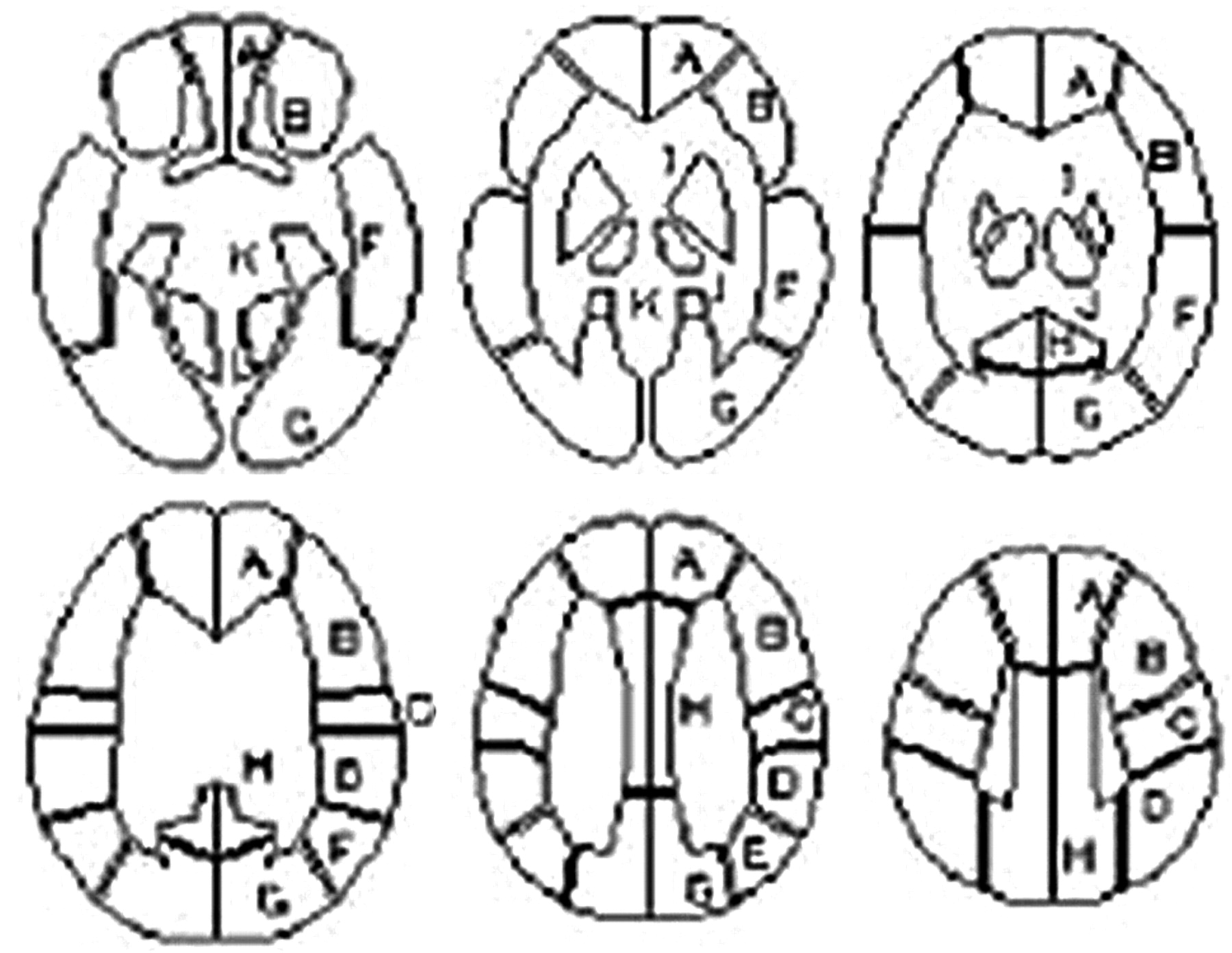
  • Fig 3.
    • Download figure
    • Open in new tab
    • Download powerpoint
    Fig 3.

    The template of constant regions of interest for the 3D-SRT. A representative 6 sections of the template are shown: A, callosomarginal; B, precentral; C, central; D, parietal; E, angular; F, temporal; G, posterior cerebral; H, pericallosal; I, basal ganglia; J, thalamus; K, hippocampus; L, cerebellum. Adapted from Takeuchi R et al.11

  • Fig 4.
    • Download figure
    • Open in new tab
    • Download powerpoint
    Fig 4.

    Bar graph shows the ivy sign score of each type of hemispheric symptom. The ivy sign scores show significant positive correlation with the grades of clinical types of hemispheric symptoms (P < .001).

  • Fig 5.
    • Download figure
    • Open in new tab
    • Download powerpoint
    Fig 5.

    Bar graph shows the ivy sign score according to 4 cortico-subcortical regions, each color indicating a different grade. Each color represents the percentage of the score in the hemisphere compared with all 96 hemispheres for each ivy sign score. The number along the each color represents the score.

  • Fig 6.
    • Download figure
    • Open in new tab
    • Download powerpoint
    Fig 6.

    A 47-year-old woman with minor CSs with left motor weakness and sensory disturbance. The type of hemisphere was CS on the right side and AS on the left side. A, Axial FLAIR image shows the ivy sign over the right hemisphere (arrows). The ivy sign score is 2 in the ACA, ant-MCA, and post-MCA regions, respectively, and zero in PCA region on the right side. There is no ivy sign on the left. The ivy sign score in the individual hemispheres is 6 on the right side and zero on the left side. B, In resting SPECT, CBF is reduced in the ACA, ant-MCA, and post-MCA regions on the right side (arrowheads). C, In ACZ-SPECT, CBF in the left hemisphere is markedly elevated, whereas in the right hemisphere it is not and shows relative depression in the ACA, ant-MCA, and post-MCA regions, which indicates the decreased CVR in the right side (arrowheads). The calculated CVR (%) is 13.7, 8.7, 12.4, and 57.4 from the ACA, ant-MCA, post-MCA, and PCA regions, respectively on the right, whereas it is 46.5, 51.3, 72.1, and 73.9, respectively, on the left. The CVR is relatively preserved in the right PCA region and in all 4 regions on the left side. Note that the areas with decreased CVR correspond to the regions with a prominent ivy sign on the FLAIR image (A). D, Axial FLAIR image obtained at 3 months after revascularization surgery, when her symptoms were alleviated, shows almost complete disappearance of the ivy sign, which was noted over the right hemisphere.

  • Fig 7.
    • Download figure
    • Open in new tab
    • Download powerpoint
    Fig 7.

    A 36-year-old man with Moyamoya disease. Extremely dilated fine pial vasculature and opaque thickened leptomeninges are seen on the brain surface when a craniotomy is performed.

Tables

  • Figures
  • The relationship between the ivy sign score and SPECT findings*

    Ivy Sign Score (No. of Regions)Resting CBF (mL/100 g/min), Median (25th Percentile, 75th Percentile)CVR (%), Median (25th Percentile, 75th Percentile)
    0 (115)33.3 (30.4, 37.2)9.1 (1.8, 30.1)
    1 (37)32.4 (28.6, 35.0)4.3 (−5.7, 21.5)
    2 (40)31.6 (28.2, 33.6)−0.2 (−7.4, 5.3)
    • Note:—SPECT indicates single-photon emission CT; CBE, cerebral blood flow; CVR, cerebral vascular reserve.

    • * As the ivy sign score increased, the median value of the resting CBF decreased slightly (P = .0034). On the other hand, as the ivy sign score increased, the median value of the CVR decreased more obviously (P < .001).

PreviousNext
Back to top

In this issue

American Journal of Neuroradiology: 30 (5)
American Journal of Neuroradiology
Vol. 30, Issue 5
May 2009
  • Table of Contents
  • Index by author
Advertisement
Print
Download PDF
Email Article

Thank you for your interest in spreading the word on American Journal of Neuroradiology.

NOTE: We only request your email address so that the person you are recommending the page to knows that you wanted them to see it, and that it is not junk mail. We do not capture any email address.

Enter multiple addresses on separate lines or separate them with commas.
The Leptomeningeal “Ivy Sign” on Fluid-Attenuated Inversion Recovery MR Imaging in Moyamoya Disease: A Sign of Decreased Cerebral Vascular Reserve?
(Your Name) has sent you a message from American Journal of Neuroradiology
(Your Name) thought you would like to see the American Journal of Neuroradiology web site.
CAPTCHA
This question is for testing whether or not you are a human visitor and to prevent automated spam submissions.
Cite this article
N. Mori, S. Mugikura, S. Higano, T. Kaneta, M. Fujimura, A. Umetsu, T. Murata, S. Takahashi
The Leptomeningeal “Ivy Sign” on Fluid-Attenuated Inversion Recovery MR Imaging in Moyamoya Disease: A Sign of Decreased Cerebral Vascular Reserve?
American Journal of Neuroradiology May 2009, 30 (5) 930-935; DOI: 10.3174/ajnr.A1504

Citation Manager Formats

  • BibTeX
  • Bookends
  • EasyBib
  • EndNote (tagged)
  • EndNote 8 (xml)
  • Medlars
  • Mendeley
  • Papers
  • RefWorks Tagged
  • Ref Manager
  • RIS
  • Zotero
0 Responses
Respond to this article
Share
Bookmark this article
The Leptomeningeal “Ivy Sign” on Fluid-Attenuated Inversion Recovery MR Imaging in Moyamoya Disease: A Sign of Decreased Cerebral Vascular Reserve?
N. Mori, S. Mugikura, S. Higano, T. Kaneta, M. Fujimura, A. Umetsu, T. Murata, S. Takahashi
American Journal of Neuroradiology May 2009, 30 (5) 930-935; DOI: 10.3174/ajnr.A1504
del.icio.us logo Twitter logo Facebook logo Mendeley logo
  • Tweet Widget
  • Facebook Like
  • Google Plus One
Purchase

Jump to section

  • Article
    • Abstract
    • Materials and Methods
    • Results
    • Discussion
    • Conclusions
    • References
  • Figures & Data
  • Info & Metrics
  • Responses
  • References
  • PDF

Related Articles

  • No related articles found.
  • PubMed
  • Google Scholar

Cited By...

  • Reply:
  • The Possible Difference of Underlying Pathophysiologies between "Ivy Sign" on Contrast-Enhanced MRI and FLAIR
  • Ivy Sign in Moyamoya Disease: A Comparative Study of the FLAIR Vascular Hyperintensity Sign Against Contrast-Enhanced MRI
  • FLAIR vascular hyperintensities predict early ischemic recurrence in TIA
  • Elevated Cerebral Blood Volume Contributes to Increased FLAIR Signal in the Cerebral Sulci of Propofol-Sedated Children
  • De Novo Ivy Sign Indicates Postoperative Hyperperfusion in Moyamoya Disease
  • Decrease in Leptomeningeal Ivy Sign on Fluid-Attenuated Inversion Recovery Images after Cerebral Revascularization in Patients with Moyamoya Disease
  • Crossref (104)
  • Google Scholar

This article has been cited by the following articles in journals that are participating in Crossref Cited-by Linking.

  • Imaging of Moyamoya Disease and Moyamoya Syndrome: Current Status
    Jincheng Li, Meiyu Jin, Xiaoyan Sun, Jingying Li, Yumei Liu, Yanli Xi, Qiang Wang, Wenzhuo Zhao, Yanzhi Huang
    Journal of Computer Assisted Tomography 2019 43 2
  • Arterial spin-labeling MR imaging in moyamoya disease compared with SPECT imaging
    Tomoyuki Noguchi, Masatou Kawashima, Hiroyuki Irie, Takateru Ootsuka, Masashi Nishihara, Toshio Matsushima, Sho Kudo
    European Journal of Radiology 2011 80 3
  • Serum miRNA Signature in Moyamoya Disease
    Dongwei Dai, Qiong Lu, Qinghai Huang, Pengfei Yang, Bo Hong, Yi Xu, Wenyuan Zhao, Jianmin Liu, Qiang Li, Balraj Mittal
    PLoS ONE 2014 9 8
  • Clinical implications of the cortical hyperintensity belt sign in fluid-attenuated inversion recovery images after bypass surgery for moyamoya disease
    Eika Hamano, Hiroharu Kataoka, Naomi Morita, Daisuke Maruyama, Tetsu Satow, Koji Iihara, Jun C. Takahashi
    Journal of Neurosurgery 2017 126 1
  • Hyperintense vessels on FLAIR: A useful non-invasive method for assessing intracerebral collaterals
    Wenhua Liu, Gelin Xu, Xuanye Yue, Xiaoliang Wang, Minmin Ma, Renliang Zhang, Handong Wang, Changsheng Zhou, Xinfeng Liu
    European Journal of Radiology 2011 80 3
  • Neuroimaging findings in sickle cell disease
    S C Thust, C Burke, A Siddiqui
    The British Journal of Radiology 2014 87 1040
  • Decrease in Leptomeningeal Ivy Sign on Fluid-Attenuated Inversion Recovery Images after Cerebral Revascularization in Patients with Moyamoya Disease
    M. Kawashima, T. Noguchi, Y. Takase, Y. Nakahara, T. Matsushima
    American Journal of Neuroradiology 2010 31 9
  • Postoperative Cerebral Infarction Risk Factors and Postoperative Management of Pediatric Patients with Moyamoya Disease
    Shinsuke Muraoka, Yoshio Araki, Goro Kondo, Michihiro Kurimoto, Yoshiki Shiba, Kenji Uda, Shinji Ota, Sho Okamoto, Toshihiko Wakabayashi
    World Neurosurgery 2018 113
  • Arterial spin-labeling MR imaging in moyamoya disease compared with clinical assessments and other MR imaging finings
    Tomoyuki Noguchi, Masatou Kawashima, Masashi Nishihara, Tetsuyoshi Hirai, Toshio Matsushima, Hiroyuki Irie
    European Journal of Radiology 2013 82 12
  • De Novo Ivy Sign Indicates Postoperative Hyperperfusion in Moyamoya Disease
    Nobutaka Horie, Minoru Morikawa, Youichi Morofuji, Takeshi Hiu, Tsuyoshi Izumo, Kentaro Hayashi, Izumi Nagata
    Stroke 2014 45 5

More in this TOC Section

  • Usefulness of Quantitative Susceptibility Mapping for the Diagnosis of Parkinson Disease
  • White Matter Alterations in the Brains of Patients with Active, Remitted, and Cured Cushing Syndrome: A DTI Study
  • Qualitative and Quantitative Analysis of MR Imaging Findings in Patients with Middle Cerebral Artery Stroke Implanted with Mesenchymal Stem Cells
Show more BRAIN

Similar Articles

Advertisement

Indexed Content

  • Current Issue
  • Accepted Manuscripts
  • Article Preview
  • Past Issues
  • Editorials
  • Editor's Choice
  • Fellows' Journal Club
  • Letters to the Editor
  • Video Articles

Cases

  • Case Collection
  • Archive - Case of the Week
  • Archive - Case of the Month
  • Archive - Classic Case

Special Collections

  • AJNR Awards
  • ASNR Foundation Special Collection
  • Most Impactful AJNR Articles
  • Photon-Counting CT
  • Spinal CSF Leak Articles (Jan 2020-June 2024)

More from AJNR

  • Trainee Corner
  • Imaging Protocols
  • MRI Safety Corner

Multimedia

  • AJNR Podcasts
  • AJNR Scantastics

Resources

  • Turnaround Time
  • Submit a Manuscript
  • Submit a Video Article
  • Submit an eLetter to the Editor/Response
  • Manuscript Submission Guidelines
  • Statistical Tips
  • Fast Publishing of Accepted Manuscripts
  • Graphical Abstract Preparation
  • Imaging Protocol Submission
  • Evidence-Based Medicine Level Guide
  • Publishing Checklists
  • Author Policies
  • Become a Reviewer/Academy of Reviewers
  • News and Updates

About Us

  • About AJNR
  • Editorial Board
  • Editorial Board Alumni
  • Alerts
  • Permissions
  • Not an AJNR Subscriber? Join Now
  • Advertise with Us
  • Librarian Resources
  • Feedback
  • Terms and Conditions
  • AJNR Editorial Board Alumni

American Society of Neuroradiology

  • Not an ASNR Member? Join Now

© 2025 by the American Society of Neuroradiology All rights, including for text and data mining, AI training, and similar technologies, are reserved.
Print ISSN: 0195-6108 Online ISSN: 1936-959X

Powered by HighWire